Bone-Making Ability of Amniotic Epithelial Cells


Human amniotic epithelial cells (hAECs) develop from the embryo as early as eight days after fertilization. hAECs have been shown to possess remarkable plasticity, or the ability to form many cell types. Mattioli and others showed that AECs from sheep have capacity to differentiate into bone cells (see Cell Biol Int 2012;36:7-19). Therefore, it is possible that these cells could be developed into a source of cells for regenerative treatments.

Steve Shen from the Shanghai Jiao Tong University School of Medicine and his coworkers have examined the ability of cultured hAECs to make bone in culture, and then applied their techniques to an animal system to test the ability of cultured hAECs to restore tooth sockets and facial bone.

To begin, hAECs were isolated from healthy mothers who had undergone childbirth by cesarean section. These cells were cultured in a standard cell culture medium (DMEM/F12 for those who are interested), expanded in culture, and then subjected to flow cytometry to isolate those cells with all the right cell surface markers.

These cells wee then placed into a specialized culture medium designed to convert mesenchymal stem cells into bone-making cells. This osteogenic induction medium was changed every three days for 21 days.  The figure below shows how well the bone-induction worked.  A red stain called alizarin red binds to calcium and therefore stains forming bone matrices rather well.  As you can see in the panel labeled “G” below, the hAECs grown in the osteogenic medium stain red rather well, which shows that they are making bone.
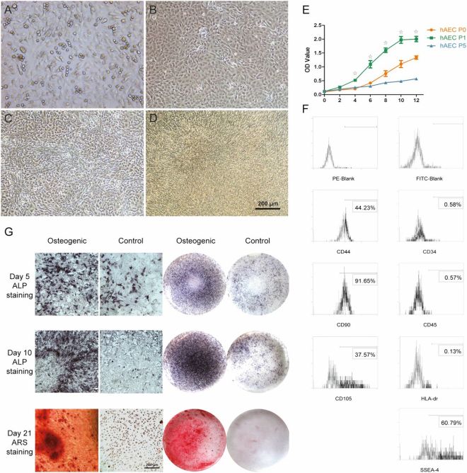
Characterization of hAECs in vitro. (A, B): hAECs at passages 0 and 1 displayed a cobblestone-like morphology. (C): Some hAECs changed into fibroblast-like cells after 7 days of osteoblastic culture. (D): hAECs showed significant morphological changes and settled on superimposed layers after 21 days of osteoblastic culture. (E): Cell proliferation of hAECs at passage 1 was significantly higher than at passages 0 and 5 from day 4 to day 12; hAECs at passage 5 displayed the lowest proliferation rate from day 6 to day 14 (☆, p < .05). (F): Flow cytometry analysis showed that hAECs expressed CD44, CD90, CD105, and SSEA-4 and did not express CD34, CD45, and HLA-DR. Values represent the percentages of all assessed cells positively stained by the indicated antigens (bottom of each graph). Nonspecific fluorescence was determined as the blank control using isotype-matched monoclonal antibodies (PE blank, FITC blank). (G): Representative images of microscopic and general photographs for ALP and ARS staining in osteogenic and control groups indicated the osteogenic differentiation of hAECs in vitro. Scale bar = 200 μm. Abbreviations: ALP, alkaline phosphatase; ARS, alizarin red S; FITC, fluorescein isothiocyanate; hAEC, human amniotic epithelial cell; OD, optical density; p, passage; PE, phycoerythrin.

When hAECs were subjected to gene expression assays, it was clear that the cells grown in the osteogenic medium expressed a whole host of bone-specific genes.  Therefore, the formation of bone was not a fluke, since these cells not only concentrated calcium, but also increased their expression of bone-specific genes (these include osterix, Runx2, Alkaline phosphatase, Collagen I, and osteoprotegerin).

Additionally, those hAECs grown in osteogenic medium stopped forming sheets of cells and began to grow as individual cells.  This is an important transformation for the synthesis of bone because bone-making cells secrete a bone-specific protein matrix upon which calcium phosphate-based crystals deposit to form bone.  If the cells were organized in sheets, then bone deposition would not be possible.  When isolated from amniotic membranes, hAECs grow as sheets of cells that are known as epithelia.  In order to become bone-making cells, the hAECs must undergo an “epithelial-to-mesenchymal transformation” or EMT.  This requires turning on new genes and turning off others.  Gene expression assays of hAECs grown in the osteogenic medium, show extensive evidence of EMT.  For example, a protein called E-cadherin is essential for cells growing in sheets, because it helps cells stick to each other.  However, in hAECs grown in the osteogenic medium, E-cadherin expression was quite low.  Also, a protein called vimentin is highly expressed during EMT, and the hAECs grown in osteogenic medium showed high expression of vimentin.  Thus, these hAECs were undergoing all the necessary changes in order to become bone-making cells,, making all the right genes, and made bone in culture to boot.

This is certainly interesting, but can these hAECs repair bone in a living animal?  Shen’s group tried that very experiment.  The hAECs that were grown in the osteogenic medium were loaded on tricalcium phosphate scaffolds and implanted into rodents with tooth socket lesions.  Control animals were implanted with tricalcium phosphate scaffolds without cells.  The scaffold with cells significantly increased bone formation in the rodents, and showed much more infilling with mineralized tissue.  There was also extensive evidence of the formation of new vasculature and wandering cells called macrophages that are important for the degradation of the implanted scaffold required for new bone formation.  Tissue samples were examined 4 to 8 weeks after the implants were placed.

In vivo healing process in alveolar defect at 4 and 8 weeks postoperatively. (A): Representative three-dimensional micro-computed tomography (CT) reconstruction images of hAECs+β-TCP scaffold (EXP) and β-TCP scaffold (CTR) at 4 and 8 weeks postoperatively. Scale bar = 1 mm. (B): Micro-CT parameters acquired among β-TCP scaffold in vitro, hAECs+β-TCP scaffold in vivo, and β-TCP scaffold alone in vivo at 4 and 8 weeks postoperatively (☆, p < .05). (C): Hematoxylin and eosin staining of the rat alveolar defect at 4 and 8 weeks postoperatively revealed more active new bone formation in the EXP group than in the CTR group (×50 and ×200 magnification). Alveolar defect treated with hAECs+β-TCP scaffold exhibited a more mature lamellae-bone formation at 8 weeks postoperatively. Scale bar = 200 μm. (D): ANA-positive cells, visible as green fluorescence in the nuclei, were observed within the newly deposited OCN, and OPN-positive bone tissue, visible as red fluorescence, in the EXP group at 4 weeks postoperatively, indicating a mature osteoblastic function of these hAEC-derived cells. Scale bar = 50 μm. (E): Representative images of immunohistochemical staining of sections with anti-VEGF antibody and anti-CD68 antibody in hAECs+β-TCP scaffold (EXP) and β-TCP scaffold (CTR) at 4 and 8 weeks postoperatively. Scale bar = 200 μm. (F): The histomorphometric quantification of the relative new bone area, VEGF-positive area, and CD68-positive area showed that more bone tissue regeneration was observed in the EXP group than in the CTR group at 4 and 8 weeks postoperatively. The positive signal of VEGF and CD68 in the EXP group was much weaker at 4 weeks postoperatively and became more intense at 8 weeks postoperatively compared with the CTR group (☆, p < .05). Abbreviations: ANA, anti-nuclear antibody; BV/TV, bone volume/tissue volume ratio; BMD, bone mineralization density; hAECs, human amniotic epithelial cells; OPN, osteopontin; post op, postoperatively; SMI, structure model index; Tb.Th., trabecular thickness; Tb.N., trabecular number; Tb.Sp., trabecular separation; β-TCP, β-tricalcium phosphate; VEGF, vascular endothelial growth factor.

hAECs are typically not recognized by the immune system as foreign and they also have anti-scarring capabilities.  Not only can they effectively form bone in culture, but they can also repair an alveolar defect in a rodent model.  Clearly these cells show promise for clinical applications.  However, before they can be used in the clinic more has to be known about their culture conditions, how many cells are required for transplantation, the best cell dose, at what passages they need to be used, and so on.  Thus, this paper represents a good start for hAECs, but they have to be better understood before they can come to the clinic.