Yale Scientists Find Marker for High-Quality Induced Pluripotent Stem Cells


Pluripotent stem cells can be made by genetically engineering adult cells into less mature cells that have pluripotency. These induced pluripotent stem cells or iPSCs can potentially differentiate into any cell type in the adult body and because they are made from the patient’s own cells, they have a lower risk of being rejected by the patient’s immune system.

However, iPSCs suffer from an increased mutation rate when they are made and these increased mutation rate increases their risk of causing tumors and being rejected by the patient’s immune system. Having said that, not all iPSCs are created equal, and the safety of iPSCs seems to be very line-specific. Thus, how do you know a good stem cell from a bad one?

Yale Stem Cell Center researchers led by Andrew Xiao Yale have published a report in the Sept. 4 issue of Cell Stem Cell in which they describe an indicator that seems to predict which batch of personalized stem cells will differentiate into patient-specific tissue types and which will develop into unusable placental or tumor-like tissues.

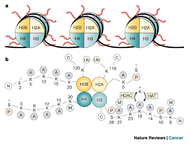
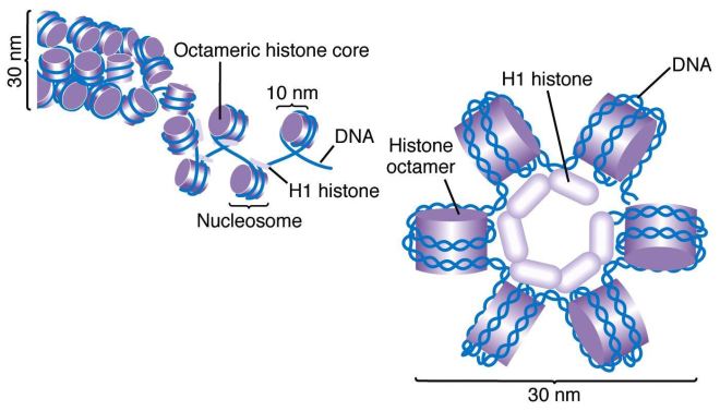
Xiao’s group identified a variant histone protein called H2A.X that seems to predict the developmental path of iPSC cells in mice. Histone proteins assemble into tiny spools around which DNA winds. This DNA spooling allows cells to tightly package their DNA into a tight, compact structure that is easily stored called “chromatin.” Histones that are commonly used include histones H2A, H2B, H3 and H4.

Core histones

Two copies of each of these proteins assemble into a globular structure called a core histone and the DNA of the cell winds around this core histone to form a “nucleosome.” Then linker histones (H1 or H5) take these nucleosomes package them into spiraled coils.

DNA solenoids

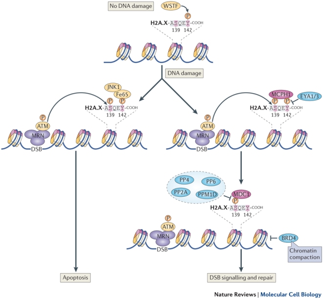
H2A.X is a variant version of histone H2A is modified when DNA damage occurs. Modified H2A.X signals to the DNA repair machinery to fix the broken DNA (see TT Paull, and others, Curr. Biol. 10(15):886–95).

nrm3659-f3

According to the data from Xiao’s research team, in pluripotent stem cells, H2A.X is specifically targeted to those genes typically expressed in cells used to make the placenta, and it helps suppress differentiation of pluripotent stem cells into cells of the placental lineage. Given this distribution in mouse embryonic stem cells, H2A.X deposition pattern is a functional marker of the quality of iPSCs. Conversely, defective H2A.X deposition predisposes iPSCs toward differentiating into placental-type cells and tumors.

“The trend is to raise the standards and quality very high, so we can think about using these cells in clinic,” Xiao said. “With our assay, we have a reliable molecular marker that can tell what is a good cell and what is a bad one.”
Histones Might Hold the Key to the Generation of Totipotent Stem Cells


Reprogramming adult cells into pluripotent stem cells remains a major challenge to stem cell research. The process remains relatively inefficient and slow and a great deal of effort has been expended to improve the speed, efficiency and safety of the reprogramming procedure.

Researchers from RIKEN in Japan have reported one piece of the reprogramming puzzle that can increase the efficiency of reprogramming. Shunsuke Ishii and his colleagues from RIKEN Tsukuba Institute in Ibaraki, Japan have identified two variant histone proteins that dramatically enhance the efficiency of induced pluripotent stem cell (iPS cell) derivation. These proteins might be the key to generating iPS cells.

Terminally-differentiated adult cells can be reprogrammed into a stem-like pluripotent state either by artificially inducing the expression of four factors called the Yamanaka factors, or as recently shown by shocking them with sublethal stress, such as low pH or pressure. However, attempts to create totipotent stem cells capable of giving rise to a fully formed organism, from differentiated cells, have failed.  However, a paper recently published in the journal Nature has shown that STAP or stimulus-triggered acquisition of pluripotency cells from mouse cells have the capacity to form placenta in culture and therefore, are totipotent.

The study by Shunsuke Ishii and his RIKEN colleagues, which was published in the journal Cell Stem Cell, attempted to identify molecules in mammalian oocytes (eggs) that induce the complete reprograming of the genome and lead to the generation of totipotent embryonic stem cells. This is exactly what happens during normal fertilization, and during cloning by means of the technique known as Somatic-Cell Nuclear Transfer (SCNT). SCNT has been used successfully to clone various species of mammals, but the technique has serious limitations and its use on human cells has been controversial for ethical reasons.

Ishii’s research group focused on two histone variants named TH2A and TH2B, which are known to be specific to the testes where they bind tightly to DNA and influence gene expression.

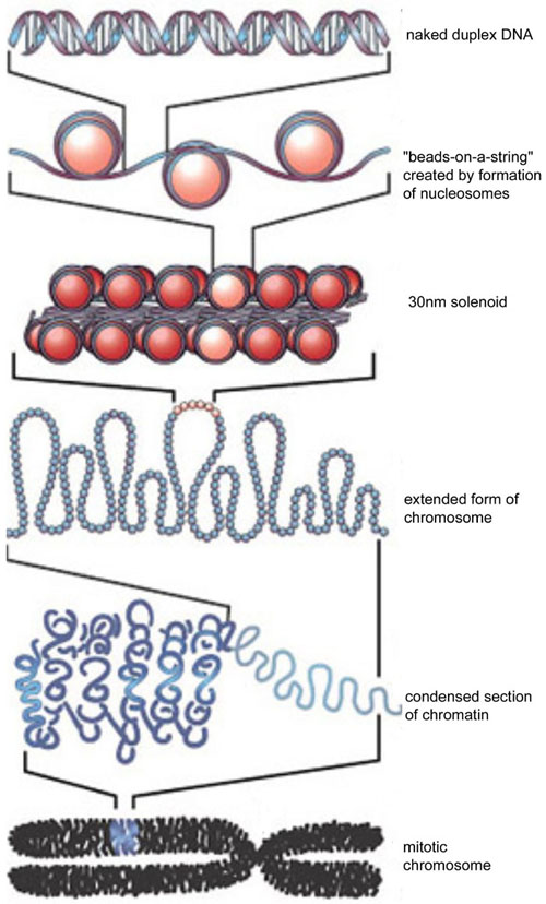
Histones are proteins that bind to DNA non-specifically and act as little spool around which the DNA winds.  These little wound spools of DNA then assemble into spirals that form thread-like structures.  These threads are then looped around a protein scaffold to form the basic structure of a chromosome.  This compacted form of DNA is called “chromatin,” and the DNA is compacted some 10,000 to 100,000 times.  Histones are the main arbiters of chromatin formation.  In the figure below, you can see that the “beads on a string” consist of histones with DNA wrapped around them.

DNA_to_Chromatin_Formation

There are five “standard” histone proteins: H1, H2A, H2B, H3, and H4.  H2A, H2B, H3 and H4 form the beads and the H1 histone brings the beads together to for the 30nm solenoid.  Variant histones are different histones that assemble into beads that do not wrap the DNA quite as tightly or wrap it differently than the standard histones.  Two variant histones in particular, TH2A and TH2B, tend to allow DNA wrapped into chromatin to form and more loosely packed structure that allows the expression of particular genes.

When members of Ishii’s laboratory added these two variant histone proteins, TH2A/TH2B, to the Yamanaka cocktail (Oct4, c-Myc, Sox2, and Klf4) to reprogram mouse fibroblasts, they increased the efficiency of iPSC cell generation about twenty-fold and the speed of the process two- to threefold. In fact, TH2A and TH2B function as substitutes for two of the Yamanaka factors (Sox2 and c-Myc).

Ishii and other made knockout mice that lacked the genes that encoded TH2A and TH2B. This work demonstrated that TH2A and TH2B function as a pair, and are highly expressed in oocytes and fertilized eggs. Furthermore, these two proteins are needed for the development of the embryo after fertilization, although their levels decrease as the embryo grows.

Graphical Abstract1 [更新済み]

In early embryos, TH2A and TH2B bind to DNA and induce an open chromatin structure in the paternal genome (the genome of sperm cells), which contributes to its activation after fertilization.

These results indicate that TH2A/TH2B might induce reprogramming by regulating a different set of genes than the Yamanaka factors, and that these genes are involved in the generation of totipotent cells in oocyte-based reprogramming as seen in SCNT.

“We believe that TH2A and TH2B in combination enhance reprogramming because they introduce a process that normally operates in the zygote during fertilization and SCNT, and lead to a form of reprogramming that bears more similarity to oocyte-based reprogramming and SCNT” explains Dr. Ishii.

Polycomb Proteins Pave the Way for Proper Stem Cell Differentiation


Embryonic stem cells have the ability to differentiate into one of the more than 200 cell types. Differentiation requires a strictly regulated program of gene expression that turns certain genes on at specific times and shuts other genes off. Loss of this regulatory circuit prevents stem cells from properly differentiating into adult cell types, and an inability to differentiate has also been linked to the onset of cancer.

Researchers at the BRIC, University of Copenhagen have identified a crucial role of the molecule Fbx110 in embryonic stem cell differentiation. Kristian Helin from the BRIC said, “Our new results show that this molecule is required for he function of one of the most important molecular switches that constantly regulated the activity of our genes. If Fbx110 is not present in embryonic stem cells, the cells cannot differentiate properly and this can lead to developmental defects.”

What is the function of Fbx110? Fbx110 recruits members of the “Polycomb” gene family to DNA. Polycomb proteins, in particular PRC1 & 2, are known to modulate the structure of chromatin, even though they do not bind DNA. Fbx110,, however binds DNA, but it also binds PRC1 . Therefore, Fbx110 seems to serve as an adapter that recruits Polycomb proteins to DNA.

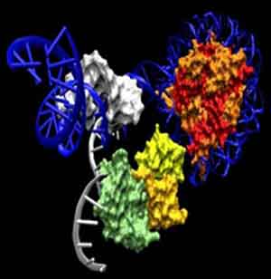
Polycomb proteins bound to nucleosomes

Postdoctoral fellow Xudong Wu, who led the experimental part of this investigation, said, “Our results show that Fbx110 is essential for recruiting PRC1 to genes that are to be silenced in embryonic stem cells. Fbx110 binds directly to DNA and to PRC1, and this way it serves to bring PRC1 to specific genes. When PRC1 is bound to DNA it can modify the DNA associated proteins, which lead to silencing of the gene to which it binds.”

Timing of gene activity is crucial during development and must be maintained throughout the lifespan of any cell. Particular genes are active at a certain times and inactive at other times, and PRC1 seems to be part of the reason for this coordination of gene activity. PRC1 is dynamically recruited to and dissociates from genes according to the needs of the organism.

When cancer arises, this tight regulation of gene activity is often lost and the cells are locked in an inchoate state. This loss of terminal differentiation causes increases cell proliferation and the accumulation of other mutations that allow the cancer cells to undergo continuous self-renewal through endless cell divisions. Such an ability is denied to mature cells because of their tightly controlled programs of gene expression.

Wu added, “Given the emerging relationship between cancer and stem cells, our findings may implicate that an aberrant activity of Fbx110 can disturb PRC function and promote a lack of differentiation in our cells. This makes it worth studying whether blocking the function of Fbx110 could be a strategy for tumor therapy.”

In collaboration with a biotech company called EpiTherapeutics, the BRIC researchers want to develop Fbx110 inhibitors as potential novel therapies for cancer.

A Gene that Prevents Induced Pluripotent Stem Cell Formation Linked to Cancer Severity


A Mount Sinai research team has published some remarkable observations in the journal Nature Communications. Emily Bernstein, PhD, and her team at Mount Sinai have discovered a particular protein that prevents normal cells from being reprogrammed into induced pluripotent stem cells (iPSCs). Since iPSCs resemble embryonic stem cells, these data might provide significant insights into how cells lose their plasticity during normal development, which has wide-reaching implications for how cells change during both normal and disease development.

Previously, Bernstein and others showed that during the formation of particular tumors known as melanomas in mice and human patients, the loss of a specific histone variant called macroH2A (a protein that helps package DNA) correlated rather strongly to the growth and metastasis of the tumor. In this current study, Bernstein and her team determined if macroH2A acted as a barrier to cellular reprogramming during the derivation of iPSCs (see Costanzi C, Pehrson JR (1998). “Histone macroH2A1 is concentrated in the inactive X chromosome of female mammals”. Nature 393 (6685): 599–601).

In collaboration with researchers at the University of Pennsylvania, Bernstein evaluated mice that had been genetically engineered to lack macroH2A. When skin cells were used from macroH2A(-) mice were used to make iPSCs and compared with skin cells from macroH2A(+) mice, the cells from macroH2A(-) mice that lacked macroH2A were much more plastic and were much more easily reprogrammed into iPSCs compared to the wild-type or macroH2A(+) mice. This indicates that macroH2A may block cellular reprogramming by silencing genes required for plasticity.

Bernstein, who is an Assistant Professor of Oncological Sciences and Dermatology at the Graduate School of Biomedical Sciences at Mount Sinai, and corresponding author of the study, said: “This is the first evidence of the involvement of a histone variant protein as an epigenetic barrier to induced pluripotency (iPS) reprogramming.” She continued: “These findings help us to understand the progression of different cancers and how macroH2A might be acting as a barrier to tumor development.”

In their next group of experiments, Bernstein and her team plan to create cancer cells in a culture dish by inducing mutations in genes that are commonly abnormal in particular types of cancer cells and then couple those mutations to the removal of macroH2A to examine whether the cells are capable of forming tumors.