Stem Cells from Baby Teeth Regenerate Dental Pulp after Implantation into Injured Teeth


Going to the dentist is usually not anyone’s idea of fun.  In particular, root canals are no fun.  However, if you have an abscessed tooth that hurts like the dickens, then a root canal may be your best bet for resuming normalcy.

In younger patients, the innermost part of the tooth, the pulp, may die off for a variety of reasons.  This phenomenon, known as “pulp necrosis” arrests root development and may result in tooth loss.  Injury to the pulp as a result of trauma, inflammation, tooth decay, or infection can result in the irreversible loss of teeth.

Tooth-Anatomy

Regenerating dental pulp has proven to be a bear.  Getting the mass of blood vessels and nerves to regrow is not straight forward. However, teeth, fortunately, are blessed with a host of stem cell populations.  This includes the pulp, which contains “human deciduous pulp stem cells” or hDPSCs.  These cells can be extracted from baby teeth.  Can they be tamed to regenerate the pulp?

A new paper from Science Translational Medicine by Kun Xuan and others have used hDPSCs to regenerate the pulp in two different animals.  However, this Chinese team did not stop there, since they turned around and tried hDPSCs in human patients.

In their animal model, implantation of hDPSCs into damaged teeth regenerated dental pulp with blood vessels and nerves.  However, it also with a layer that deposited dentine. In short, the regenerated pulp saved the damaged teeth.

On the strength of these results, Xuan and others enrolled 40 patients with pulp necrosis after traumatic dental injuries in a randomized, controlled clinical trial. In this trial, Xuan and his colleagues randomly assigned 30 patients to the hDPSC group and 10 to the group that received a traditional dental treatment called apexification.   Apexification materials like calcium hydroxide and mineral trioxide aggregates to form a calcified barrier in the lower parts of the tooth root to seal it and prevent it from further degradation. 

apexification.png

They lost four patients, whose teeth experienced new trauma and were lost.  In the 26 patients they examined after hDPSC implantation and 10 patients (10 teeth) after apexification treatment, Xuan and others found that hDPSC implantation, but not apexification treatment, regenerated the pulp tissue complete with blood vessels and sensory nerves at 12 months after treatment. hDPSC implantation also led to regeneration of sensory nerves in the pulp.

They further followed 20 of the hDPSC-implanted patients for 24 months to determine safety risks.  In these observations Xuan and others did not observe any adverse events.

This is a small study, but it is a very encouraging study.  It suggests that hDPSCs can regenerate whole dental pulp and may potentially revolutionize the treatment of tooth injuries due to trauma.  Larger studies are needed and all of this must be verified before commercialization of this treatment is possible, but it seems like a great start.

Building a Better CAR-T cell to Attack Solid Tumors


Chimeric Antigen Receptor T-cells or CAR T-cells are genetically engineered white blood cells that have been taken from a patient’s own blood, genetically engineered to express a receptor that can tightly bind to cancer cells, expanded in culture, and then re-administered into the patient’s blood.  These CAR T-cells then act like guided anti-cancer missiles that find, attack, and kill cancer cells.  The results of CAR T-cell treatments have been astoundingly successful, and the FDA has just approved several such treatments for specific cancers.  On May 1, the Food and Drug Administration (FDA) approved the CAR T-cell therapy tisagenlecleucel (Kymriah) for adults with certain types of non-Hodgkin lymphoma.  Last year, FDA approved another CAR T-cell therapy, axicabtagene ciloleucel (Yescarta), for the treatment of diffuse large B-cell lymphoma (DLBCL).

car-t20diagram20v2

CAR-T cell therapies are effective against blood cancers like lymphoma and leukemia, but when it comes to solid tumors, this treatment has been less effective.  Can CAR T-cell treatments be improved to attack solid tumors?  Research teams at Memorial Sloan-Kettering Cancer Center (MSKCC) and Eureka Therapeutics have bet that they can.  In a proof-of-concept study, MSKCC and Eureka Therapeutics scientists designed tumor-specific T-cells that express “checkpoint inhibitor” antibodies that protect the T-cell and allow it to evade the immunosuppressive tumor microenvironment found within a solid tumor.  The results of this study were published in the journal Nature Biotechnology.

Checkpoint inhibitors are monoclonal antibodies bind to cell surface proteins on white blood cells that are used by cancer cells to disengage the white cells from the tumor.  These checkpoint inhibitors, such as PD-1, when bound by cancer cells, cause the white blood cells to “forget” that they ever encountered the tumor.  This effectively permits the tumor to hide from the immune system.  Checkpoint inhibitors have successfully treated some solid tumors. In this experiment, the engineered CAR-T cells produced a single-variable fragment (scFv) PD-1 blocking antibody that is similar to already commercially available checkpoint inhibitor drugs.  This checkpoint inhibitor antibody innocuously binds to PD-1 and prevents the cancer cell engaging it.  This pulls back the tumor’s invisibility cloak and the CAR T cell recruits other neighboring immune cells to gang up on the tumor and kill it.

By using a mouse model, this study examined two different types of the anti-PD-1-expressing CAR-T cells; one of which targeted B-cell cancers and another that targeted solid tumors from in the ovary and the pancreas. ovarian and pancreatic cancer.  These groups discovered that their anti-PD-1-expressing CAR-T cells stayed near the tumor site longer and, once inside the tumor, they recruited other neighboring tumor-fighting cells to wake up and sock it to the tumor.

This very exciting finding may be foundational for future targeted therapies. “We can build CAR-T cells to secrete a variety of different molecules, tailored to the needs of the patient,” he says. “It’s not just limited to this one drug,” said Renier Brentjens, Director of the Cellular Therapeutics Center at MSK and one of the pioneers of CAR therapy.


Cynata’s Cymerus MSC Treatment Improves Cardiac Function Recovery in Preclinical Heart Attack Study


Cynata Therapeutics Limited, an Australian stem cell and regenerative medicine company, has announced the results of a preclinical trial in which it tests its Cymerus mesenchymal stem cell treatment in a preclinical heart attack model.

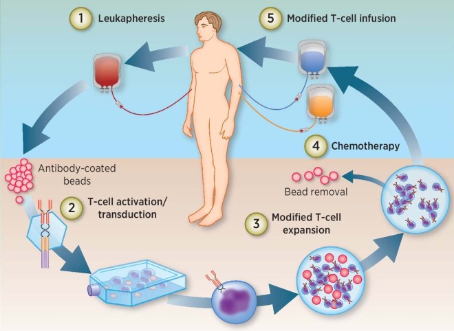
Cynata uses induced pluripotent stem cell technology to make large numbers of mesenchymal stem cells for therapeutic treatments.  To review, induced pluripotent stem cells (iPSCs) are made from body cells by means of genetic engineering and cell culture techniques.  Briefly, four different genes – OCT4, KLF4, SOX2, and c-MYC – are transfected into bodily cells.  Transient expression of these genes in regular body cells drives a small proportion of them (0.1%-0.5%) to undergo dedifferentiation into developmentally immature cells that resemble embryonic stem cells.  These embryonic stem cell-like cells, or iPSCs, will outgrow the other cells and can be successfully cultured in cell culture systems.  Once purified, cloned, and established as a viable cell line, the iPSC cell line can be grown to large numbers and differentiated into specific cell types.

Cynata has differentiated iPSCs derived from cells provided a healthy donor to make a recently identified precursor cell, known as a mesenchymoangioblast (MCA).  MCAs were first identified by Professor Igor Slukvin and his coworker from the University of Wisconsin-Madison. MCAs are early clonal mesoendodermal precursor cells that are the common precursor for both mesenchymal stem cells (MSCs) and endothelial cells, which are the main component of blood vessels. MCAs can also differentiate into pericytes and smooth muscle cells.

By making MCAs from iPSCs, Cynata seeks to overcome some of the largest hurdles for adult stem cell-based treatments, those being the dependence on donors for a steady supply of stem cells, the extensive variability of donated adult stem cells, the contamination of isolated adult stem cells with other cell types that do not have established therapeutic activities, and, probably most importantly, the limited scalability of stem cells from donors. Thus, in principle, Cynata’s Cymerus technology incorporates iPSC-derived MCA, which are used to make MCA-derived MSCs. This platform can potentially address the shortcomings of adult stem cell-based treatments, since iPSCs can proliferate indefinitely, and MCAs themselves can expand into extremely large quantities of MSCs. To quote the Cyanata web site; “Cynata should be able to manufacture all of the MCAs that it will ever need from a single Master Cell Bank of iPSCs – derived from a single donor.”

This most recent experiment was conducted by James Chong, Associate Professor at the Westmead Institute for Medical Research in Sydney, Australia, and colleagues. Chong and his coworkers induced heart attacks in three groups of rats (15 groups per group) and treated them four days later.  All the laboratory animals were assessed for 28 days after the heart attack (I wish they had observed them for longer periods of time). The rat groups consisted of three treatment groups: Group 1 received infusions of Cymerus MSCs, which were derived from iPSC-derived MCAs; Group 2 received MSCs from bone marrow; Group 3 was a placebo group that received infusions of buffer. All post-heart attack assessments were performed in a blinded manner, which simply means that the staff conducting the assessments had no idea which treatment the animal they tested had received.

The results of this study were rather encouraging.  The first category examined was “fractional shortening,” which is an estimate of the ability of the heart to contract effectively. Improvements in fractional shortening is indicative of recovery of the pumping function of the heart after a heart attack. Treatment with Cynata’s Cymerus MSCs resulted in an improvement in fractional shortening 28 days after the heart attack. Statistical comparisons showed that the Cymerus MSC-induced improvements were better than those observed in the placebo group (p=0.013) and the BM-MSC group (p=0.003).

The next assessment examined the effects of these treatments on left ventricular end-systolic diameter (LVESD). If, after a heart attack, the LVESD values are higher, then the heart is not contracting well.  However, reduced LVESD values are associated with improved cardiac function and correlated quite well with a reduced risk of further cardiac events. In this study, LVESD was lower in the Cymerus MSC group compared to those rats that had been treated with the placebo (p=0.054) and bone marrow-derived MSCs (p<0.001).

Finally, Chong and his gang examined the scar size as a proportion of the left ventricle size. For all three groups, there were no statistically significant differences between groups when it came to scar size as a proportion of the size of the left ventricle. It is possible that 28 weeks is far too short a time for the stem cells to produce any reduction in the size of the cardiac scar.  Therefore, further assessments of scar are ongoing.

To summarize, the Cymerus MSC treatments improved recovery of cardiac function post heart attack and reduced left ventricular end-systolic diameter (LVESD) compared to either placebo or bone marrow-derived MSCs, and further demonstrates the broad applicability of the Cymerus cell-manufacturing platform for clinical treatments.

In response to these positive results, Cynata’s Vice President of Product Development, Dr. Kilian Kelly said: “These very encouraging results add to a growing body of evidence showing that Cymerus MSCs may have an important role to play in the treatment of a wide range of diseases. There is still a huge unmet medical need associated with heart attacks, which cause over 8,000 deaths and more than 50,000 hospitalizations each year in Australia alone. We are optimistic about the potential benefits that Cymerus MSCs could bring to patients who experience these life-changing events.”