A Powerful Tool For Repairing Damaged Hearts


A new report from Johns Hopkins University researchers indicates that a particular stem cells that helps build mouse hearts can self-renew. This discovery, which might very well apply to humans as well, could potentially open inroads to use these cells to repair hearts damaged by disease, or, perhaps, even grow new heart tissue for transplantation.

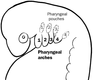
This study is slated for publication in the journal eLife. Chulan Kwon, Ph.D., an assistant professor of cardiology and member of the Institute for Cell Engineering at the Johns Hopkins University School of Medicine and his team, found that during heart formation, these so-called cardiac progenitor cells or CPCs proliferate, but do not differentiate into heart cells in an embryonic structure known as the second pharyngeal arch. This insight into the biology of CPCs may contribute to better understanding of how to prevent and treat congenital heart defects.

Kwon noted that, “Our finding that CPCs are self-renewing—that they can keep dividing to form new CPCs—means they might eventually be maintained in a dish and used to make specific types of heart cells.”

Kwon continued: “Growing such cells in a dish would be an enormous step toward better treatment for heart disease.”

Kwon’s laboratory initially tackled the elucidating the contribution of two genes, Numb and Numbl, in the CPC biology. Other studies have shown that these two genes are required to guide stem and progenitor cells to their fully mature, specialized functions. Numb and Numbl are highly conserved in mice and humans (i.e. the proteins encoded by these genes in mice and humans are nearly identical). This conservation indicates that Numb and Numbl are probably doing something very important the lives of CPCs.

As a first step, Kwon and others made loss-function mutations in Numb and Numbl. The results were striking. According to Kwon, embryos that lacked functional Numb and Numbl protein, “failed to develop normal hearts and died at an early stage of development, showing us that Numb and Numbl are needed for CPCs to build the heart.”

With the crucial role of Numb and Numbl in the lives of CPCs in mind, Kwon and his colleagues tried to determine the location of CPCs in the developing embryo. For these experiments, they used mouse embryonic stem cells that lacked functional Numb and Numbl, and expressed a glowing red protein in all CPC cells. Such a glowing red protein would instantly give away the CPCs’ location. These embryonic stem cells have the ability to integrate into a growing embryo, but the absence of functional Numb and Numble proteins in these cells prevents them from growing into a viable embryo.

Next, Kwon’s group injected these engineered embryonic stem cells into viable mouse embryos at the blastocysts stage. The blastocyst stage forms early during mammalian development, and it consists of the two cell populations that will form the embryo (inner cell mass cells) and the placenta (trophoblast cells). “The normal cells in these blastocysts compensated for those that lacked Numb and Numbl, allowing the resulting embryos to survive,” Kwon says.

Once these chimeric embryos began to grow, Kwon’s group examined them for red-glowing cells. They found the glowing red cells in the second pharyngeal arch, which is known for forming parts of the neck and face. Kwon says their study is the first to identify the second pharyngeal arch as the home of the CPCs.

pharyngeal_arches


The cells of the second pharyngeal arch go on to form the stapes in the middle ear and the stapedius muscle that attaches to the stapes.

Pharyngeal_arch_cartilages

Additionally, Kwon’s group cultured second pharyngeal arch cells with CPCs. They discovered that the cultured CPCs self-renewed without developing into specialized heart cells. This is potentially an important step toward using CPCs to treat heart disease.

The next step, he says, is to direct the laboratory-grown CPCs to form new heart tissue that could be used to regenerate disease-damaged heart tissue. “Eventually, we might even be able to deliver cells to damaged hearts to repair heart disease,” Kwon says.
Mouse Blood Cells Reprogrammed into Blood Cell Stem Cells


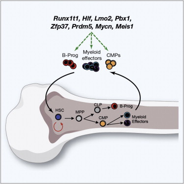
Boston Children’s Hospital researchers have directly reprogrammed mature blood cells from mice into blood-forming hematopoietic stem cells by using a cocktail of eight different transcription factors.

These reprogrammed cells have been called induced hematopoietic stem stem cells or iHSCs. These cells have all the hallmarks of mature mouse HSCs and they have the capacity to self-renew and differentiate into all the blood cells that circulate throughout the body.

These findings are highly significant from a clinical perspective because they indicate that it might be entirely possible to directly reprogram a patient’s existing, mature blood cells into a hematopoietic stem cell for transplantation purposes. Such a procedure, known as hematopoietic stem cells transplantation or HSCT, is a common treatment for patients whose bone marrow has suffered irreparable damage due to environmental causes (heavy metals, chloramphenicol, etc) or disease (cancer). The problem with HSCT is finding a proper match for the patient and then procuring clinically significant quantities of high-quality bone marrow for HSCT.

Derrick J. Rossi, an investigator in the Program in Cellular and Molecular Medicine at Boston Children’s Hospital and Assistant Professor in the Department of Stem Cell & Regenerative Biology, explained: “HSCs comprise only about one in every 20,000 cells in the bone marrow. If we could generate autologous (a patient’s own) HSCs from other cells, it could be transformative for transplant medicine and for our ability to model diseases of blood development.”

Rossi and his collaborators have screened genes that are expressed in 40 different types of blood progenitor cells in mice. This screen identified 36 different genes that control the expression of the other genes. These 36 genes encode so-called “transcription factors,” which are proteins that bind to DNA and turn gene express on or shut it off.

Blood cell production tends to go from the stem cells to progeny cells called progenitor cells that can still divide to some limited extent, and to effector cells that are completely mature and, in most cases, do not divide (the exception is lymphocytes, which expand when activated by specific foreign substances called antigens).

Further work by Rossi and others identified six transcription factors (Hlf, Runx1t1, Pbx1, Lmo2, Zfp37, and Prdm5) of these 36 genes, plus two others that were not part of their original screen (N-Myc and Meis1) that could robustly reprogram myeloid progenitor cells or pro/pre B lymphocytes into iHSCs.

Print

To put these genes into these blood cells, Rossi and others uses souped-up viruses that introduced all either genes under the control of sequences that only allowed expression of these eight genes in the presence of the antibiotic doxycycline. Once these transfected cells were transplanted into mice, the recipient mice were treated with doxycycline, and the implanted cells formed HSCs.

When this experiment utilized mice that were unable to make their own blood cells, because their bone marrow had been wiped out, the implanted iHSCs reconstituted the bone marrow and blood cells of the recipient mice.

To further show that this reconstituted bone marrow was normal, high-quality bone marrow, Rossi and others used these recipient mice as bone marrow donors for sibling mice whose bone marrow had been wiped out. This experiment showed that the mice that had received the iHSCs had bone marrow that completely reconstituted the bone marrow of their siblings. This established that the iHSCs could completely reestablish the bone marrow of another mouse.

Thus Rossi and others had established that iHSCs could in fact created HSCs from progenitor cells, but could they do the same thing with mature blood cells that were not progenitor cells? Make that another yes. When Rossi and others transfected their eight-gene cocktail into mature myeloid cells, these mature cells also made high-quality iHSCs.

Rossi noted that no one has been able to culture HSCs in the laboratory for long periods of time. A few laboratories have managed expand HSCs in culture, but only on a limited basis for short periods of time (see Aggarwal R1, Lu J, Pompili VJ, Das H. Curr Mol Med. 2012 Jan;12(1):34-49).  In these experiments, Rossi used his laboratory mice as living culture systems to expand his HSCs, which overcomes the problems associated with growing these fussy stem cells in culture.

Gene expression studies of his iHSCs established that, from a gene expression perspective, the iHSCs were remarkably similar to HSCs isolated from adult mice.

This is certainly an exciting advance in regenerative medicine, but it is far from being translated into the clinic.  Can human blood progenitor cells also be directly reprogrammed using the same cocktail?  Can mature myeloid cells be successfully reprogrammed?  Will some non-blood cell be a better starting cell for iHSC production in humans?  As you can see there are many questions that have to be satisfactorily answered before this procedure can come to the clinic.

Nevertheless, Rossi and his team has succeeded where others have failed and the results are remarkable.  HSCs can be generated and transplanted with the use of only a few genes.  This is certainly the start of what will hopefully be a fruitful regenerative clinical strategy.

On a personal note, my mother passed about almost a decade ago after a long battle with myelodysplastic syndrome, which is a pre-leukemic condition in which the bone marrow fails to make mature red blood cells.  Instead the bone marrow fills up with immature red blood cells and the patient has to survive on blood transfusions.

The reason for this condition almost certainly stems from defective HSCs that do not make normal progeny.  Therefore the possibility of using a patient’s own cells to make new HSCs that can repopulate the bone marrow is a joyful discovery for me to read about, even though it is some ways from the clinic at this point.

Skin Tissue Grown From Human Stem Cells


A research team from King’s College, London, in collaboration with the San Francisco Veteran Affairs Medical Center has succeeded in growing the epidermal layer of skin in culture, this cultured skin has many of the mechanical and biological properties of actual human skin.

The outermost layer of the skin, known as the epidermis forms a protective barrier between the external environment and the body. It protects against water loss and prevents the entry of microorganisms.

Tissue engineers have been able to grow skin cells (keratinocytes) in culture, but getting them to organize into an organ that resembles biological skin has proven rather difficult. However, the ability to test drugs on cultured skin that greatly mimics human skin has been the goal of such research for several years.

For this present project, keratinocytes were made from induced pluripotent stem cells that were derived from skin cells obtained from biopsies. These keratinocytes made from induced pluripotent stem cells (iPSCs) were very similar to keratinocytes made from embryonic stem cells and primary keratinocytes isolated from skin biopsies.

To form a three-dimensional structure like skin, the keratinocytes were cultured in a high-to-low humidity environment and they assembled into a layer structure that looked like human skin. When this cultured skin was compared with skin made from embryonic stem cell-derived keratinocytes or from keratinocytes isolated from skin biopsies, there were no significant structural differences.

Scientists hope to use this cultured skin to study congenital skin diseases like ichthyosis (characterized by dry, flaky skin) or atopic dermatitis. Growing large quantities of skin in culture will also allow drugs and cosmetics to be effectively tested for safety without the use of expensive and sometimes highly variable animal models.

This technology would also allow different laboratories to grow skin from different ethnic groups that have distinct types of skin with variable biological properties.

Mesenchymal Stem Cells Reduce Scarring of Intervertebral Discs and Facilitate Healing


Intervertebral disc degeneration causes substantial back pain and associated pain that shoots down the legs (radiculopathy). Back issues associated with bad intervertebral discs are a leading cause of disability. Such disability costs employers millions of dollars of lost man and woman power and employees extensive loss of wages. Chronic back pain can also seriously compromise the quality of life and presents a large societal burden.

To date, surgery is the only effective treatment option, but surgical interventions sometimes leave patients worse off than before. Thus there is presently no effective intervention for this disease.

However, in a recent paper, Victor Y.L. Leung and his colleagues from the University of Hong Kong and several other institutions as well have used human mesenchymal stem cells from bone marrow to treat damaged intervertebral discs in rabbits. The results, published in the journal Stem Cells, are quite hopeful

Leung and others discovered that by puncturing the intervertebral discs of rabbits with a syringe needle, they could induce damage to the disc that mimics disc degeneration in humans.

Next, they implanted human bone marrow-derived mesenchymal stem cells (MSCs) into the damaged discs. Such implantations prevented scarring of the disc in the center of the disc. The center of the disc, the nucleus pulposus, is more gel-like than the surrounding annulus fibrosus. Scarring of the nucleus pulposus stiffens it and prevents it from moving with stress. An inability to bend with stress causes the disc to become brittle with time and herniate. However, implantation of mesenchymal stem cells preserved the mechanical properties of the disc and benefitted overall spinal function.

By looking more deeply at the mechanism by which mesenchymal stem cells preserve disc function, Leung and others showed that MSCs suppress abnormal deposition of collagen I in the nucleus pulposus. Since collagen is made during scarring, suppression of collagen I synthesis suppressed scarring. Secondly, implanted MSCs decreased the expression of two molecules that promote the synthesis of collagen I. By suppressing the expression of MMP12 and HSP47, the implanted MSCs also reduced collagen aggregation and maintained the microarchitecture of the disc and its mechanical properties.

This  study supports the ability of MSCs to stimulate resident stem cell activities and disc healing. The implanted MSCs seem to do so by means of down-regulating collagen  fibril formation. This provides the basis for the MSC‐based disc therapies.

Researchers Transplant Regenerated Esophagus


Paolo Macchiarini and a research consortium from the Karolinska Institutet in Stockholm, Sweden have made a tissue engineered scaffold for the esophagus from esophagi that were extracted from rats.

After the cells were stripped from the rat esophagi, bone marrow mesenchymal stromal cells were seeded onto the decellularized esophagi and grown in a perfusion bioreactor. A variety of experiments demonstrated that these mesenchymal stromal cells differentiated into esophageal epithelial cells and smooth muscle cells. Macchiarini and his group used several gene expression and functional assays to confirm that these cells had in fact differentiated into these esophageal-specific cell types.

Next, Maccharini and others transplanted these esophagi into rats. The transplanted rats survived 14 days after the transplantations and ate and gain weight. Because the cells used to reconstitute the esophagi came from the rats into which they were transplanted, immunosuppressive drugs were neither used nor needed.

When the recipients of the transplanted esophagi were sacrificed after fourteen days, tissue examinations showed that all the major cell and tissue components of the esophagus including the inside covering of the esophagus (epithelium), muscle fibers, nerves, and blood vessels had nicely regenerated.

This successful bioproduction and transplantation of a tissue-engineered esophagus represents a significant step towards the clinical application of bioengineered esophagi.

Think of it: children and adults with tumors, congenital malformations of the esophagus of traumatic injuries to the esophagus may have new hope and possibilities because of advances in tissue engineering like this.

Meta Study Shows that Mesenchymal Stem Cells Promote Healing in Animal Models of Stroke


Two scientists from my alma mater, UC Irvine, have examined experiments that treated stroke with bone marrow-derived stem cells. Their analysis has shown that infusions of these stem cells trigger repair mechanisms and limit inflammation in the brains of stroke patients.

UC Irvine neurologist Dr. Steven Cramer and biomedical engineer Weian Zhao identified 46 studies that examined the use of a specific type of bone marrow stem cells called mesenchymal stromal cells to treat stroke. Mesenchymal stromal cells are a type of multipotent adult stem cells that are found in many locations in the body. The best-known examples of mesenchymal stem cells are from bone marrow. When purified from whole bone marrow and used to treat stroke in animal models of stroke, Cramer and Zhao found that mesenchymal stromal cells (MSCs) were significantly better than control therapy in 44 of the 46 studies that were examined.

Further data culling of these studies showed that functional recovery from stroke were robust regardless of the MSC dosage or the time when MSCs were administered relative to the onset of the stroke, or the method of administration (whether introduced directly into the brain or injected via a blood vessel).

“Stroke remains a major cause of disability, and we are encouraged that the preclinical evidence shows [MSCs’] efficacy with ischemic stroke,” said Cramer, a professor of neurology and leading stroke expert. “MSCs are of particular interest because they come from bone marrow, which is readily available, and are relatively easy to culture. In addition, they already have demonstrated value when used to treat other human diseases.”

Another theme of these studies is that MSCs do not differentiate into brain-specific. MSCs have the capacity to differentiate into bone, cartilage and fat cells. “But they do their magic as an inducible pharmacy on wheels and as good immune system modulators, not as cells that directly replace lost brain parts,” he said.

In an earlier Cramer and Zhao examined the mechanism by which MSCs promote brain repair after stroke. These cells have the ability to home to the damages areas in the brain and release chemicals that stimulate healing. By releasing their cornucopia of healing-promoting molecules, MSCs orchestrate blood vessel creation to enhance circulation, the protection of moribund cells on the verge of death, and the growth of existing brain cells. Additionally, when MSCs reach the bloodstream, they settle in those parts of the body that control the immune system and they suppress the inflammatory response that can augment tissue damage. In this way, MSCs foster an environment more conducive to brain repair.

“We conclude that MSCs have consistently improved multiple outcome measures, with very large effect sizes, in a high number of animal studies and, therefore, that these findings should be the foundation of further studies on the use of MSCs in the treatment of ischemic stroke in humans,” said Cramer, who is also clinical director of the Sue & Bill Gross Stem Cell Research Center.

Long-Term Survival of Transplanted Human Neural Stem Cells in Primate Brains


A Korean research consortium has transplanted human neural stem cells (hNSCs) into the brains of nonhuman primates and ascertained the fate of these cells after being inside the brains of these animals for 22 and 24 months. They discovered that the implanted hNSCs had not only survived, but differentiated into neurons and never caused any tumors.

This important study is slated to be published in the journal Cell Transplantation.

To properly label the hNSCs so that they were detectable inside the brains of the animals, Lee and others loaded them with magnetic nanoparticles to enable them to be followed by magnetic resonance imaging (MRI). Also, they did not use immunosuppressants when they transplanted their hNSCs into the animals. This study is the first to examine the long-term survival and differentiation of hNSCs without the need for immunosuppression.

“Stroke is the fourth major cause of death in the US behind heart failure, cancer, and lower respiratory disease,” said study co-author Dr. Seung U. Kim of University of British Columbia Hospital’s department of neurology in Canada. “While tissue plasminogen activator (tPA) treatment within three hours after a stroke has shown good outcomes, stem cell therapy has the potential to address the treatment needs of those stroke patients for whom tPA treatment was unavailable or did not help.”

Based on the ability of hNSCs to differentiate into a variety of types of nerves cells, Lee and his colleagues thought that these cells have remarkable potential to treat damaged brain tissue and replace what was lost after a stroke, head injury or other type of brain trauma. Cell regeneration therapy can potentially treat brain-specific diseases like Parkinson’s disease, Huntington’s disease, Alzheimer’s disease, amyotrophic lateral sclerosis (ALS), spinal cord injury and stroke.

Dr. Kim and colleagues in Korea grafted magnetic particle-labeled hNSCs into the brains of laboratory primates and evaluated their performance to assess their survival and differentiation over 24 months. Of particular interest was determining their ability to differentiate into neurons and to determine whether the cells caused tumors.

“We injected hNSCs into the frontal lobe and the putamen of the monkey brain because they are included in the middle cerebral artery (MCA) territory, which is the main target in the development of the ischemic lesion in animal stroke models,” commented Dr. Kim. “Thus, research on survival and differentiation of hNSCs in the MCA territory should provide more meaningful information to cell transplantation in the MCA occlusion stroke model.”

Lee’s team said that they chose NSCs for transplantation because the existence of multipotent NSCs “has been known in developing rodents and in the human brain with the properties of indefinite growth and multipotent potential to differentiate” into the three major CNS cell types – neurons, astrocytes and oligodendrocytes.

“The results of this study serve as a proof-of-principle and provide evidence that hNSCs transplanted into the non-human primate brain in the absence of immunosuppressants can survive and differentiate into neurons,” wrote the researchers. “The study also serves as a preliminary study in our planned preclinical studies of hNSC transplantation in non-human primate stroke models.”

“The absence of tumors and differentiation of the transplanted cells into neurons in the absence of immunosuppression after transplantation into non-human primates provides hope that such a therapy could be applicable for use in humans.” said Dr. Cesar V. Borlongan, Prof. of Neurosurgery and Director of the Center of Excellence for Aging & Brain Repair at the University of South Florida. “This is an encouraging study towards the use of NSCs to treat neurodegenerative disorders”.

Reversing Age-Related Tendon Damage with Reprogrammed Tendon Stem Cells


Adults over 60 are much more likely to experience tendon injuries. Since tendons connect muscles to bones, tendon injuries can lead to substantial restrictions in movement and movement-associated pain.

Injured tendons tend to be left on their own to heal and, as I can attest to from personal experience, injured tendons can take a very long time to heal.

I new study, however, shows that tendon damage can be reversed with stem cell therapies.

Hui Sun from Albert Einstein College of Medicine found that reprogramming the CITED2 in aged tendon stem cells can not only reverse age-related dysfunction of these cells, but when implanted into injured, aged tendons, these engineered tendon stem cells healed and rejuvenated the tendons.

The CITED2 gene encodes a protein that inhibits transactivation of HIF1A-induced genes. It does so by competing with binding of hypoxia-inducible factor1α to its co-factor p300-CH1.

With therapies like this, older adults, even though they may always have to contend with gray hair and wrinkles, might not be concerned about decreased mobility in their advanced years after suffering an injury.

“We are developing a novel patient- and surgeon-friendly intervention for tendon tissue repair, especially in aged individuals,” Sun said. “We’re also working on strategies to rejuvenate aged tendon and other muscuoskeletal tissues based on this discovery.”

Proteins that Control Energy Metabolism Necessary to Form Stem Cells


University of Washington scientists have discovered that the way cells degrade sugars plays a rather central role in reprogramming adult cells into pluripotent stem cells.

Julie Mathieu, a postdoctoral research fellow at the University of Washington, Wenyu Zhou, a postdoctoral scholar at Stanford University, and Hannele Ruhola-Baker, UW professor of Biochemistry, teamed up to address this problem. Their findings might have implications for the way pluripotent stem cells are made from adult cells for regenerative purposes in the future.

Reprogramming adult cells to induced pluripotent stem cells requires genetically engineering the cells with four different genes (now known as the Yamanaka factors after Shinya Yamanaka who discovered them). These four genes – Oct4, Klf4, Sox2 and c-Myc – causes the cells to de-differentiate into embryonic stem cell-like cells.

However, during reprogramming, cells change the way they make their energy. The reprogramming cells shut down the oxygen-utilizing part of metabolism and switch to a fermentative kind of metabolism that does not require the presence of oxygen.

This metabolism shift seems to mimic, in part, the metabolism of embryonic cells, which have to survive and grow in low-oxygen environments. Cancer cells also perform a similar shift in their metabolism.

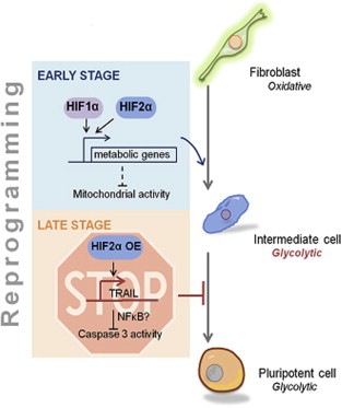
Ruhola-Baker and her gang examined the function of two proteins that are known to control the use of oxygen: HIF or hypoxia-induced factor-1α and -2α. These two proteins regulate several genes involved in energy metabolism. When Ruhola-Baker and her colleagues made adult cells that lacked a functional copy of either HIF-1α or HIF-2α, these cells were unable to be reprogrammed into induced pluripotent stem cells.

When Ruhola-Baker and her team used these proteins to examine gain-of-function experiments, the results were very different. Stabilization of HIF-1α throughout the reprogramming process increased the formation of induced pluripotent stem cells from adult cells. However, stabilization of HIF-2α during the later stages of reprogramming inhibited the reprogramming process. The HIF-2α-mediated inhibition of reprogramming occurs in the presence or absence of extra HIF-1α. Thus HIF-1α increases reprogramming at all stages, but HIF-2α increases the efficiency of the early stages of reprogramming but inhibits reprogramming at the later stages.

HIF function during reprogramming

“HIF-2α is like Darth Vader, originally a Jedi who falls to the dark side,” said Ruhola-Baker. “HIF-1α, the good guy, is beneficial for reprogramming throughout the process. HIF-2α, if not eliminated, turns bad in the middle and represses pluripotency.”

How does HIF-2α repress reprogramming to pluripotency? By increasing the production of a protein called TRAIL, which stands for Tumor necrosis factor-related apoptosis-inducing ligand. TRAIL has been known to cancer researchers for some time, since it causes some cancer cells to self-destruct.  TRAIL has to be inhibited in order for reprogramming to occur.  When HIF-2α activates TRAIL, it inhibits reprogramming.

Zhoi said that their data suggests that TRAIL and other members of this protein family might be alternate between playing good cop/bad cop during stem cell development.

Practically speaking, it might be possible to use HIF-1α to increase the number of stem cells derived from adult cells in a particular experiment. This might decrease the degree to which the cells are genetically manipulated in order to form induced pluripotent stem cells.

Alternatively, since activated HIF-1α is a marker for aggressive cancers, these data might also have implications for treating cancer. Blocking HIF-1α function or stimulating HIF-2α might interfere with particular cancers. The treatment possibilities are very intriguing.

Scientists Make Cloned Stem Cells from Adult Cells


For the first time, stem cell scientists have derived stem cells from cloned human embryos that were made from adult cells.  This brings them closer to developing patient-specific lines of cells that can be used to treat a whole host of human maladies, but at a cost.  This research was described in the April 17th online edition of the journal Cell Stem Cell.

In May of last year, Shoukhrat Mitalipov from the Oregon Health and Science University, reported the derivation of human embryonic stem cells from cloned human embryos.  However, these cloned were made using cells that came from infants.  Miltalipov worked out a new protocol for cloning human embryos by using nonhuman primate embryos, in particular those from a Rhesus monkey.

In this study, the donor cells came from two men, a 35-year-old and a 75-year-old.  By using the protocol developed by Mitalipov and his group, Robert Lanza, Young Gie Chung, and Dong Ryul Lee and their colleagues made personalized embryonic stem cells from these two men.

Stem cell biologist Paul Knoepfler, an associate professor at the University of California at Davis who runs the widely read Stem Cell Blog, called the new research “exciting, important, and technically convincing.”  He continued: “In theory you could use those stem cells to produce almost any kind of cell and give it back to a person as a therapy.”

In their paper, Young Gie Chung from the Research Institute for Stem Cell Research for CHA Health Systems in Los Angeles, Robert Lanza from Advanced Cell Technology in Marlborough, Mass., and their co-authors pointed out the potential promise of this technology for new regenerative therapies.  However, their work is also an important discovery for human cloning, since it shows that age-associated changes are not necessarily an impediment to SCNT-based nuclear reprogramming of human cells.

Even though it was the intent of Chung and others to gestate these cloned embryos to form cloned children, this work could be the first step toward creating a baby with the same genetic makeup as a donor.  Thus, this technology presents a so-called “dual-use dilemma.”

Marcy Darnovsky, executive director of the Berkeley, Calif.-based Center for Genetics and Society, explained that many technologies developed for good can be used in ways that the inventor may not have intended and may not like.

“This and every technical advance in cloning human tissue raises the possibility that somebody will use it to clone a human being, and that is a prospect everyone is against,” Darnovsky said.

This paper represents a collaboration between members of academic laboratories and industry.  Funding for this work came from a private medical foundation and South Korea’s Ministry of Science.

Technically, the somatic-cell nuclear transfer protocols used in paper are still somewhat inefficient.  Chung’s team had to attempt 39 times to produce only two blastocyst-stage embryos.  Their first attempts were complete failures, but when they modified the Mitalipov protocol and activated the cloned embryos 2 hours after fusion rather than 30 minutes after fusion, the embryos grew successfully.

“We have reaffirmed that it is possible to generate patient-specific stem cells using [this] technology,” Chung said.

Shoukhrat Mitalipov, director of the Center for Embryonic Cell and Gene Therapy at Oregon Health & Science University, who developed the method that Chung’s group built upon, said that this work involves eggs that have not been fertilized.

“There will always be opposition to embryonic research, but the potential benefits are huge,” Mitalipov said.

Yes, there will be opposition to destructive research on embryos because they are the youngest among us.  No they do not have the right to vote, drive a car, or buy a hunting license, but they have the right to not be harmed.  To deny them that right because they cannot presently exercise particular capacities assumes that the embryo undergoes essential changes as it develops.  But human embryos develop into the kinds of entities they become because of their intrinsic human nature that drives them to do so.  Yes development is a progressive program that causes the embryo to acquire new structures and capabilities that it previously did not have, but what kind of entity can develop into a human adult that is not itself human?  It takes a human embryo to make a human fetus, which makes a human new-born baby, which makes a human toddler, and do on.  This continuum or development and change occurs throughout or lives and this continuum begins at the end of fertilization.

Cloned embryos begin this continuum at the completion of somatic cell nuclear transfer (SCNT).  SCNT works as a stand-in for fertilization, but the result is still the same – a human embryo.  It also should have the right not to be harmed, but instead she is being produced solely for the purpose of being dismembered.  Is this the way we should treat the smallest and most defenseless among us? surely not.  All this talk about, “well we did not form a fully human being” is a crock.  Yes you did.  You formed a fully formed human embryo.  We were all human embryos at one time and these embryos developed into you and me.  We were inarticulate and incapable at the time, but we gained those capacities over time.  Again, how can something that gives rise to a human child not be human?  The embryo is a human being, but it is a very young human being.  Youth should not disqualify it from being able to live.

Seventeen years ago, when Ian Wilmut from the Roslin Institute in Edinburgh, Scotland announced news about the birth of the first sheep cloned from somatic cells named Dolly, several legislators called for a ban on human cloning.  Several countries took measures to limit or outlaw such work, but in the United States.  The cloning issue was obfuscated by dividing it into “reproductive cloning” for the purposes of making cloned children, and “therapeutic cloning” for the development of new therapies.  Unfortunately, this dichotomy is slightly disingenuous since the techniques for both of these procedures are exactly the same except that reproductive cloning uses a surrogate mother to gestate the cloned embryo and bring her to term.  Both of these procedures produce human embryos, but one uses them to make a baby and the other destroys them before they can do so.

President George W. Bush tried to split the difference by restricting federal funding for stem cell research that harms to a human embryo.  This led to talk of Bush’s “embryonic stem cell ban,” which was inaccurate and was used unfairly used to paint Bush as an idiot.  However, some 15 states have laws addressing human cloning, and about half of them ban both reproductive and therapeutic cloning.

Embryonic stem cell research has typically used embryos that are left over from the fertility industry.  However, some religious groups such as the U.S. Conference of Catholic Bishops and others as well  objected to this, since it destroys a very young human being.

However, about seven years ago, Shinya Yamanaka and his colleagues discovered a way to make induced pluripotent stem cells from mature adult cells.  Genetic engineering techniques could convert ordinary cells into pluripotent stem cells without the need for human eggs.  While this technique did not present the same ethical issues, some induced pluripotent stem cells lines contain significant genetic abnormalities and there is still debate over how safe these cells are for clinical use.

The research conducted by Mitalipov and Chung provides a second way of producing pluripotent cells through laboratory techniques that is, in my view, far less ethical and will almost certainly also have unintended consequences as well.

An Improved Way to Make Motor Neurons in the Laboratory from Stem Cells


A research team from the University of Illinois at Urbana-Champaign has reported that they can produce human motor neurons from stem cells much more quickly and efficiently than previous methods allowed. This finding was published in the journal Nature Communications and it will almost certainly provide new ways to model human motor neuron development, diseases of the nervous system, and ways to treat spinal cord injuries.

The new protocol described in the Nature Communications paper includes adding critical signaling molecules to precursor cells a few days earlier than specified by previous methods. This innovation increases the proportion of healthy motor neurons derived from stem cells from 30 to 70 percent. It also cuts in half the time required to make motor neurons.

“We would argue that whatever happens in the human body is going to be quite efficient, quite rapid,” said University of Illinois cell and developmental biology professor Fei Wang, who led the study with visiting scholar Qiuhao Qu and materials science and engineering professor Jianjun Cheng. “Previous approaches took 40 to 50 days, and then the efficiency was very low – 20 to 30 percent. So it’s unlikely that those methods recreate human motor neuron development.”

The new method designed by Qu generated a larger population of mature, functional motor neurons in 20 days. According to Wang, this new approach will allow scientists to induce mature human motor neuron development in cell culture, and to identify the factors that drive this process

Because stem cells can differentiate into a wide variety of cell types, they are unique compared to mature, adult cells. Making neurons from either embryonic stem cells or induced pluripotent stem cells requires the addition of signaling molecules to the cells at critical moments in culture.

Previously, Wang and his colleagues discovered a molecule called compound C that converts stem cells into “neural progenitor cells,” or NPCs. NPCs represent an early stage in neuronal development, and further manipulation of NPCs can drive them to become neurons, but differentiating NPCs into motor neurons presents another set of problems.

Other published studies have established that the addition of two important signaling molecules, six days after exposure to compound C, to NPCs in culture can generate motor neurons, but at rather poor efficiencies. In this newly published study, Qu showed that adding the signaling molecules at Day 3 worked better: The NPCs differentiated into motor neurons quickly and efficiently. Thus, Day 3 represents a previously unrecognized NPC cell stage.

This new approach has immediate applications in the laboratory. Amyotrophic lateral sclerosis or ALS is a neurological disease that causes motor neurons to die off. By using Wang and Qu’s cell culture system to make neurons from the skin cells of ALS, and watching them develop into motor neurons, scientists and physicians will divine other new insights into disease processes. Therefore, any method that improves the speed and efficiency of generating the motor neurons will be a boon to neuroscientists. These cells can also be used to screen for drugs to treat motor neuron diseases, and might even be used to therapeutically restore lost function in patients someday.

“To have a rapid, efficient way to generate motor neurons will undoubtedly be crucial to studying – and potentially also treating – spinal cord injuries and diseases like ALS,” Wang said.

A Protein that Signals Neurons to Regenerate


Presently damaged nerve fibers (or axons) from spinal nerves do not regenerate and there are no good ways to repair them. However, a new set of experiments suggest that the repair of these nerves might be possible after spinal cord injury or brain trauma.

Researchers from Imperial College London and the Hertie Institute, University of Tubingen, Germany, have identified a possible mechanism for re-growing damaged central nervous system nerve fibers. Such damage causes loss of sensation and permanent paralysis. However, regenerating nerve fibers is one of the best hopes for those suffering from CNS damage to recover.

This work was published in the journal Nature Communications, and this work trades on the function of a protein called P300/CBP-associated factor (PCAF). PCAF seems to be essential for the cellular pathway that damaged neurons use to properly regenerate. When PCAF was injected into mice with damage to their central nervous systems, the injected mice showed significant increases in the number of nerve fibers that regenerated. This indicates that it might be possible to chemically control the regeneration of nerves in the CNS.

“The results suggest that we may be able to target specific chemical changes to enhance the growth of nerves after injury to the central nervous system,” said lead study author Professor Simone Di Giovanni, from Imperial College London’s Department of Medicine. “The ultimate goal could be to develop a pharmaceutical method to trigger the nerves to grow and repair and to see some level of recovery in patients. We are excited about the potential of this work but the findings are preliminary.

“The next step is to see whether we can bring about some form of recovery of movement and function in mice after we have stimulated nerve growth through the mechanism we have identified. If this is successful, then there could be a move towards developing a drug and running clinical trials with people. We hope that our new work could one day help people to recover feeling and movement, but there are many hurdles to overcome first,” he added.

Of particular interest to Dr. Di Giovanni and his colleagues was how axons in the peripheral nervous system (PNS) make a concerted effort to grow back when they are damaged, whereas CNS axons mount little or no effort. Damage to the peripheral nervous system is followed by regeneration of about 30% of the damaged nerves, accompanied by recovery of some movement and function. Could neurons in the central nervous system be coaxed into a similar behavior?

Co-author Dr. Radhika Puttagunta from the University of Tubingen said: “With this work we add another level of understanding into the specific mechanisms of how the body is able to regenerate in the PNS and have used this knowledge to drive regeneration where it is lacking in the CNS. We believe this will help further our understanding of mechanisms that could enhance regeneration and physical recovery after CNS injury.”

To investigate damage and regeneration in central and peripheral nervous systems, Di Giovanni and his group examined mouse models and cells in culture. They compared the responses to PNS damage and CNS damage in a type of neuron called a dorsal root ganglion, which connects to both the CNS and the PNS.

Interestingly, they discovered that epigenetic mechanisms were at the core of the regeneration capacity of these cells. Epigenetic mechanisms are processes that, without altering our DNA, manage to activate or deactivate genes in response to the environment, and are linked to changes in the way DNA is packaged within the cell. Epigenetic considerations control genes that influence the onset of diseases such as cancer and diabetes. However this is the first demonstration of a specific epigenetic mechanism responsible for nerve regeneration.

When nerves are damaged in the PNS, the damaged nerves send ‘retrograde’ signals back to the cell body to switch on an epigenetic program to initiate nerve growth. Very little was previously known about the mechanism which allows this ‘switching on’ to occur. When DiGiovanni’s group identified the signal transduction pathway that led to the ‘switching on’ of the program to initiate nerve regrowth, they discovered that PCAF was central to this process. Furthermore when they injected PCAF into mice with damage to their central nervous system, there was a significant increase in the number of nerve fibers that grew back.

Thus, PCAF is necessary for conditioning-dependent axonal regeneration and also promotes regeneration after spinal cord injury. Thus, PCAF is a part of a specific epigenetic mechanism that regulates axonal regeneration of CNS axons, which also makes it and the protein with which it associates a novel target for clinical application.

Fat-Based Stem Cells in the PRECISE Trial Stabilizes Exercise Performance in Chronic Heart Disease Patients


Cytori Therapeutics has announced the publication of safety and efficacy data from a 36-month European clinical trial of Cytori Cell Therapy in patients with chronic ischemic heart failure. Final data from the Company’s PRECISE trial, a 27-patient, prospective, randomized, double-blind, placebo-controlled, feasibility trial (Phase I/IIA), demonstrated statistically significant differences in cardiac functional capacity between treated and placebo groups.

Their research will appear in the upcoming issue of the American Heart Journal. Cytori Cell Therapy is a mixed population of adipose derived regenerative cells (ADRCs™) extracted from a patient’s own adipose tissue using Cytori’s proprietary Celution® System.

“The PRECISE trial is the first-in-man trial involving the myocardial injection of ADRCs for heart disease,” said Dr. Emerson Perin , Co-Principal Investigator of the trial. “By demonstrating a strong safety profile and suggesting that the use of ADRCs may preserve functional capacity, the data indicates that this therapy may have meaningful impacts on the lives of these very sick patients.”

This particular publication was co-authored by trial investigators Drs. Emerson C. Perin at Texas Heart Institute, Francisco Fernández-Avilés at Hospital Universitario Gregorio Marañón and others. This clinical trial shows that the procedure was safe, feasible and showed indications of a favorable benefit to the patients who received it. The study demonstrated that fat harvest through liposuction could be performed safely in cardiac patients. Exercise capacity as reflected by maximum oxygen consumption (MVO2) during treadmill testing, a reflection of cardiac functional capacity, was sustained in the ADRC treated group but declined in the placebo group at 6 and 18 months. Statistically significant differences were observed between the two groups.

“These results supported the design of the ongoing U.S. Phase II ATHENA trial that is evaluating a similar patient population,” said Steven Kesten , M.D., Chief Medical Officer for Cytori. “We are encouraged by the sustained effects in functional endpoints, particularly MVO2, which is a relevant clinical endpoint in heart disease, and is an aid in directing treatment options, such as assist devices or heart transplant. We look forward to reporting the initial six-month results from the ATHENA trial.”

Additionally, other data trends in this study suggest that ADRC therapy may have a modest beneficial effect in stabilization of the heart scar tissue. To understand the meaning of this benefit, remember that ischemic heart disease might also be known as coronary artery disease (CAD), atherosclerotic heart disease, or coronary heart disease. Ischemic Heart Disease is the most common type of heart disease and cause of heart attacks. This disease is typically caused by plaque build up along the inner walls of the arteries of the heart, which leads to narrowing of the arteries and reduction of blood flow to the heart. After a heart attack, the region of the heart that was deprived of oxygen for a period time dies and the dead heart muscle tissue is replaced by scar tissue that contracts over time, but does not contract or conduct heartbeat impulses. In this study, the scar mass of the left ventricle remained consistent in ADRC-treated patients at six months compared to an increase in control patients. This suggests that ADRCs may prevent scar tissue from increasing. Other endpoints such as ventricular volumes and ejection fraction showed inconsistent findings.

In the PRECISE trial, all patients were treated with standard-of-care and subsequently underwent a liposuction procedure. Each patient’s adipose tissue was processed using Cytori’s proprietary Celution® System to prepare the cell therapy. Cells (n=21) or placebo (n=6) were injected into areas of the heart muscle that were severely damaged but still viable and reversible using the NOGA XP System.

Cytori is currently enrolling patients in the U.S. ATHENA and ATHENA II trials, both 45 patient prospective, randomized, double-blind, placebo-controlled trials investigating a lower and a higher dose, respectively, of Cytori Cell Therapy in a similar patient population as PRECISE.

The PRECISE study is a small study, but the fact that it was double-blinded and placebo controlled makes it an important study. The experimental group showed a clear stabilization of maximum oxygen consumption as opposed to the control group, whose exercise tolerance decreased during the course of the trial. This is potentially significant.  The ADRCs could be preventing the heart from enlarging as a result of working harder.

Questions, however, remain.  For example, is this a short-term effect or does it maintain its effect over the long-term period? To answer that, patient follow-up is necessary. Second, the other physiological parameters showed confusing outcomes (ejection fraction, end-diastolic volume, and so on).  If the ADRCs are truly helping the heart function better, then why don’t the physiological parameters used to measure heart function show some semblance of improvement?  The stabilization of the maximum oxygen consumption stabilization might not mean much in retrospect if it is short-term.

A larger trial like the ATHENA study will be more powerful. Hopefully these PRECISE patients will be followed and examined several years after the treatment to determine the duration of the ADR-provided benefits.

Repairing Muscles in Muscular Dystrophy Depends on the Degree of Muscle Deterioration


Pier Lorenzo Puri, M.D., an associate professor at Sanford-Burnham Medical Research Institute (Sanford-Burnham), has led a research team that work in collaboration with Fondazione Santa Lucia in Rome, Italy, to characterize the mechanism by which a class of drugs called “HDACis” drive muscle-cell regeneration in the early stages of dystrophic muscles, but fail to work in late stages. These findings are integral for designing HDACis drugs for Duchenne muscular dystrophy (DMD), which presently, is an incurable muscle-wasting disease.

Puri’s research was published April 15th, 2014 edition of the journal Genes and Development. In their paper, Puri and his colleagues used mouse models of DMD to show how special cells known as “fibro-adipogenic progenitor cells” or FAPs, direct muscle regeneration. FAPs reside in the spaces between muscle fibers and detect those cues that indicate that muscles have been damaged. In response to muscle damage, FAPs direct muscle stem cells, known as satellite cells, to rebuild muscle.

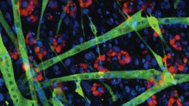
 HDAC inhibitors (HDACi) promote muscle regeneration in a mouse model of Duchenne Muscular Dystrophy at early stages of disease by targeting fibro-adipogenic progenitors (FAPs). Staining of FAPs from muscles of HDACi-treated young mdx mice reveals presence of differentiated muscle cells (green) at the expense of fat cells (red). Nuclei are stained in blue. Image: Lorenzo Puri, M.D.

HDAC inhibitors (HDACi) promote muscle regeneration in a mouse model of Duchenne Muscular Dystrophy at early stages of disease by targeting fibro-adipogenic progenitors (FAPs). Staining of FAPs from muscles of HDACi-treated young mdx mice reveals presence of differentiated muscle cells (green) at the expense of fat cells (red). Nuclei are stained in blue. Image: Lorenzo Puri, M.D.

“HDACis create an environment conducive for FAPs to direct muscle regeneration—but only during the early stages of DMD progression in mice,” said Puri. “At some point, DMD progresses to a pathological point of no return and become permanently resistant to muscle-regeneration cures and to HDACis.”

Indeed, Puri’s research showed exactly that; namely that FAPs embedded in muscle that was in the earlier stages of muscular dystrophy responded robustly to HDACis and upregulated a wide range of muscle-specific genes. In contrast, FAPs from late-stage dystrophic muscles were resistant to HDACi-induced muscle-specific gene expression and failed to activate satellite cells.

HDACis stands for histone deacetylase inhibitors. These are epigenetic drugs that regulate the accessibility of those genes that code for muscle proteins. HDACis ensure that the DNA within cells is open and easily accessible to the gene expression machinery. In the presence of FAPs, in particular, rev up their support for muscle regeneration. Under conditions of normal wear and tear, FAPs direct stem cells within the muscle to regenerate and repair damaged muscle. However in patients with DMD, the persistent breakdown of muscle cells creates a chaotic environment that overwhelms the ability of the FAP’s to direct muscle regeneration.

Puri collaborated with Italian colleagues at Fondazione Santa Lucia, Italfarmaco, and Parent Project Muscular Dystrophy, an advocacy association. The goal of this research is to develop HDACis for the treatment of DMD. To that end, Puri and others have launched a clinical trial with DMD boys.

“Our study is important because it provides the rationale for the clinical development of HDACis to treat DMD,” said Puri. “And, now that we understand the mechanics and sensitivities of the muscle-regeneration system, we have the rationale and can use new tools to select patients most likely to benefit from HDACIs based on their FAP profile, predict outcomes, and see how long patients should remain on the therapy.”

“Duchenne muscular dystrophy patients and their families rely on important research such as that performed by Dr. Puri,” said Debra Miller, Founder of Cure Duchenne, a patient advocacy group. “Our efforts at Cure Duchenne are to support leading scientists in the world to bring life-saving drugs to help this generation of Duchenne boys, and our vision is to cure Duchenne muscular dystrophy. Every added piece of knowledge about the disease brings us closer to realizing our goals.”

The Puri paper also shows why trying to regenerate muscle cells in severely affected individuals is not feasible, since the dystrophic muscles have deteriorated to the point of no return. This will definitely influence the construction of treatment strategies for patients with muscular dystrophy.

Lead Author On STAP Papers Publicly Apologizes in Press Conference


On April 9th, the Japanese scientist at the center of a controversy over studies purporting to turn mature cells to stem cells simply by bathing them in acid or subjecting them to mechanical stress, Haruko Obokata, publicly apologized for her errors associated with the published work.

In a press conference in Osaka, Japan, with a crowd of voracious reporters flashing their cameras, Obokata blamed her scientific immaturity and lack of awareness of research protocols for the errors that were found in her two high-profile papers on the studies that were published in the journal Nature in January, which included the use of a duplicated image. With Obokata were two lawyers who are representing her.

To her credit, Obokata took full responsibility for the errors in the papers and apologized to her co-authors for the messy situation in which they presently find themselves. She also apologized to the RIKEN Center for Developmental Biology, where she did her work, for the embarrassing press this ordeal had brought upon it. Additionally, she sought forgiveness from the RIKEN committee whose report earlier this month found her guilty of scientific misconduct. At the time, Obokata had attacked the report.

This is Obokata’s first public statement in more than two months, and she held the press conference to apologize for the errors and to make the case that her research, despite the caveats and mistakes, was still valid. Also, Obokata wanted to establish that the inaccuracies in the papers were not deliberate. The day before the press conference, Obokata submitted a formal appeal to RIKEN for their committee to retract its misconduct findings. She insisted that the “stimulus-triggered activation pluripotency” or STAP phenomenon, as it has been dubbed, exists. RIKEN has 50 days to respond to her appeal.

In the STAP work, lead author Obokata, along with Japanese and US colleagues, described remarkable experiments in which she reprogrammed mature mouse cells to an embryonic state merely by stressing them. Unfortunately, she her two papers soon fell under suspicion and last month a RIKEN-appointed investigative committee found in a preliminary report that they contained numerous errors. A further report on 1 April by the RIKEN committee concluded that two of the errors in this paper constituted a case of scientific misconduct. Obokata aggressively responded on the same day in a written statement in which she expressed “shock and anger” at these conclusions. She also thought that the committee had unfairly come to their conclusions without giving her a chance to explain herself. On this day, however, Obokata’s seemed to sing a very different tune in which she pleaded for forgiveness and presented several apologies. However, she steadfastly maintains that her primary findings are true.

Obokata continues to insist that the two problems that the committee declared cases of scientific misconduct (the duplicated image and the swapping of a diagram of an electrophoresis gel) were honest mistakes, and that she had not been given enough time to explain her side to the committee.

After her brief introductory remarks, Obokata’s lawyer gave a 20-minute presentation to make the case that neither problem constituted misconduct. Defining fraud as fabrication, he countered that in both cases Obokata had the original data that should have been used but merely added the wrong data by mistake. For the more damning finding — an image of teratomas that had appeared in her doctoral dissertation and then again in the recent papers — the committee had found that she had changed a caption, which made it look intentional. The lawyer however traced the image back to a slide, part of a presentation that Obokata had continually updated and reused, until its origin became obscured. In one of her many apologies, Obokata said, “If I had gone back to carefully check the original data, there wouldn’t have been this problem.”

After the lawyer’s presentation, Obokata responded to journalists’ questions for more than 2 hours. Why had she only handed two laboratory notebooks over to the committee looking into her research? She said that she said she had four or five more that the committee hadn’t requested. Obokata denied that she ever agreed to retract the papers. Had she asked to retract her PhD dissertation? No, she merely sought advice on how to proceed Obokata’s dissertation is under investigation at Waseda University, where she studied for her doctorate).

Obokata also denied the possibility that the STAP cells had resulted from contamination from embryonic stem cells, saying that she had not allowed embryonic cells in the same laboratory and that she had carried out tests which precluded that possibility.

She said that she had created STAP cells more than 200 times, adding that she knows someone who has independently achieved it but refused to give the name (citing privacy). She believes that a RIKEN group trying to demonstrate STAP cells will help her. She has not, she said, been asked to participate in those efforts. She added that she would consider doing a public replication experiment but that it was not up to her whether she could.

Two hours into the questioning, her lawyer cut off journalists, citing concern for Obokata’s frail emotional state, and said she had to return to the hospital where she has been staying. She bowed, apologized, then bowed again and left with the reporter’s cameras flashing away as she retreated.

I feel genuinely sorry for this young lady.  Her career in science is essentially over.  It is within the realm of possibility that her mistakes were unintentional and were the result of a hurry to publish.  In this case, her adviser does bear some of the blame for her mistakes.  However, at this point it seems more likely that her mistakes were probably intentional.  If that is the case she should have known that such a high-prolife paper describing such a novel finding would be subjected to intense scrutiny and repeated attempts to verify it.  I am reminder of Moses’ admonition to the tribes that had settled on the East side of the Jordan River if they do not help the other tribes fight for their lands.  In Numbers 32:22-24, Moses said, “then when the land is subdued before the Lord, you may return and be free from your obligation to the Lord and to Israel. And this land will be your possession before the Lord.  But if you fail to do this, you will be sinning against the Lord; and you may be sure that your sin will find you out. 24 Build cities for your women and children, and pens for your flocks, but do what you have promised.”

Indeed your sin will find you out, and if Ms. Obakata intentionally attempted to deceive her colleagues, then it would appear that her sin has found her out.  At the moment I am willing to give her the benefit of the doubt, but if further evidence emerges that the whole thing is bogus, then I will retract my half-hearted support.  It is entirely possible that she found something novel and interesting that happens to cells when they are stresses.  However, it seems equally clear that a conversion into an embryonic stem cell-like state is probably not one of these things.  I reiterate my original belief – the original STAP paper should be retracted.

Testing Cord Blood Stem Cells as a Treatment for Cerebral Palsy


The Cord Blood Registry (CBR) has announced partnerships with the University of Texas Health Science Center at Houston and Georgia Regents University to establish FDA-regulated clinical trials to test the efficacy of intravenous infusions of umbilical cord blood in children with cerebral palsy.

According to statistics from the Center for Disease Control (CDC), one in every 323 children in the United States has been diagnosed with cerebral palsy or related disorders that affect movements, balance, and posture.

In these proposed clinical trials, a child who has been diagnosed with cerebral palsy-type disorders will receive intravenous infusions of their own umbilical cord blood that was banked at the time of their birth.

Because cerebral palsy results from abnormal brain development or brain damage to the motor centers of the developing brain, umbilical cord blood treatments might provide the means to help the brain heal itself. These umbilical cord blood treatments will take place along side more traditional treatments such as surgery, medications, orthopedic braces, and physical, occupational, and speech therapies.

Encapsulated Stem Cells to Treat Diabetes


A research group from the Sanford-Burnham Medical Research Institute in La Jolla, San Diego, California has used pluripotent stem cells to make insulin-secreting pancreatic beta cells that are encapsulated in a porous capsule from which they secrete insulin in response to rising blood glucose levels.

“Our study critically evaluates some of the potential pitfalls of using stem cells to treat insulin-dependent diabetes,” said Pamela Itkin-Ansari, an adjunct assistant professor with a joint appointment at UC San Diego. “We have shown that encapsulated hESC-derived pancreatic cells are able to produce insulin in response to elevated glucose without an increase in the mass or their escape from the capsule. This means that the encapsulated cells are both fully functional and retrievable.”

For this particular study, Itkin-Ansari and her colleagues used glowing cells to ensure that their encapsulated cells stayed in the capsule. To encapsulate the cells, this group utilized a pouch-like encapsulation device made by TheraCyte, Inc. that features a bilaminar polytetrafluoroethylene (PTFE) membrane system. This pouch surrounds the cells and protects from the immune system of the host while giving cells access to nutrients and oxygen.

With respect to the cells, making insulin-secreting beta cells from embryonic stem cell lines have met with formidable challenges. Not only are beta cells differentiated from embryonic stem cells poorly functional, but upon transplantation, they tend to be fragile and poorly viable.

To circumvent this problem, encapsulation technology was tapped to protect donor cells from the ravages of the host immune system. However, an additional advance made by Itkin-Ansari and her colleagues is that when they encapsulated islet-precursor cells, derived from embryonic stem cells, these cells survived and differentiated into pancreatic beta cells. In fact, islet progenitor cells turn out to be the ideal cell type for encapsulation, since they are heartier, and differentiate into beta cells quite efficiently when encapsulated.

In their animal model tests, these cells remained encapsulated for up to 150 days. Also, as an added bonus, because the progenitor cells develop glucose responsiveness without significant changes in mass, they do not outgrow their capsules.

In order to properly get this protocol to work in humans, Itkin-Ansari and her group has to scale up the size of their capsules and the number of cells packaged into them. Another nagging question is, “How long will an implanted capsule last in a human patient?

“Given the goals and continued successful results, I expect to see the technology become a treatment option for patients with insulin-dependent diabetes,” said Itkin-Ansari.

To date, Itkin-Ansari and others have been able to successfully treat diabetic mice. The problem with these experiments is that they mice were made diabetic by treatment with a drug called beta-alloxan, which destroys the pancreatic beta cells. Human type 1 diabetic patients have an immune system that is sensitized to beta cells. Even though the encapsulation shields the beta cells from contact with the immune system, will this last in human patients with an aggressive immune response against their own beta cells? It seems to me that induced pluripotent cells made from the patient’s own cells would be a better choice in this case than an embryonic stem cell line.

Nevertheless, this is a fine piece of research for diabetic patients.

Scar-less Healing in the Fetus


In early fetal development, skin wounds undergo regeneration and healing without scar formation. Unfortunately, this wound healing mechanism later disappears, but by studying the fetal stem cells capable of this scarless wound healing, researchers may be able to apply these mechanisms to develop cell-based approaches able to minimize scarring in adult wounds.

Michael Longaker, Peter Lorenz, and co-authors from Stanford University School of Medicine and John A. Burns School of Medicine, University of Hawaii, Honolulu, describe a new stem cell that has been identified in fetal skin and blood that may have a role in scarless wound healing. In the article “The Role of Stem Cells During Scarless Skin Wound Healing,” the authors propose future directions for research to characterize the differences in wound healing mechanisms between fetal and adult skin-specific stem cells.

“This work comes from the pioneers in the field and delineates the opportunities towards scarless healing in adults,” says Editor-in-Chief Chandan K. Sen, PhD, Professor of Surgery and Director of the Comprehensive Wound Center and the Center for Regenerative Medicine and Cell-Based Therapies at The Ohio State University Wexner Medical Center, Columbus, OH.

New Method Derived Skeletal Muscle Cells from Pluripotent Stem Cells


A University of Wisconsin research team led by Masatoshi Suzuki has devised a new protocol for the production of large quantities of skeletal muscle cells from pluripotent stem cells.

Suzuki and his team used embryonic stem cells lines and induced pluripotent stem cells to generate large quantities of muscles and muscle progenitor.

Suzuki adapted a technique used to make brain cells to derive his muscle cells in culture. He grew the stem cells as floating spheres in high concentrations of two growth factors: fibroblast growth factor-2 (FGF2) and epidermal growth factor (EGF). This combination of growth factors directed the stem cells to differentiate into skeletal muscle cells and muscle progenitors.

To replace damaged or diseased muscles in the clinic, physicians will require large quantities of muscle cells. Therefore, there was an ardent search to design a technique that was efficient, but also fast and relatively simple. Even though several protocols have been devised to differentiate pluripotent stem cells into muscle cells, not all of these protocols are practical for clinical use. For example, some protocols are simply too cumbersome for clinical use. Still others make use of genetically engineered cells that have not been approved for clinical use.

Earlier, Suzuki transplanted lab-engineered skeletal muscle into mice that had a form of amyotrophic lateral sclerosis. These animals had better muscle function and survived better than the control animals.

The muscle progenitors generated in Suzuki’s laboratory could potentially play a similar role in human patients with Lou Gehring’s disease. Suzuki’s method can grow muscle progenitor cells, which can grow in culture, from induced pluripotent stem cells, which are derived from the patient’s own cells. Such cells could be used as a model system to study the efficacy of particular treatments on the patient’s muscles, or they could be used to treat patients who have muscle defects.

“Our protocol can work in multiple ways and so we hope to provide a resource for people who are exploring specific neuromuscular diseases in the laboratory,” said Suzuki.

The advantages of Suzuki’s protocol are manifold. First, the cells are grown in a defined medium devoid of animal products. Secondly, the stem cells are grown as spheres, and these grow faster when grown as spheres than they do with other techniques. Third, 40-60 percent of the cells grown in this culture system differentiate into skeletal muscle cells or muscle progenitor cells. This is a very high proportion of muscle cells when compared to other protocols.

Suzuki hopes that by toying with the culture system, he and his colleagues can increase this proportion of muscle cells that form from the initial stem cell culture. This would enhance the potential of using these cells for clinical purposes.

RIKEN Institute Investigation into STAP Paper Concludes Misconduct was Committed


The STAP paper that generated so much excitement in Nature has been subjected to some pretty substantial knocks. Several labs have tried to replicate the experiments from this paper, and no one has consistently succeeded. Also, a detailed protocol was released, but the claims of this protocol contradict those in the published paper. Also, one of the authors on the original STAP paper, has even said that he no longer believes the results of his own paper.

The RIKEN institute, where this research was conducted, convened an internal investigation to determine what went wrong. Even though they do not call for the paper to be retracted, they do conclude that deliberate falsification did occur in the paper. There report can be read, in English, here.

The report examines six problems with the original paper:
1. Unnatural appearance of colored cell parts shown by arrows in d2 and d3 images of Figure 1f.
2. In Figure 1i, lane 3 appears to have been inserted later.
3. A part of the Methods section on karyotyping appears to have been copied from another paper.
4. A part of the procedures described in the Methods section on karyotyping appears to be different from the actual procedures used in the experiment.
5. The images for Figures 2d and 2e appear to be incorrect, and closely resemble images in Dr. Obokata’s PhD dissertation.

The first problem is chalked up to what happens to microscope pictures when they are compressed into JPEG files and sent with an electronic copy of a manuscript. Having had figures sliced, diced, shrunk and compressed, blurred, and converted to black and white after submitting them to journals, I can vouch for Dr. Obokata on this one. Therefore, they do think that this one is a problem.

Problem 2 they think is due to true tampering. Lanes in gels, western, southern and northern blots are sometimes cut and pasted in papers, but Nature, apparently has a policy about this and their policy is that this is a no-no. Also, they conclude that the gel lane pasting “created the illusion that the data of two different gels belonged to only one gel, but may also lead to the danger of misinterpretation of the data.” I think they are completely correct on this one.

Problem 3 was probably a dunderheaded mistake. They think that Dr. Obokata plagiarized the protocol, but in all honesty, it could have simply been the result of being in a hurry and having a deadline that you have to meet to finish your Ph.D. and get your papers submitted by a certain date. To my reading, this one sounds like a lack of sleep and being in a hurry. But honestly lady, couldn’t you have at least cited the other paper from which you took the protocol in the first place?

Problem 4 they think is a simple case of someone else did the work and you didn’t check with them first before including it in the paper.  Thus it is an oversight and not a case of falsification. On this one, I think the senior author has to bear a lot of the blame. It’s his butt on the line if the paper has anything wrong in it, and he simply did not read the submitted paper carefully enough before submitting it.

Problem 5 is a pretty flagrant case of bait-and-switch. The original figure in the paper was supposed to be STAP cells made from spleen. However, Dr. Obokata said that these were pictures of bone marrow blood cell-making stem cells instead of spleen stem cells. Also the pictures she substituted came from her Ph.D. dissertation, and were of cells that had not been treated with acid, but had been subjected to shear forces by forcing them through a narrow pipette. This is a different experiment than the one she reported. Also, her statements that she had forgotten that these figures of cells treated completely differently are hard to believe. I think we are justified in calling this one a whopper.

Problem 6 is a mislabeling of two figures of cells that came from the same experiment. It is a classic case of the paper being rewritten before publication, the figures being completely reworked, and the labeling getting all messed up. This one is not falsification but it is negligence.

All in all, the paper is a mess. Whatever might have been observed has been fogged over by fraud, negligence, and too many cooks in the paper-writing kitchen. This sounds like too many people were involved in the preparation of the paper and they did not properly talk to each other. This is a black eye for the Riken Institute, which has done so much very fine work. They are to be commended for speedily convening the investigation and for expeditiously examining the evidence. However, large efforts need to have one clearing house for data and all that data needs to be checked, checked and rechecked after every rewrite and before submission.

I think the papers clearly need to be retracted. The investigation does not make that recommendation, but it is the honorable thing to do under the circumstances.