Betatrophin, a New Liver Protein that Increases the Number of Insulin-Making Cells


Douglas Melton’s laboratory at the Harvard University Stem Cell Institute in Cambridge, Massachusetts has discovered a liver hormone that stimulates the growth of insulin-secreting beta cells in the pancreas. This discovery could very well lead to new treatments for diabetes.

This hormone, betatrophin, was induced in mice by treating them with a peptide that binds to insulin receptors. The insulin-occupied insulin receptors were unable to bind insulin, and that caused the animals to be resistant to insulin. Under these conditions, the livers of these mice produced betatrophin, which caused the animals’ insulin-secreting pancreatic β cells to proliferate. Melton and others searched for genes that showed increased activity under these insulin-resistant conditions, which allowed Melton and colleagues to isolate and identify betatrophin.

According to Melton and his co-workers, “Transient expression of betatrophin in mouse liver significantly and specifically promotes pancreatic β cell proliferation, expands β cell mass, and improves glucose tolerance” (from the abstract of the paper).

Further experiments showed that when eight-week-old mice injected with betatrophin there was an average 17-fold rise in the proliferation of their insulin-secreting pancreatic β cells. Melton and others published these results in the journal Cell. Fortunately, betatrophin is also found in the human liver, according to Melton and others.

“It’s rare that one discovers a new hormone, and this one is interesting because it’s so specific,” says Melton. “It works only on β cells and it’s so robust and so potent.”

Pancreatic β cells replicate rapidly during embryonic and neonatal stages in both mice and humans, but beta cell growth decreases dramatically in adults. A decrease in the function of beta cells late in life is the main cause of type 2 diabetes. Type 2 diabetes is a metabolic disorder that affects more than 300 million people worldwide. In the United States alone, the two forms of diabetes — type 2 and type 1— account for US$176 billion in direct medical costs each year.

Melton hypothesized that injections of betatrophin once a month, or even once a year, could potentially induce enough activity in pancreatic β cells to provide the same level of blood-sugar control for people with type 2 diabetes as do daily insulin injections. According to Melton, betatrophin would cause fewer complications, since the body would make its own insulin. He also hopes that betatrophin will be able to help people with type 1 diabetes.

Matthias Hebrok, director of the University of California, San Francisco, Diabetes Center, says that the work “is a great advance”. “The findings are very interesting,” he says, although he would like to see the experiments repeated in older mice. “Do mice that are on their way to becoming diabetic at an advanced age truly have an increase in proliferative capacity upon treatment with betatrophin?” he asks. This is a fair question.

Henrik Semb, managing director of the Danish Stem Cell Center in Copenhagen, says that “the identification of a factor, betatrophin, that stimulates mouse β-cell replication with remarkable efficiency is a very important discovery, because it provides the starting point for further studies to elucidate the underlying mechanism of β-cell replication.”

β-cell replication has proved difficult to control in humans, but producing enough betatrophin for testing in human clinical trials will take about two years, according to Melton, who is also working to identify the hormone’s receptor and its mechanism of action.

References: Yi, P., Park, J.-S. & Melton, D. A. Cell http://dx.doi.org/10.1016/j.cell.2013.04.008 (2013).

No Evidence of Regeneration of Insulin-Making Cells in the Pancreas


Type 1 diabetics and severe type 2 diabetics show reduction of insulin secretion as a result of destruction of the specific cells in the pancreas that produce insulin. These cells, the so-called beta cells, suffer destruction from the patient’s immune system (type 1 diabetes) or from overwork (type 2 diabetes). The holy grail of diabetes treatment is the regeneration of lost beta cells.

pancreas beta cells

Several reports have marshaled evidence that the pancreatic beta cells do regenerate, but the constant assault by the immune system eventually destroys all the beta cells. Other reports have argued that a stem cell population in the ductal system of the pancreas can replenish the beta cells. Thus, augmenting beta cell regeneration seemed to be simply a matter of employing the already-present regenerative properties of the pancreas.

Unfortunately, a recent study seems to put the kibosh on any hope that the pancreatic beta cells regenerate. This new study was published in the Journal of Clinical Investigation. In this paper, researchers at Children’s Hospital of Pittsburgh report were unable to find signs of new beta cell production in several common models of pancreatic injury (see Xiao, X., et al. 2013. No evidence for beta-cell neogenesis in murine adult pancreas. J Clin Invest., 123(5):2207-17).

“Overall, the paper puts one more nail in what was already becoming an increasingly tight coffin for what had been the prevailing hypothesis about β-cell neogenesis in adult mice,” said Fred Levine, who studies β-cell regeneration at Sanford Burnham Medical Research Institute in La Jolla, California and was not involved in the study. Still, Levine cautions that this negative result does not completely rule out adult regeneration of β-cells in other injury models.

To detect the formation of new beta cells, George Gittes and his colleagues used an old cell tracking method, but applied it in a different manner. They used two fluorescent tags in transgenic mice: a red tag that targets a protein in the cell membrane of most cells in the body, except for insulin producing cells, and a green tag that only tagged pancreatic beta cells. Gittes team looked for cells that turned on their insulin genes for the first time during a 40–48 hour window. The cells, therefore, would express both tags and, as a result, appeared yellow.

The yellow transition was detected in embryonic mice, where neogenesis (new beta cell production) is expected to occur. But when the researchers examined adult cells, they saw no yellow cells—meaning no evidence of neogenesis. They repeated this experiment in several common models of pancreatic damage. For example, the pancreatic duct ligation model (PDL damages other pancreatic cell types but not β-cells. The absence of detectable neogenesis in these models “puts pressure on us to find models in which there is neogenesis,” said Gittes. But he remains “very confident” that there are other models in which neogenesis occurs.

In fact, several models not tested in this paper have shown evidence of neogenesis, including one of Gittes’ own. In 2011, in which his team found evidence of neogenesis in a mice who beta cells were engineered to express diphtheria toxin receptors, that led to their death (see Criscimanna, A., et al. 2011. Duct cells contribute to regeneration of endocrine and acinar cells following pancreatic damage in adult mice. Gastroenterology, 141(4):1451-62). In 2010, two other research groups, including one headed by Levine, also demonstrated neogenesis in adult mice through trandifferentiation of preexisting α-cells in pancreatic islets into β-cells (Thorel, F., et al. 2010. Conversion of adult pancreatic alpha-cells to beta-cells after extreme beta-cell loss. Nature,464(7292):1149-54 and Chung, C.H., et al. 2010. Pancreatic β-cell neogenesis by direct conversion from mature α-cells. Stem Cells, 28(9):1630-8).

“Overall, I believe that the pathway by which β-cell regeneration occurs…is likely to vary depending on the stimulus for regeneration,” said Levine. Therefore, the current work does not rule out neogenesis, even from duct cells, in other models. “I would argue that the old cliché, ‘Absence of evidence is not evidence of absence’ should be kept in mind when evaluating studies like this.”

Stem Cells For Better Drug Assays


Moving a drug from the laboratory to the clinic is terrifically expensive and slow. Even after extensive tests in cultured cells and laboratory animals, the drug may still fail in its clinical tests. Such failures cost drug companies massive amounts of money, and this drives up the cost of those drugs that succeed in clinical trials and secure FDA approval. If scientists could design drug assays that better predict whether a compound will succeed in human trials could help pharmaceutical companies identify the most promising drugs in which to invest their resources.

Fortunately, a study published this week in Cell Stem Cell might represent such a breakthrough. In this paper, researchers reported that a new stem cell-based assay was actually able to pinpoint a potential small molecule treatment for amyotrophic lateral sclerosis (ALS, also known as known as Lou Gehrig’s disease). In follow-up experiments, the drug promoted better cell survival better than two other drug candidates that had recently failed in phase III clinical trials.

One of the study’s co-authors, Clifford Woolf, the director of the F. M. Kirby Neurobiology Center at Boston Children’s Hospital, said that this new strategy “could either be used in the late preclinical stage to confirm the cellular actions of particular leads, or even better as a driver of early exploratory preclinical testing, revealing new targets and pathways.”

The laboratory of Lee Rubin at Harvard Medical School developed this new cell-bases assay by using embryonic stem cells derived from both healthy mice and those with a mutation in the gene SOD1. Mutations in SOD1 are known to cause ALS in people. After differentiating the stem cells into motor neurons, which are the cells that die off in ALS patients — the group exposed these embryonic stem cell-derived motor neurons to 5000 different small molecular-weight compounds. The cultured cells were also deprived of essential chemicals from the culture medium in order to accelerate their death.

In these experiments, a molecule called kenpaullone, which is an inhibitor of the enzyme GSK-3, stood out in their initial screen. GSK-3 controls cell growth and death, and kenpaullone strongly promoted the survival of both normal and mutated motor neurons and kept them morphologically healthy. In a different experiment, the group showed that the drug decreased levels of SOD1, which is thought to aggregate in the motor neurons of people with the disorder.

In further tests, Rubin and his colleagues treated motor neurons made from induced pluripotent stem cells derived from adult cells that had been taken from two ALS patients with kenpaullone. One of these ALS patients had mutations in the SOD1 gene, while the other harbored mutations in the TDP-43 gene. TDP-43 is yet another gene associated with ALS, since mutations in it also cause ALS. Rubin’s team found that the small molecule substantially boosted motor neuron survival by some 2- to 4-fold, and this effect was dose-dependent and was observed in both healthy and diseased cells.

In contrast, two drug candidates that recently failed phase III clinical trials were less effective when tested in the same assay. The drug dexpramipexole had no effect on patient-derived motor neurons, and olesoxime had a variable but only moderately positive effect.

It’s not clear how the SOD1 mutation causes the degeneration of motor neurons. According to Alysson Muotri, assistant professor of pediatrics and cellular and molecular medicine at the University of California, San Diego, who was not involved in the new study; knowing the pathological mechanism of inactive SOD1 on motor neurons could inform additional endpoints for the stem cell assays
.
To date, kenpaullone has only been tested in on cultured neurons and not in living mice to date. And, as with all cell culture assays, it is an open question as to “the extent to which changes in neurons in a dish phenocopy complex diseases that may take many years to manifest, and if rescue of the phenotype by a hit in a screen will translate into therapeutic benefit in patients,” Woolf noted.

However Muotri sees great potential for stem cell-based assays and their use for drug discovery. “Stem cell based screens will definitely speed up drug discovery, bringing more powerful candidates to clinical trial,” said Muotri. “I can see this going into personalized medicine—we will be testing drugs and doses in motor neurons derived from each patient to personalize treatment.”

Reference
Yang, Y. M., S. K. Gupta, K. J. Kim, B. E. Powers, A. Cerqueira, B. J. Wainger, H. D. Ngo, K. A. Rosowski, P. A. Schein, C. A. Ackeifi, et al. 2013. A small molecule screen in Stem-Cell-derived motor neurons identifies a kinase inhibitor as a candidate therapeutic for ALS. Cell Stem Cell (April).

A New Automated Protocol to Prepare and Purify Induced Pluripotent Stem Cell Lines


Induced pluripotent stem cells (iPSCs) come from adult cells and not embryos. By genetically engineering adult cells to express a cadre of genes that are normally found in early embryonic cells, scientists can de-differentiate the adult cells into cells that resemble embryonic stem cells in many (although not all) ways.

Generating iPSCs from human adult cells is tedious and not terribly efficient, but there are ways to increase the efficiency of iPSC generation (see here). Additionally, iPSCs can show a substantial tendency to form tumors, but this tendency is cell line-specific (see here and here). Furthermore, there are ways to screen iPSC lines for tumorgenicity.

Because iPSCs are directly from the patient’s cells, the chances of rejection by the immune system are less likely (see here). Therefore, many stem cells scientists believe that iPSCs may represent one of the best future possibilities for regenerative medicine. However, a hurdle in iPSC development is the ability to generate and evaluation iPSC lines in a rapid, but reliable manner. Once adult cells are induced to become iPSCs, the iPSC cultures are a mixed bag of iPSCs, undifferentiated adult cells that failed to make the transition to iPSCs, and partially reprogrammed cells. Selecting the iPSCs by merely eye-balling the cells through the microscope is tricky and fraught with errors. If the scientist wants to select iPSCs for toxicity studies and not partially differentiated cells, selecting the wrong cells for the experiment can be fatal to the experiment itself.

Scientists from the New York Stem Cell Foundation (NYSCF) Research Institute have developed a protocol for iPSC generation and evaluation is automated and efficient, and may bring us closer to the goal of using iPSCs in the clinic some day. This protocol is the culmination of three and a half years of work. This protocol uses a technology called “fluorescence activated cell sorting” or FACS to identify fully reprogrammed cells. FACS sorts the cells according to their expression of two specific cell surface molecules and the absence of another cell surface molecule. This negative selection for a cell surface molecule found in partially reprogrammed cells but not iPSCs is a very powerful technique for purifying iPSCs.

David Kahler, the NYSCF director of laboratory automation, said, “To date, this protocol has enabled our group to derive (and characterize over) 228 individual iPS cell lines, representing one of the largest collections derived in a single lab.” Kahler continued: “This standardized method means that these iPS cells can be compared to one another, an essential step for the use in drug screens and the development of cell therapies.”

This particular cell selection technique provides the basis for a new technology developed by NYSCF, the Global Stem Cell Array, which is a fully automated, robotic platform to generate cell lines in parallel.

Underway at the NYSCF Laboratory, the Array reprograms thousands of adult cells from kin and blood samples taken from healthy donors and diseased patients into iPSC lines. Sorting and characterizing cells at an early stage of reprogramming allows efficient development of iPSC clones and derivation of adult cell types.

“We are excited about the promise this protocol holds to the field. As stem cells move towards the clinic, Kahler’s work is a critical step to ensure safe, effective treatments for everyone.” said Susan L. Solomon, who is the Chief Executive Officer of NYSCF.

Using Hydrogels to Direct Stem Cell Differentiation and Proliferation


Stem cells are incredible healers that can undergo self-renewal. However, getting them to do this in culture is often tedious and difficult. Stem cells requires their own specific set of surroundings to grow and self renew and recapitulating those surroundings takes a deep understanding of stem cell niches.

Fortunately there is a way to grow stem cells in a three-dimensional culture system that more closely resembles their native milieu, and this system utilizes biomaterials. One such biomaterial is a hydrogel; a water-loving material that consists of a polymer that is highly dissolvable in water. By varying the degree of cross-links between polymer chains, the properties of the hydrogel can vary and these diverse characteristics can adapt the hydrogel to particular stem cell cultures systems.

A research team and Case Western Reserve University in Cleveland, Ohio has created several different hydrogels.  Some consist of molecules that compose a highly crosslinked three-dimensional checkerboard, while others are less highly crosslinked.  The different degree of crosslinking changes the spaces within the hydrogel, and when stem cells are grown in these hydrogels, the spacing differences affect stem cell behaviors such as proliferation and differentiation.

Eben Alsberg, associate professor of biomedical engineering, said this about his research project; “We think that control over local biomaterial properties may allow us to guide the formation of complex tissues.  With this system, we can regulate cell proliferation and cell-specific differentiation into, for example bone-like or cartilage-like cells.”

The degree of crosslinking between the soluble molecules of a hydrogel influences the rigidity of the hydrogel.  Also the porosity of the hydrogel is decreased when the degree of crosslinking increases.  Alsberg used a hydrogel of oxidized methacrylated alginate with an 8-arm (polyethylene glycol) amine .  When the methacrylated alginate was reacted with the polyethylene glycol, it generated crosslinks that conveyed an organized internal structure to the hydrogel.

Methacrylated alginate
Poly (ethylene glycol) amine

By playing with the mixture and the proportion of methacrylated alginate to polyethylene glycol, Alsberg and his coworkers made a second set of crosslinks that were light-activated.  when they made these molecules in checkerboard masks, patterns of alternating single and double crosslinked spaces emerged that ranged, in size, from 25 to 200 micrometers across.  These little molecule cubbyholes resulted from molecules that were evenly singly and doubly crosslinked.

When human stem cells isolated from fat were grown in the singly and doubly-crosslinked regions of the hydrogel, the cells grew better and differentiated better in the hydrogels with larger spaces.  The larger the spaces, the better the growth and the clusters they formed, and the more efficiently the cells differentiated.

Alsberg commented on his results: “Potentially, what’s happening is the single-crosslinked regions allow better nutrient transport and provide more space for cells to interact and, because it’s less restrictive, there’s space for new cells and matrix production.  CLuster formation, in turn, may influence proliferation and differentiation.  Differences in mechanical properties between regions likely also regulate the cell behaviors.

Alsberg and his team would ultimately like to understand how micropatterning influences stem cell fate decisions.  By using biomaterials to  produce particular micropatterns, Alsberg and his colleagues hope to engineer multiple tissues composed of multiple cell types by using a single stem cell source whose differentiation is directed and control by the structures into which the cells grow and differentiation.

Treating Heart Patients with “Smart” Stem Cells


By aggressively treating heart attack patients soon after their episodes, clinicians have been able to reduce early mortality from heart attacks. However, the survival of these patients tends to create a whole new set of issues for them and their hearts. Chronic heart failure is a common aftermath of a heart attack for heart attack survivors. (see Kovacic JC and Fuster V., Clin Pharmacol Ther 2011;90:509-18).

Since the heart muscle (myocardium) has only a limited capacity to regenerate after a heart attack, multifaceted treatments have emerged that are designed to relieve symptoms and improve the patient’s clinical status. In particular, therapies target impaired contractility of the heart and the ability of the heart to handle the workload without enlarging. However, these treatments do not address the loss of heart muscle that underlies all heart attacks (see McMurray JJ. Systolic heart failure. N Engl J Med 2010;362:228-38). To address the loss of contracting heart tissue, stem cells, traditionally isolated from bone marrow, have been used in several clinical trials. However, the results of these studies have been highly variable, since most bone marrow stem cells placed in a heart after a heart attack, die soon after implantation.

To improve the ability of bone marrow stem cells to repair the heart, Andre Terzic from the Mayo Clinic Center for Regenerative Medicine has designed a special cocktail to induce mesenchymal stem cells from bone marrow to become more heart-friendly. This cocktail consisted of the following growth factors: TGFβ1, BMP-4, Activin-A, retinoic acid, IGF-1, FGF-2, α-thrombin and IL-6. Mesenchymal stem cells were cultured for 10 days in this cocktail and then tested for heart-specific genes.

Terzic calls this procedure “cardiopoiesis,” and when he subjected bone marrow mesenchymal stem cells (BM-MSCs) to this procedure, they expressed a cadre of genes that is normally found in developing heart cells (Nkx2-5, MEF2C, GATA4, TBX5, etc.). In an earlier publication, Terzic and his colleagues transplanted BM-MSCs from heart patients into the hearts of mice that had suffered a heart attack and compared the effects of these cells on the heart, with BM-MSCs that had undergone this guided cardiopoiesis protocol. The results were astounding. Not only did the function of the hearts that had received the guided cardiopoiesis M-MSCs much more normal than those had had received the untreated BM-MSCs, but post-mortem examination of the hearts showed that the hearts that had received guided cardiopoiesis BM-MSCs contained human heart muscle cells integrated into the heart muscle tissue (Atta Behfar, et al., J Am Coll Cardiol. 2010 August 24; 56(9): 721–734). Therefore, this procedure, cried out for a clinical trial, and data from such a trial has already been reported.

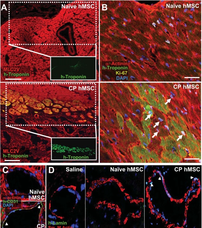
A, Human-specific troponin-I (green) in the anterior wall of naive- versus CP-treated hearts, respectively, co-localized with ventricular myosin light chain (MLC2v, red). Bar, 100 μm. B, Human troponin-I staining of naïve versus CP hMSC treated hearts, counterstained with α-Actinin (red), demonstrated engraftment of human cells. Cell cycle activation, documented by Ki-67 expression (yellow, arrows), noted in human troponin positive and endogenous cardiomyocytes. C, Confocal evaluation of collateral vessels from CP hMSC treated hearts demonstrated human-specific CD-31 (PECAM-1) staining. D, Human lamin staining (arrows) co-localized with nuclei of smooth muscle in vessels from CP hMSC treated but not saline or naïve treated hearts. Bar, 20 μm for B-D.
A, Human-specific troponin-I (green) in the anterior wall of naive- versus CP-treated hearts,
respectively, co-localized with ventricular myosin light chain (MLC2v, red). Bar, 100 μm.
B, Human troponin-I staining of naïve versus CP hMSC treated hearts, counterstained with
α-Actinin (red), demonstrated engraftment of human cells. Cell cycle activation,
documented by Ki-67 expression (yellow, arrows), noted in human troponin positive and
endogenous cardiomyocytes. C, Confocal evaluation of collateral vessels from CP hMSC
treated hearts demonstrated human-specific CD-31 (PECAM-1) staining. D, Human lamin
staining (arrows) co-localized with nuclei of smooth muscle in vessels from CP hMSC
treated but not saline or naïve treated hearts. Bar, 20 μm for B-D.

In a paper from February 2013 (Bartunek J, et al., Journal of the American College of Cardiology (2013), doi: 10.1016/j.jacc.2013.02.071), Terzic and his team has reported on the administration of BM-MSCs into the hearts of 34 heart patients. Of these patients, 21 were implanted with their own BM-MSCs that had undergone guided cardiopoiesis and the other 12 received standard therapy for heart patients with no transplanted cells.

The results from this study were striking to say the least. According to Terzic, “The benefit to patients who received cardiopoietic stem cell delivery was significant.” Cardiologist Charles Murry wrote in an editorial, “Six months after treatment, the cell therapy group had a seven percent absolute improvement in EF (ejection fraction) over baseline, versus a non-significant change in the control group. The improvement in EF is dramatic, particularly given the duration between the ischemic injury and cell therapy. It compared favorably with our most potent therapies in heart failure.”

This clinical trial, known as the C-CURE trial, which stands for Cardiopoietic Stem Cell Therapy in Heart Failure. was an international, multi-center trial that treated enrolled patients from hospitals in Belgium, Serbia, and Switzerland. This trial represents the culmination of almost a decade of work by Terzic and others. “Discovery of rare stem cells that could inherently promote heart regeneration provided a critical clue. In following this natural blueprint, we further developed the know-how needed to convert patient-derived stem cells into cells that can reliably repair a failing heart.”

For this trial, Mayo Clinic partnered with Cadio3 Biosciences, which is a bio-science company in Mort-Saint-Guilbert, Belgium. This company provided advance product development, manufacturing scale-up, and clinical trial execution.  Adaptation of this exciting new technology to the clinic could mean a new exciting fix for heart patients.

Allergy-Associated White Blood Cell Triggers Stem Cell-Mediated Muscle Repair


White blood cells help our bodies ward off invasions from microorganisms, but they serve other purposes too. Once white blood cell in particular, the “eosinophil” helps us when we are infected by multicellular parasites (worms and the like). Eosinophils, however, also play a more unpleasant role, and that is in allergies. When we suffer from allergies, eosinophils multiply and move to our lungs and other places, where they mediate inflammation and tissue damage. Thus eosinophils are the white blood cells we all love to hate.

However, researchers at the University of California, San Francisco (UCSF) have generated new data that, in their view, suggests that eosinophils also play an integral role in muscle regeneration.

Ajay Chawla, Associate Professor at the Cardiovascular Research Institute at UCSF and

Eosinophil
Eosinophil

lead researcher for this study, said, “Eosinophils are needed for the rapid clearance of necrotic debris, a process that is necessary for timely and complete regeneration of tissues.”

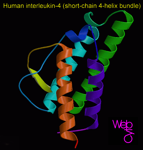
Chawla’s laboratory showed that eosinophils serve double duty when it comes to muscle repair. First, they remove the cellular debris that results from damaged tissues. Secondly, eosinophils secrete a protein called “Interleukin 4” or IL-4. IL-4 triggers a specific type of stem cell to replicate and repair muscle tissue.

Interleukin-4
Interleukin-4

According to Chawla, “Without eosinophils you cannot regenerate muscle.”

These eosinophil-activated stem cells are known as “fibro/adipogenic progenitors” (FAP). Until recently, the general thinking surrounding FAPs was that they could only form fat tissue (see Natarajan A., Lemos DR, and Rossi FM, Cell Cycle 2010 9(11): 2045-6).  However, when FAPs are exposed to IL-4, they begin to differentiate into muscle fibers.

“They wake up the cells in muscle that divide and form muscle fibers,” said Chawla

“Bites from venomous animals, many toxicants, and parasitic worms all trigger somewhat similar immune responses that cause injury. We want to know if eosinophils and FAPs are universally employed in these situations as a way to get rid of debris without triggering severe reactions such as anaphylactic shock,” said Chawla.

Making New Neurons When You Need Them


Western societies are aging societies, and the incidence of dementias, Alzheimer’s disease, and other diseases of the aged are on the rise. Treatments for these conditions are largely supportive, but being able to make new neurons to replace the ones that have died is almost certainly where it’s at.

At INSERM and CEA in Marseille, France, researchers have shown that chemicals that block the activity of a growth factor called TGF-beta improves the generation of new neurons in aged mice. These findings have spurred new investigations into compounds that can enable new neuron production in order to mitigate the symptoms of neurodegenerative diseases. Such treatments could also restore the cognitive abilities of those who have suffered neuron loss as a result of radiation therapy or a stroke.

The brain forms new neurons regularly to maintain our cognitive abilities, but aging or radiation therapy to treat tumors can greatly perturb this function. Radiation therapy is the adjunctive therapy of choice for brain tumors in children and adults.

Various studies suggest that the reduction in our cache of neurons contributes to cognitive decline. For example, exposure of mice to 15 Grays of radiation is accompanied by disruption to the olfactory memory and reduction in neuron production. A similar event occurs as a result of aging, but in human patients undergoing radiation treatment, cognitive decline is accelerated and seems to result from the death of neurons.

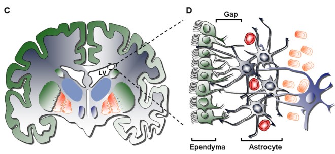
How then, can we preserve the cache of neurons in our brains? The first step is to determine the factors responsible for the decline is neuron production. In contrast to contemporary theory, neither heavy doses of radiation nor aging causes completely destruction of the neural stem cells that can replenish neurons. Even after doses of radiation and aging, neuron stem cell activity remains highly localized in the subventricular zone (a paired brain structure located in the outer walls of the lateral ventricles), but they do not work properly.

Subventricular Zone
Subventricular Zone

Experiments at the INSERM and CEA strongly suggest that in response to aging and high doses of radiation, the brain makes high levels of a signaling molecule called TGF-beta, and this signaling molecule pushes neural stem cell populations into dormancy. This dormancy also increases the susceptibility of neural stem cells into apoptosis.

Marc-Andre Mouthon, one of the main authors of this research, explained his results in this manner: “Our study concluded that although neurogenesis is reduced in aging and after a high dose of radiation, many stem cells survive for several months, retaining their ‘stem’ characteristics.”

Part two of this project showed that blocking TGFbeta with drugs restored the production of new neurons in aging or irradiated mice.

Thus targeted therapies that block TGFbeta in the brains of older patients or cancer patients who have undergone high dose radiation for a brain tumor might reduce the impact of brain lesions caused by such events in elderly patients who show distinct signs of cognitive decline.

Adult Stem Cells Isolated From Human Intestines


A laboratory at the University of North Carolina at Chapel Hill has, for the first time, isolated adult stem cells from human intestinal tissue. This achievement should provide a much-needed resource for stem cells researchers to examine the nuances of stem cell biology. Also, these new stem cells should provide stem cell researchers a new tool to treat inflammatory bowel diseases or to mitigate the side effects of chemotherapy and radiation, which often damage the gut.

Scott T. Magness, assistant professor in the departments of physiology at UNC, Chapel Hill, said, “Not having these cells to study has been a significant roadblock to research. Until now, we have not had the technology to isolate and study these stem cells – now we have the tools to start solving many of these problems.”

The study represents a leap forward for a field that for many years has had to resort to conducting experiments with mouse stem cells. While significant progress has been made using mouse models, differences in stem cell biology between mice and humans have kept researchers from investigating new therapeutics for human afflictions.

Adam Grace, a graduate student in Magness’ lab, and one of the first authors of this publication, noted, “While the information we get from mice is good foundational mechanistic data to explain how this tissue works, there are some opportunities that we might not be able to pursue until we do similar experiments with human tissue”

This study from the Magness laboratory was the first in the United States to isolate and grow single intestinal stem cells from mice. Therefore, Magness and his colleagues already had experience with the isolation and manipulation of intestinal stem cells. In their quest to isolate human intestinal stem cells, Magness and his colleagues also procured human small intestinal tissue for their experiments that had been discarded after gastric bypass surgery at UNC.

To develop their technique, Magness and others simply tried to recapitulate the technique they had developed in used to isolate mouse intestines to isolate stem cells from human intestinal stem cells. They used cell surface molecules found on in the membranes of mouse intestinal stem cells. These proteins, CD24 and CD44, were also found on the surfaces of human intestinal stem cells. Therefore, the antibodies that had been used to isolate mouse intestinal stem cells worked quite well to isolate human intestinal stem cells. Magness and his co-workers attached fluorescent tags to the stem cells and then isolated by means of fluorescence-activated cell sorting.

This technique worked so well, that Magness and his colleagues were able to not only isolated human intestinal stem cells, but also distinct types of intestinal stem cells. These two types of intestinal stem cells are either active stem cells or quiescent stem cells that are held in reserve. This is a fascinating finding, since the reserve cells can replenish the stem cell population after radiation, chemotherapy, or injury.

“Now that we have been able to do this, the next step is to carefully characterize these populations to assess their potential, said Magness. He continued: “Can we expand these cells outside the body to potentially provide a cell source for therapy? Can we use these for tissue regeneration? Or to take it to the extreme, can we genetically modify these cells to cure inborn disorders or inflammatory bowel disease? Those are some questions that we are going to explore in the future.”

Certainly more papers are forthcoming on this fascinating and important topic.

The Surprising Ability of Blood Stem Cells to Respond to Emergencies


A research team from Marseille, France has revealed an unexpected role for hematopoietic stem cells (the cells that make blood cells): not only do these cells continuously renew our blood cells, but in emergencies these cells can make white blood cells on demand. that help the body deal with inflammation and infection. This stem cell-based activity could be utilized to protect against infection in patients who are undergoing a bone marrow transplant.

The research team that discovered this previously unknown property of hematopoietic stem cells were from INSERM, CNRS and MDC led by Michael Sieweke of the Centre d’Immunologie de Marseille Luminy and the Max Delbruck Centre for Molecular Medicine, Berlin-Buch.

Cells in our blood feed, clean, and defend our tissues, but their lifespan is limited. The life expectancy of a red blood cell rarely exceeds three months, our platelets die after ten days and the vast majority of our white blood cells survive only a few days.

Therefore, our bodies must produce replacements for these dying cells in a timely manner and in the right quantities and proportions. Blood cells replacement is the domain of the hematopoietic stem cells, which are nested in the bone marrow; that soft tissue inside long bones of the chest, spine, pelvis, upper leg and shoulder. Bone marrow produces and releases billions of new cells into out blood every day. To do this, hematopoietic stem cells must not only divide but their progeny must also differentiate into specialized cells, such as white blood cells, red blood cells, platelets, and so on.

For several years, researchers have been interested in how the process of differentiation and specialization is triggered in stem cell progeny. Sieweke and his colleagues discovered in previous work that hematopoietic stem cell progeny are not preprogrammed to assume a particular cell fate, but respond to environmental cues that direct them to become one cell type or another.

Nevertheless, it is still unclear how stem cells respond during emergencies? How are hematopoietic stem cells able to meet the demand for white blood cells during an infection? Recently, the answer was considered clear: the stem cells neither sensed nor responded to the signals sent to induce their progeny to differentiate into particular cell types. They merely proliferated and their progeny responded to the available signals and differentiated into the necessary cell fates. However, Sieweke’s research team has found that rather than being insensitive to these inductive signals meant for their progeny, hematopoietic stem cells perceive these environmental signals and, in response to them, manufacture the cells that are most appropriate for the danger faced by the individual.

Dr. Sandrine Sarrazin, INSERM researcher and co-author of the publication, said, “We have discovered that a biological molecule produced in large quantities by the body during infection or inflammation directly shows stem cells the path to take.”

Sieweke added, “Now that we have identified this signal, it may be possible in the future to accelerate the production of these cells in patients facing the risk of acute infection.” He continued: “This is the case for 50,000 patients worldwide each year who are totally defenseless against infections just after bone marrow transplantation. Thanks to M-CSF [monocyte-colony stimulating factor], it may be possible to stimulate the production of useful cells while avoiding to produce those that can inadvertently attack the body of these patients. They could therefore protect against infections while their immune system is being reconstituted.”

To reach their conclusions the team had to measure the change of state in each cell. This was a terrifically difficult challenge since the stem cells in question are very rare in the bone marrow: only one cell in 10,000 in the bone marrow of a mouse. Furthermore, the hematopoietic stem cells are, by appearance, indistinguishable from their progeny, the hematopoietic progenitor cells. Therefore, this experiment was tedious and difficult, but it proved that M-CSF could instruct single hematopoietic stem cells to differentiate into the monocyte lineage.

The clinical use of M-CSF will hopefully follow in the near future, but for now, this is certainly an exciting finding that may lead to clinical trials and applications in the future.

The Transformation of Ordinary Skin Cells into Functional Brain Cells


A paper in Nature Biotechnology from research groups at Case Western Reserve School of Medicine describes a technique that directly converts skin cells to the specific type of brain cells that suffer destruction in patients with multiple sclerosis, cerebral palsy, and other so-called myelin disorders. This particular breakthrough now enables “on demand” production of those cells that wrap or “myelinate” the axons of neurons.

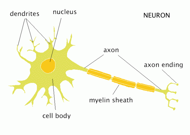
Myelin is a sheath that wraps the extension of neurons called the axons. Neurons are the conductive cells that initiate and propagate nerve impulses. Neurons contain cell extensions known as axons that connect with other neurons. The nerve impulse runs from the base of the cell body of the neurons, down the axon, to the neuron to which it is connected. An insulating myelin sheath that surrounds the axon increases the speed at which nerve impulses move down the axon. When this myelin sheath is damaged, nerve impulse conduction goes awry as does nerve function. For example, patients with multiple sclerosis (MS), cerebral palsy (CP), and rare genetic disorders called leukodystrophies, myelinating cells are destroyed are not replaced.

neuron

The new technique discussed in this Nature Biotechnology paper, directly converts skin cells called fibroblasts, which are rather abundant in the skin and most organs, into oligodendrocytes, the type of cell that constructs the myelin sheath in the central nervous system.

Oligodendrocyte

“Its ‘cellular alchemy,'” explains Paul Tesar, PhD, assistant professor of genetics and genome sciences at Case Western Reserve School of Medicine and senior author of the study. “We are taking a readily accessible and abundant cell and completely switching its identity to become a highly valuable cell for therapy.”

Tesar and his group used a technique called “cellular reprogramming,” to manipulate the levels of three naturally occurring proteins to induce the fibroblasts to differentiate into the cellular precursors to oligodendrocytes (called oligodendrocyte progenitor cells, or OPCs).

OPCs

Led by Case Western Reserve researchers and co-first authors Fadi Najm and Angela Lager, Tesar’s research team rapidly generated billions of these induced OPCs (called iOPCs). They demonstrated that iOPCs could regenerate new myelin coatings around nerves after being transplanted to mice—a result that offers hope the technique might be used to treat human myelin disorders.

Demyelinating diseases damage the oligodendrocytes and cause loss of the insulating myelin coating. A cure for these diseases requires replacement of the myelin coating by replacement oligodendrocytes.

Until now, OPCs and oligodendrocytes could only be obtained from fetal tissue or pluripotent stem cells. These techniques have been valuable, but have distinct limitations.

“The myelin repair field has been hampered by an inability to rapidly generate safe and effective sources of functional oligodendrocytes,” explains co-author and myelin expert Robert Miller, PhD, professor of neurosciences at the Case Western Reserve School of Medicine and the university’s vice president for research. “The new technique may overcome all of these issues by providing a rapid and streamlined way to directly generate functional myelin producing cells.”

Even though this initial study used mouse cells, the next critical next step is to demonstrate feasibility and safety of human cells in a laboratory setting. If successful, the technique could have widespread therapeutic application to human myelin disorders.

“The progression of stem cell biology is providing opportunities for clinical translation that a decade ago would not have been possible,” says Stanton Gerson, MD, professor of Medicine-Hematology/Oncology at the School of Medicine and director of the National Center for Regenerative Medicine and the UH Case Medical Center Seidman Cancer Center. “It is a real breakthrough.”

The Nooks and Crannies in Bone Marrow that Nurture Stem Cells


Stems cells in our bodies often require a specific environment to maximize their survival and efficiency. These specialized locations that nurture stem cells is called a stem cell niche. Finding the right niche for a stem cell population can go a long way toward growing more stem cells in culture and increasing their potency.

To that end, a recent discovery has identified the distinct niches that exist in bone marrow for hematopoietic stem cells (HSCs), which form the blood cells in our bodies.

A research team from Washington university School of Medicine in St. Louis has shown that stem niches in bone marrow can be targeted, which may potentially improve bone marrow transplants and cancer chemotherapy. Drugs that support particular niches could encourage stem cells to establish themselves in the bone marrow, which would greatly increase the success rate of bone marrow transplants. Alternatively, tumor cells are known to hide in stem cell niches, and if drugs could disrupt such niches, then the tumor cells would be driven from the niches and become more susceptible to chemotherapeutic agents.

Daniel Link, the Alan A. and Edith L. Wolff Professor of Medicine at Washington University, said, “Our results offer hope for targeting these niches to treat specific cancers or to impress the success of stem cell transplants. Already, we and others are leading clinical trials to evaluate whether it is possible to disrupt these niches in patients with leukemia or multiple myeloma.”

Working in mice, Link and his colleagues deleted a gene called CXCL12, only in “candidate niche stromal cell populations.” CXCL12 which encodes a receptor protein known to be crucial for maintaining HSC function, including retaining HSCs in the bone marrow, controlling  HSCs activity, and repopulating the bone marrow with HSCs after injury.

CXCL12 crystal structure

CXCL12 signaling pathways

In bone marrow, HSCs are surrounded by a whole host of cells, and it is difficult to precisely identify which type of cells serve as the niche cells. These bone marrow cells are known collectively as “stroma,” but there are several different types of cells in stroma. Cells that have been implicated in the HSC niche include endosteal osteoblasts (osteoblasts are bone-making cells and the endosteum in the layer of connective tissue that lines the inner cavity of the bone), perivascular stromal cells (cells that hang out around blood vessels), CXCL12-abundant reticular cells, leptin-receptor-positive stromal cells, and nestin–positive mesenchymal progenitors. Basically, there are a lot of cells in the stroma and figuring out which one is the HSC niche is a big deal.

bone marrow stromal cells

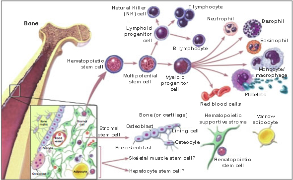
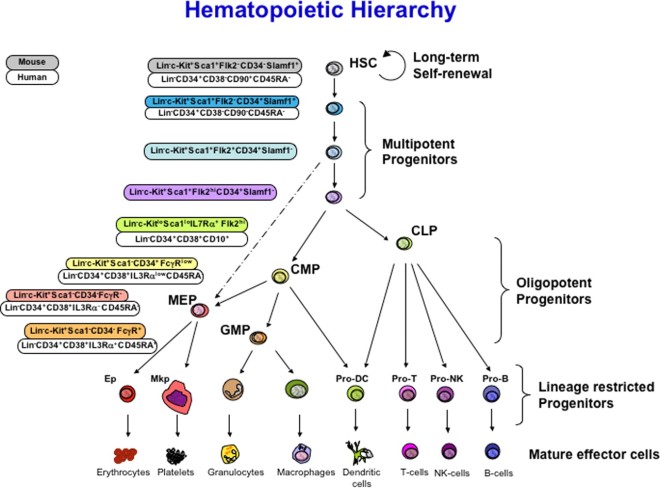
When HSCs divide, they form two cells, one of which replaces the HSC that just divided and a new cells called a hematopoietic progenitor cell (HPC), which can divide and differentiate into either a lymphoid progenitor or a myeloid progenitor. The lymphoid progenitor differentiates into either a B or T lymphocyte and the myeloid progenitor differentiates into a red blood cell, or other types of white blood cells (neutrophil, basophil, macrophage, platelet or eosinophil). As the cells become more differentiated, they lose their capacity to divide.

HSC differentiation

Deleting CXCL12 from mineralizing osteoblasts (bone making cells) did nothing to the HSCs or those cells that form lymphocytes (lymphoid progenitors). Deletion of Cxcl12 from osterix-expressing stromal cells, which include CXCL12-abundant reticular cells and osteoblasts, causes mobilization of hematopoietic progenitor cells (HPCs) from the bone marrow into the bloodstream, and loss of B-lymphoid progenitors, but HSC function is normal. Cxcl12 deletion from blood vessel cells causes a modest loss of long-term repopulating activity. Deletion of Cxcl12 from nestin-negative mesenchymal progenitors causes a marked loss of HSCs, long-term repopulating activity, and lymphoid progenitors. All of these data suggest that osterix-expressing stromal cells comprise a distinct niche that supports B-lymphoid progenitors and retains HPCs in the bone marrow. Also, the expression of CXCL12 from stromal cells in the perivascular region, including endothelial cells and mesenchymal progenitors, supports HSCs.

Link summarized his results this way: “What we found was rather surprising. There’s not just one niche for developing blood cells in the bone marrow. There’s a distinct niche for stem cells, which have the ability to become any blood cell in the body, and a separate niche for infection-fighting cells that are destined to become T cells and B cells.”

These data provide the foundation for future investigations whether disrupting these niches can improve the effectiveness of cancer chemotherapy.

In a phase 2 study at Washington University, led by oncologist Geoffrey Uy, assistant professor of medicine, Link and his team are evaluating whether the drug G-CSF (granulocyte colony stimulating growth factor) can alter the stem cell niche in patients with acute lymphoblastic leukemia and whose disease is resistant to chemotherapy or has recurred. The FDA approved this drug more than 20 years ago to stimulate the production of white blood cells in patients undergoing chemotherapy, who have often weakened immune systems and are prone to infections.

Uy and his colleagues want to evaluate G-CSF if it is given prior to chemotherapy. Patients enrolled at the Siteman Cancer Center will receive G-CSF for five days before starting chemotherapy, and the investigators will determine whether it can disrupt the protective environment of the bone marrow and make cancer cells more sensitive to chemotherapy.

This trial is ongoing, and the results are not yet in, but Link’s work has received a welcome corroboration of his work. A companion paper was published in the same issue of Nature by Sean Morrison, the director of the Children’s Medical Center Research Institute at the University of Texas Southwestern Medical Center in Dallas. Morrison and his team used similar methods as Link and his colleagues and came to very similar conclusions.

Link said, “There’s a lot of interest right now in trying to understand these niches. Both of these studies add new information that will be important as we move forward. Next, we hope to understand how stem cells niches can be manipulated to help patients undergoing stem cell transplants.”

Increasing Reprogramming Efficiency by Turning Off One Gene


The removal of one genetic roadblock could improve the efficiency of adult cell reprogramming by some 10 to 30 fold, according to research by stem cell scientists at the Methodist Hospital Research Institute and two other institutions.

Rongfu Wang, the principal investigator and director of the Center for Inflammation and Epigenetics, said this about his group’s findings: “The discovery six years ago that scientist can convert adult cells into inducible pluripotent stem cells, or iPSCs, bolstered the dream that a patient’s own cells might be reprogrammed to make patient-specific iPSCs for regenerative medicine, modeling human diseases in Petri dishes, and drug screening. But reprogramming efficiency has remained very low, impeding its applications in the clinic.”

Wang and his group identified a protein encoded by a gene called Jmjd3, which is known as KDM6B, acts as an impediment to the reprogramming of adult cells into iPSCs. Jmjd3 is involved in several different biological processes, including the maturation of nerve cells and immune cell differentiation (Popov N, Gil J. Epigenetics. 2010 5(8):685-90).

These findings by Wang’s team are the first time anyone has identified a role for Jmjd3 in the reprogramming process. According to Wang, fibroblasts that lack functional Jmjd3 showed greatly enhanced reprogramming efficiency.

Helen Wang, one of the co-principal authors of this study, said, “Our findings demonstrate a previously unrecognized role of Jmjd3 in cellular reprogramming and provide molecular insight into the mechanisms by which the Jmjd3-PHF20 axis controls this process.’

While teasing apart the roles of Jmjd3 in reprogramming, Wang and his colleagues discovered that this protein regulates cell growth and cellular aging. These are two previously unidentified functions of Jmjd3, and Jmjd3 appears to work primarily by inactivating the protein PHF20. PHF20 is a protein that is required for adult cell reprogramming, and cells that lack PHF20 do not undergo reprogramming to iPSCs.

Rongfu Want explained it like this: “So when it comes to increasing iPSC yields, knocking down Jmjd3 is like hitting two birds with one stone.”

Jmjd3 is almost certainly not the only genetic roadblock to stem cell conversion. Wang noted, “Removal of multiple roadblacks could further enhance the reprogramming efficiency with which researchers can efficiently generate patient-specific iPSCs for clinical applications.”

While this is certainly an exciting finding, there is almost certainly a caveat that comes with it. increased reprogramming efficiency almost certainly brings the potential for increased numbers of mutations. Other studies have shown that iPSC generation is much more efficient if the protein P53 is inhibited, but P53 is the guardian of the genome. It prevents the cell from dividing if there is substantial amounts of DNA damage. Inhibiting P53 activity allows iPSC generation even if the cells have excessive amounts of DNA damage. Therefore, inhibiting those cellular processes that are meant to guard against excessive cell proliferation and growth can lead to greater numbers of mutations. Thus, before Jmjd3 inactivation is used to generate iPSCs for clinical uses, extensive animal testing must be required to ensure that this procedure does make iPSCs even less safe than they already are.

Stephen Hawking Visits UCLA Stem Cell Laboratory


Stephen Hawking
Stephen Hawking

On Tuesday, Stephen Hawking toured a stem cell laboratory where scientists are studying ways to slow the progression of Lou Gehrig’s disease, a neurological disorder that has left the British cosmologist almost completely paralyzed.

After the visit, the 71-year-old Hawking urged doctors, nurses and staff at Cedars-Sinai Medical Center to support the research.

Hawking recalled how he became depressed when he was diagnosed with the disease 50 years ago and initially didn’t see a point in finishing his doctorate. But his attitude changed when his condition didn’t progress as fast and he was able to concentrate on his studies.

“Every new day became a bonus,” he said.

The hospital last year received nearly $18 million from California’s taxpayer-funded stem cell institute to study the debilitating disease also known as amyotrophic lateral sclerosis. ALS attacks nerve cells in the brain and spinal cord that control the muscles. People gradually have more and more trouble breathing and moving as muscles weaken and waste away.

There’s no cure and no way to reverse the disease’s progression. Few people with ALS live longer than a decade.

Diagnosed at age 21 while a student at Cambridge University, Hawking has survived longer than most. He receives around-the-clock care, can only communicate by twitching his cheek, and relies on a computer mounted to his wheelchair to convey his thoughts in a distinctive robotic monotone.

A Cedars-Sinai patient who was Hawking’s former student spurred doctors to invite the physicist to glimpse their stem cell work.

“We decided it was a great opportunity for him to see the labs and for us to speak to one of the preeminent scientists in the world,” said Dr. Robert Baloh, who heads the hospital’s ALS program.

Cedar-Sinai scientists have focused on engineering stem cells to make a protein in hopes of preventing nerve cells from dying. The experiment so far has been done in rats. Baloh said he hopes to get governmental approval to test it in humans, which would be needed before any therapy can be approved.

Hawking is famous for his work on black holes and the origins of the universe. His is also famous for bringing esoteric physics concepts to the masses through his best-selling books including “A Brief History of Time,” which sold more than 10 million copies worldwide. Hawking titled his speech to Cedars-Sinai employees “A Brief History of Mine.”

Despite his diagnosis, Hawking has remained active. In 2007, he floated like an astronaut on an aircraft that creates weightlessness by making parabolic dives.

Doctors don’t know why some people with Lou Gehrig’s disease fare better than others. Dr. Baloh said he has treated patients who lived for 10 years or more.

“But 50 years is unusual, to say the least,” he said.

A Stem Cell Treatment for Stroke


A new clinical trial is enrolling people who are dealing with the disabling effects of stroke.

Every year approximately 800,000 Americans suffer a stroke. Strokes or TIAs for “trans-ischemic attacks” result from blockage of a blood vessel in the brain. The lack of blood flow to the brain results in the death of those cells that starve from oxygen, and the aftermath of a stroke is remarkably unpleasant; long-term disability, permanent brain damage, and even death. Stroke is the leading cause of adult disability and extracts an annual burden of $62 billion on the US economy. Physical therapy can improve the deficits caused by a stroke, but there are, to date, no good treatments to ameliorate the condition of a stroke patient.

In the hopes of creating new options for stroke patients, researchers at Northwestern Medicine are examining a new regenerative treatment for stroke that utilizes a novel stem cell line called SB623. This stem cell line might provide increased motor function to stroke victims.

Northwestern is only one of three sites in the nation enrolling patients in a clinical study to evaluate the efficacy and safety of adult stem cell therapy in stroke patients. Patient who have suffered from so-called “ischemic stroke” suffer from impaired bodily functions that includes such conditions as paralysis, weakness on one side, difficulty with speech and language, vision issues, and cognitive deficits.

Joshua Rosenow, the principal investigator of this clinical trial, is the director of Functional Neurosurgery at Northwestern Memorial Hospital. Rosenow had this to say of this clinical study: “Two million brain cells die each minute during a stroke making it critical to get treatment fast at the earliest sign of symptoms once brain damage occurs, there’s very little that can be done medically to reverse it. While this study is only a preliminary step towards understanding the healing potential of these cells, we are excited about what a successful trial could do for a patient population that hs very limited therapeutic options.”

The primary purpose of this study is to examine the safety of SB623 stem cells. However, there is an added motive behind this study, and that is to determine if SB623 cells are efficacious as a treatment for stroke patients. SB623 cells are genetically engineered mesenchymal stem cells from adult bone marrow.

Richard Bernstein, the director of Northwestern Memorial’s Stroke Center, weighed in: “Although not proven in humans, these stem cells (SB623) have been shown to promote healing and improve function when administered in animal models of stable stroke. The cells did not replace the neurons destroyed by stroke, but instead they appeared to encourage the brain to heal itself and promote the body’s natural regenerative process. Eventually, the implanted stem cells disappeared.”

Rosenow added, “In this study, the cells are transplanted into the brain using brain mapping technology and scans, allowing us to precisely deposit the cells in the brain adjacent to the area damaged by the stroke.”

The first participants have received injections of 25 million cells, but as the study progresses, the dose will escalate to 5 million and eventually 10 million cells. Since SB623 cells are allogeneic, which is to say that they come from someone other than the patient, a single donor’s cells can be used to treat as many other patients. All subjects in this study will be followed for up to two years with periodic evaluations for safety and effectiveness in improving motor function.

Bernstein explained, “Stroke can be a very disabling and life-changing event. Even just a slight improvement in function could make a huge difference for a person impacted [sic] by stroke. To potentially regain movement or speech is a very exciting prospect. In the animal models, the improvements appeared to remain even after the implanted stem cells disappeared.

Even at this early stage in this clinical trial, there is a great deal of excitement over the potential for stem cell therapy. Rosenow echoed this excitement when he said, “Of these cells are proven effective in improving, or even reversing brain damage, the implications of a successful outcome reach far beyond just stroke. Stem cell therapy may hold the key to treating a wide range of neurological disorders that do not have many available therapies. The Northwestern team is very excited to be a part of this groundbreaking trial.”

Participants for this trial must be between the ages of 18 and 75 years old, must have had an ischemic stroke in the last six to 36 months. They should have moderate to severe symptoms with impaired motor function. Full inclusion and exclusion criteria are available online. Full inclusion and exclusion criteria are available online. The FDA-approved phase 1-11 study is expected to enroll 18 subjects nationwide and this study is slated to last up to two years.

Other sites participating in the trial are the University of Pittsburgh Medical Center and Stanford University School of Medicine. The trial is funded by SanBio, Inc., a regenerative medicine company that developed the SB623 stem cell line.

SB623 papers:

1. Extracellular matrix produced by bone marrow stromal cells and by their derivative, SB623 cells, supports neural cell growth.  Aizman I, Tate CC, McGrogan M, Case CC.

  • J Neurosci Res. 2009, 87(14):3198-206.

2. Notch-induced rat and human bone marrow stromal cell grafts reduce ischemic cell loss and ameliorate behavioral deficits in chronic stroke animals. Yasuhara T, Matsukawa N, Hara K, Maki M, Ali MM, Yu SJ, Bae E, Yu G, Xu L, McGrogan M, Bankiewicz K, Case C, Borlongan CV.  Stem Cells Dev. 2009, 18(10):1501-14

3. Reversal of dopaminergic degeneration in a parkinsonian rat following micrografting of human bone marrow-derived neural progenitors. Glavaski-Joksimovic A, Virag T, Chang QA, West NC, Mangatu TA, McGrogan MP, Dugich-Djordjevic M, Bohn MC.  Cell Transplant. 2009, 18(7):801-14.

4.

Tate CC, Fonck C, McGrogan M, Case CC. Cell Transplant. 2010,19(8):973-84.

5. Glial cell line-derived neurotrophic factor-secreting genetically modified human bone marrow-derived mesenchymal stem cells promote recovery in a rat model of Parkinson’s disease.  Glavaski-Joksimovic A, Virag T, Mangatu TA, McGrogan M, Wang XS, Bohn MC. J Neurosci Res. 2010, 88(12):2669-81.

6. Comparing the immunosuppressive potency of naive marrow stromal cells and Notch-transfected marrow stromal cells.

  • Dao MA, Tate CC, Aizman I, McGrogan M, Case CC.

J Neuroinflammation. 2011, 8(1):133.

7.

Tate CC and Case CC.

  • Chapter in “Neurological Disorders”, InTech, 2012.

Stem Cell Therapy for Inflammatory Bowel Disease


Graca Almeida-Porada is professor of regenerative medicine at Wake Forest University in the Wake Forest Baptist’s Institute for Regenerative Medicine. Dr. Almeida-Porta and her colleagues have identified a special population of stem cells in the bone marrow that can migrate to the intestine and regenerate the intestine. Thus this stem population might provide a treatment for inflammatory bowel diseases or IBDs.

Approximately one million Americans have IBDs, and the main IBDs are ulcerative colitis, which is restricted to the large intestine, and Crohn’s disease, which involves the small and large intestine. These IBDs result from the immune system recognizing some component of the gastrointestinal system as foreign. and the immune system then attacks the gastrointestinal system as though it was a foreign invader. The result is chronic inflammation in the gastrointestinal tract, pain, bloody stools, redness and swelling of the bowel, in some severe cases, rupture of the bowel and death.

There are no cures for IDBs, but several drugs that suppress the immune response against the bowel, such as mesalamine (marketed as Asacol), sulfasalazine (Azulfidine), balsalazide (Colazal) and olsalazine (Dipentum) can reduce inflammation and assuage the symptoms of IBDs. However, there is no treatment to replace the damaged and dead cells in the bowel that result from the inflammation. Even though the bowel does regenerate to some degree, these extra bouts of cell proliferation can increase the patient’s risk of colon cancer. Is there a stem cell treatment to regenerate the bowel?

Research from Almeida-Porada’s laboratory has identified stem cells from umbilical cord blood that can make blood vessels that can also migrate to the intestine and liver (Wood JA, et al., Hepatology. 2012 Sep;56(3):1086-96). Now work in her lab has extended this original observations.

“We’ve identified two populations of human cells that migrate to the intestine – one involved in blood vessel formation and the other that can replenish intestinal cells and modulate inflammation,” said Almeida-Porada. She continued: “Our hope is that a mixture of these cells could be used as an injectable therapy to treat IBD.”

These cells would theoretically contribute cells to the intestine and facilitate and induce tissue healing and recovery. The lining of the intestine has one of the highest cellular turnover rates in the body. Intestinal cell types are being renewed weekly from this pool of intestinal cells that are in an area of the intestine known as the crypt.

In this current study, Almeida-Porada’s team used specific cell surface proteins (cell markers) to identify a stem cell population in human bone marrow that possesses the highest potential to migrate to the intestine and thrive in the intestine. These intestine-bound cells expressed high levels of a protein called ephrin type B, which is typically found on the surfaces of cells involved in tissue repair and wound closure.

Ephrin Protein Structure

When these ephrin type B-enriched bone marrow cells were injected into fetal sheep, the bone marrow-derived cells were able to migrate to the intestine and contribute to the growth and development of the sheep intestine.  Interestingly, these cells took up their positions in the intestinal crypts.

Almeida-Porada comment on her work:  “Previous studies in animals have shown that the transplantation of bone-marrow-derived cells can contribute to the regeneration of the gastrointestinal tract in IBD.  However, only small numbers of cells were successfully transplanted using this method.  Our goal with the current study was to identify populations of cells that naturally migrate to the intestine and have the intrinsic ability to restore tissue health.”

While these two studies show that the cells can migrate to and survive in a healthy intestine, the next step will be to determine whether they can survive in an inflamed intestine, like the type found in IBD patients.  In could be that preconditioning of the cells is required, as in the case of stem cell treatments for the heart after a heart attack.

Stem Cell Homing Factor Used to Treat Heart Patients


In a clinical trial that is probably one of the first of its kind, researchers from the laboratory of Marc Penn at the Summa Cardiovascular Institute in Akron, Ohio, activated the stem cells of heart failure patients by means of gene therapy.

Penn and his colleagues delivered a gene that encodes stromal-cell derived factor-1 or SDF-1. SDF-1 is a member of the chemokine family of signaling proteins, and chemokines are proteins that direct cells to get up and move somewhere. Thus, for stem cells, SDF-1 acts as a kind of “homing” signal.

Stromal-cell derived factor

In this unique study, Penn and his collaborators introduced SDF-1 into the heart in order to summon stem cells to the site of injury and enhance the body’s stem cell-based repair process. In a typical stem cell-based study, researchers extract and expand the number of cells, then deliver them back to the subject, but in this study, no stem cells were extracted. Instead they were summoned to the site of injury by SDF-1.

Marc Penn, professor of medicine at Northeast Ohio Medical University in Rootstown, Ohio and the director of research at Summa Cardiovascular Institute said of his clinical trial: “We believe stem cells are always trying to repair tissue, but they don’t do it well — not because we lack stem cells but, rather, the signals that regulate our stem cells are impaired.”

Previous research by Penn and colleagues has shown SDF-1 activates and recruits the body’s stem cells to sites of injury and this increases healing. Under normal conditions, SDF-1 is made after an injury but its effects are short-lived. For example, SDF-1 is naturally expressed after a heart attack but this augmented expression of SDF-1 only lasts only a week.

In the study, researchers attempted to re-establish and extend the time that SDF-1 could stimulate patients’ stem cells. The trial enrolled 17 NYHA Class III heart failure patients, with left ventricular ejection fractions less than 40% and an average time from heart attack of 7.3 years. Three escalating JVS-100 doses were evaluated: 5 mg (cohort 1), 15 mg (cohort 2) and 30 mg (cohort 3). The average age of the participants was 66 years old.

Researchers injected one of three doses of the SDF-1 gene (5mg, 15mg or 30mg) into the hearts of these patients, and monitored them for up to a year. Four months after treatment, they found:
1. Patients improved their average distance by 40 meters during a six-minute walking test.
2. Patients reported improved quality of life.
3. The heart’s pumping ability improved, particularly for those receiving the two highest doses of SDF-1 compared to the lowest dose.
4. No apparent side effects occurred with treatment.
According to Penn, “We found 50 percent of patients receiving the two highest doses still had positive effects one year after treatment with their heart failure classification improving by at least one level,” Penn said. “They still had evidence of damage, but they functioned better and were feeling better.”

Penn’s study suggests that our stem cells have the potential to induce healing without having to be taken out of the body. Penn said, “Our study also shows gene therapy has the potential to help people heal their own hearts.”

At the start of the study, participants didn’t have significant reversible heart damage, but lacked blood flow in the areas bordering their damaged heart tissue. The study’s results — consistent with other animal and laboratory studies of SDF-1 — suggest that SDF-1 gene injections can increase blood flow around an area of damaged tissue, which has been deemed irreversible by other testing.

In further research, Penn and his team are comparing results from heart failure patients receiving SDF-1 with patients who are not receiving SDF-1. If the trial goes well, the therapy could be widely available to heart failure patients within four to five years, Penn said.

Researchers Find a Way to Derive Sustainable Retinal Cells from Induced Pluripotent Stem Cells


Researchers from the laboratory of Jason S. Meyer have designed a protocol to generate pigmented retinal cells from induced pluripotent stem cells (iPSCs). Induced pluripotent stem cells are made from adult cells by means of genetic engineering techniques that introduce four specific transcription factors into the cells that turn on a variety of genes that dedifferentiate the adult into an embryonic stem cell-like cell. This embryonic stem cell-like cell is an iPSC. Because iPSCs are similar to embryonic stem cells, they can differentiate into any adult cell type.

Last year, a clinical trial that used embryonic stem cells to produce pigmented retinal was published. This trial injected retinal pigmented epithelial cells derived from embryonic stem cells into the retinas of two patients. Both patients suffered from retinal diseases that affected the pigmented retina. Both patients showed eyesight improvements in the injected eye, but one patient showed improvements in both eyes. Therefore, the results of this experiment are largely inconclusively. Also, the derivation of human embryonic stem cells requires the destruction of human embryos, which ends the life of a young human person. Therefore, iPSCs offer a potentially better ethical alternative to embryonic stem cells.

In Meyer’s laboratory, Meyer and his co-workers have discovered have invented a way to differentiate iPSCs from patients into retinal pigmented epithelia (RPE), and photoreceptors (the light-sensitive cells in the retina). When tested in culture, the iPSC-derived RPE cells grew and functioned just as efficiently as RPEs made from more traditional methods.

According to Meyer, assistant professor of biology in the Indiana University School of Medicine Stark Neurosciences Research Institute, “Not only were we able to develop these (hiPSC) cells into retinal cells, but we were able to do so in a system devoid of any animal cells and proteins. Since these kinds of stem cells can be generated from a patient’s own cells, there will be nothing the body will recognize as foreign.”

Meyer also noted that this research should allow scientists to better reproduce these cells because they know exactly what components were included to spur growth and minimize or eliminate any variations. Also, the cells derived from iPSCs function in a very similar fashion to cells derived from human embryonic stem cells, but they are not surrounded by the controversy that accompanies embryonic stem cells or the danger of immune rejection issues because they are derived from individual patients.

Meyer added: “This method could have a considerable impact on the treatment of retinal diseases such as age-related macular degeneration and forms of blindness with hereditary factors. We hope this will help to understand what goes wrong when diseases arise and that we can use this method as platform for the development of new treatments or drug therapies.”

Meyer continued: “We’re talking about bringing cells a significant step closer to clinical use.”

Treating Heart Failure With Menstrual Blood Stem Cells


A clinical trial that is presently being conducted in Russia is making use of a special stem cell population collected from menstrual blood. These menstrual blood stem cells are being used to treat heart failure patients and to date these stem cells appear to be quite safe for human patients.

To deliver the stem cells for this clinical trial the researchers used a catheter-based retrograde delivery technique in which simply means that angioplasty technology that is also used to place stent into coronary blood vessels was used to slowly deliver the cells to the heart. Essentially, a guidewire is inserted into the femoral artery in the patient’s groin. The guidewire is moved through the artery, all the way to the coronary sinus. There, and catheter is slipped over the wire and threaded to the end of the guidewire. While in the coronary sinus, dye is injected in order for the cardiologist to view the coronary tree of the heart. Throughout this procedure, the cardiologist is guided by a kind of live X-ray known as a fluoroscope. The guidewire is then inserted into the coronary artery that is blocked and a small balloon is threaded over the wire to expand the clogged vessel. Alternatively, a stent can be placed to prop open the vessel.

In the case of stem cell delivery, the guide wire is threaded with a small balloon filled with stem cells that are slowly released into the blood vessel. The stem cells will then move from the blood vessel into the heart muscle. This delivery methods is safe and because the cells are released slowly, they do not block the blood vessel.

In this clinical trial, the RECOVER-ERC trial, patients with congestive heart failure when treated with the menstrual blood-based stem cells and evaluated in this phase 2 study. These stem cells are the proprietary interest of Medistem Inc., and they are known as Endometrial Regenerative Cells or ERCs.

Medistem CEO, Alan Lewis, said of this trial, “To date, all stem cell trials in the cardiac space use bone marrow and adipose tissue sources. Unlike the painful and highly invasive process of collecting bone marrow and adipose stem cells, our collection processes involves [sic] extraction of a small amount of menstrual blood from young, healthy donors. In our FDA-cleared manufacturing protocol, one donor generates 20,000 doses. ERCs are administered without tissue matching or the requirement for immune suppressive drugs.”

The ERCs are delivered to the point-of-care as a cryogenically preserved allogeneic therapy that is ready to use, without the need for further manipulation by the physicians who will deliver them.

Lewis noted that, “This feature could make it practical for clinicians to effectively deliver stem cell therapy to large numbers of heart failure patients.”

Amit Patel, the director of cardiovascular regenerative medicine at the University of Utah, said, “This is the first time that the minimally-invasive catheter-based retrograde delivery technique has been used in the context of a ‘universal donor’ stem cell.” Patel noted that this particular delivery technique could be performed by any licensed cardiologist, which is in contrast to “other stem cell delivery techniques that require extensive user training and complex equipment that is not readily available.”

The RECOVER-ERC trial is a 60-person, double-blind, placebo controlled study that divides patients with congestive heart failure into three groups. One group receives 50 million ERCs, the second 100 million and the third 200 million. The main efficacy endpoints are assessed at six months after the stem cell treatment.

To date, 17 patients have been treated with the ERCs and there have been no treatment associated adverse reactions reported. The principal investigator, Leo Bockeria, is chairman of the Bakloulev Center in Russia. Safety oversight for this clinical trial is provided by the independent Data Safety Monitoring Board or DSMB.  DSMB is chaired by Warren Sherman, M.D., Director of Cardiac Cell-Based Endovascular Therapies at Columbia University.

See Bockeria, L, et al., Journal of Translational Medicine 2013, 11:56.