Musashi-2 Protein Increases Number Hematopoietic Stem Cells in Umbilical Cord Blood


Umbilical cord blood infusions save the lives of many children and adults each year. Umbilical cord blood contains hematopoietic stem cells (HSCs) that can replace those lost to anticancer treatments, chemicals, or bone marrow collapse. However, despite their advantages for transplantation, the clinical use of umbilical cord blood is limited by the fact that HSCs in cord blood are found only in small numbers.

Small molecules that enhance hematopoietic stem and progenitor cell (HSPC) expansion in culture have been identified (see Boitano, A. E. et al. Science 329, 1345–1348 (2010), and Fares, I. et al. Science 345, 1509–1512 (2014). Unfortunately, the mechanisms of action or the nature of the pathways they impinge on are poorly understood.

Now a research team from McMaster University’s Stem Cell and Cancer Research Institute have discovered a key protein in the HSC/HSPC regenerative signaling pathway.

Kristin J. Hope and her team have elucidated the role of a protein called Musashi-2 in the function and development of HSCs.

Dr. Hope says that this discovery could help the tens of thousands of patients who suffer from blood-based disorders, including leukemia, lymphoma, aplastic anemia, sickle-cell disease, and more.

“We’ve really shone a light on the way these stem cells work,” she said. “We now understand how they operate at a completely new level, and that provides us with a serious advantage in determining how to maximize these stem cells in therapeutics. With this newfound ability to control over the regeneration of these cells, more people will be able to get the treatment they need.”

Only about five percent of all umbilical cord blood samples contain enough HSCs for a transplant, which is unfortunate because umbilical cord blood is less likely to be rejected by the immune system, because of the immaturity of the cells, and is also rather abundant.

Growing HSCs in culture is a possibility, but this remains a somewhat poorly understood and ill-defined procedure.

Musashi-2 is an RNA-binding protein in cells and was actually named for the Japanese samurai who fought using two swords.

In collaboration with researchers in Dr. Gene Yeo’s lab at the University of California San Diego, Dr. Hope’s lab has found that the Musashi-2 protein plays a pivotal role in controlling stem cell production in human cord blood HSCs. When Musashi-2 levels in HSCs are the knocked down, the cod blood HSCs were no longer able to regenerate the blood system. Conversely, when the levels of Musashi-2 were increased, the number of HSCs in the cord blood sample increased significantly.

The Hope’s group new discovery has identified a new way to tightly control on the development of HSCs. Essentially, Hope and her colleagues have discovered a new way to make more cord blood stem cells in a dish.

In the past, attempts to control HSC function and development has been approached at the level of transcriptional factors. The Hope lab’s approach of directing stem cell function through manipulation of an RNA-binding protein is somewhat novel, and represents a paradigm shift in the way we think about stem cell biology.

“This discovery really highlights the underappreciated role that RNA-binding protein-mediated control has on the properties of stemness in the blood system,” explained Dr. Hope.

This paradigm shift provides new targets for pharmaceuticals that may be able to expand these cells in a safe and targeted manner.

These findings represent an important step forward in surmounting the obstacles associated with stem cell transplants. According to Dr. Hope, the ability to increase the number of available cord blood stem cells has the potential to “mitigate a lot of the problems that arise post-transplantation.” Elaborating further, Dr. Hope explained that stem cells from cord blood are a “safer and more efficient transplant product,” and detailed how their use could reduce the number of patient follow-up visits and treatments required post-transplantation. Streamlining the transplantation process could help to alleviate the stress on the healthcare system and open up space for more transplant patients.
Stem Cells that Control Skin and Hair Color


A research team at NYU Langone Medical Center has uncovered a pair of molecular signals that control the hair and skin color in mice and humans. Manipulation of these very signals may lead to therapies or even drugs to treat skin pigment disorders, such as vitiligo.

Vitiligo
Vitiligo

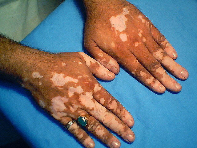
Vitiligo is somewhat disfiguring condition characterized by the loss of skin pigmentation, leaving a blotchy, white appearance. Finding ways to activate these two signaling pathways may provide clinicians with the means to mobilize the pigment-synthesizing stem cells that place pigment in skin structures and, potentially, repigment the pigment-bearing structures that were damaged in cases of vitiligo. Such treatment might also repigment grayed hair cells in older people, and even correct the discoloration that affects scars.

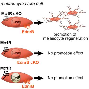
Workers in the laboratory of Mayumi Ito at the Ronald O. Perelman Department of Dermatology and the Department of Cell Biology showed that a skin-based stem cell population of pigment-producing cells, known as “melanocytes,” grow and regenerate in response to two molecular signals. The Endothelin receptor type B (EdnrB) protein is found on the surfaces of melanocytes. EdnrB signaling promotes the growth and differentiation of melanocyte stem cells (McSCs). Activation of EdnrB greatly enhances the regeneration of hair-based and epidermal-based melanocytes. However, EdnrB does act alone. Instead, the effect of EdnrB depends upon active Wnt signaling. This Wnt signal is initiated by the secretion of Wnt glycoproteins by the hair follicle cells.

This work was published in Cell Reports, April 2016 DOI: http://dx.doi.org/10.1016/j.celrep.2016.04.006.

Previous work on EdnrB has established that it plays a central role in blood vessel development. This work by Ito and his team is the first indication that pigment-producing melanocytes, which provide color to hair and skin, are controlled by this protein.

A lack of EdnrB signaling in mice caused premature graying of the hair. However, stimulating the EdnrB pathway resulted in a 15-fold increase in melanocyte stem cell pigment production, and by two months, the mice showed hyperpigmentation. In fact, wounded skin in these mice became pigmented upon healing.

Overexpression of Edn1 Promotes Upward Migration of McSCs and Generation of Epidermal Melanocytes following Wounding (A–D) Whole-mount image of X - gal - stained wound area of Dct-LacZ (control; A and B) and Tyr-CreER ; EdnrB fl/fl ; Dct-lacZ (C and D) at indicated days after re-epithelialization. (E–J) Double immunohistochemical staining of Dct and Ki67 in the bulge (E and H), upper hair follicle (F and I), and inter-follicular epidermis (G and J ) in control (E–G) and K14-rtTA ; TetO-Edn1-LacZ (Edn1; H–J) mice. (K–N) Whole - mount analyses of wound site (K and L) and de novo hair follicles (M and N) within wound site from control (K and M) and Edn1 mice (L and N) at 8 days after re-epithelialization. (O–R) Quantification of the number of Dct - LacZ+ cells in wound site (O), the percentage of Ki67+/Dct+ cells (P), the number of pigmented cells in wound site (Q), and the percentage of pigmented de novo hair (R), respectively. Dashed lines indicate periphery of wound site in (A) and (D) and boundary between epidermis and dermis in (E)–(J). Arrowheads show Dct - LacZ + cells in wound area in (A)–(D) and Ki67+/Dct+ cells (H)–(J). IFE, inter-follicular epidermis; UF, upper follicle. Data are presented as the mean ± SD. *p < 0.01; **p < 0.02; ***p < 0.05. The scale bar represents 1 mm in (A), 50 m m in (E), 200 m m in (K) and (L), and 100 m m in (M) and (N).
Overexpression of Edn1 Promotes Upward Migration of McSCs and Generation of Epidermal Melanocytes following Wounding (A–D) Whole-mount image of X-gal – stained wound area of
Dct-LacZ (control; A and B) and Tyr-CreER; EdnrB fl/fl; Dct-lacZ (C and D) at indicated days after
re-epithelialization. (E–J) Double immunohistochemical staining of Dct and Ki67 in the bulge (E and H), upper hair follicle (F and I), and inter-follicular epidermis (G and J) in control (E–G) and K14-rtTA;
TetO-Edn1-LacZ (Edn1; H–J) mice.  (K–N) Whole-mount analyses of wound site (K and L) and de novo hair follicles (M and N) within wound site from control (K and M) and Edn1 mice (L and N) at
8 days after re-epithelialization.  (O–R) Quantification of the number of Dct-LacZ+ cells in wound site (O), the percentage of Ki67+/Dct+ cells (P), the number of pigmented cells in wound site (Q),
and the percentage of pigmented de novo hair (R), respectively.  Dashed lines indicate periphery of wound site in (A) and (D) and boundary between epidermis and dermis in (E)–(J). Arrowheads show Dct-LacZ+ cells in wound area in (A)–(D) and Ki67+/Dct+ cells (H)–(J). IFE, inter-follicular epidermis; UF, upper follicle. Data are presented as the mean ± SD. *p < 0.01; **p < 0.02; ***p < 0.05.
The scale bar represents 1 mm in (A), 50 micrometer in (E), 200 micrometer in (K) and (L), and 100
micrometer in (M) and (N).

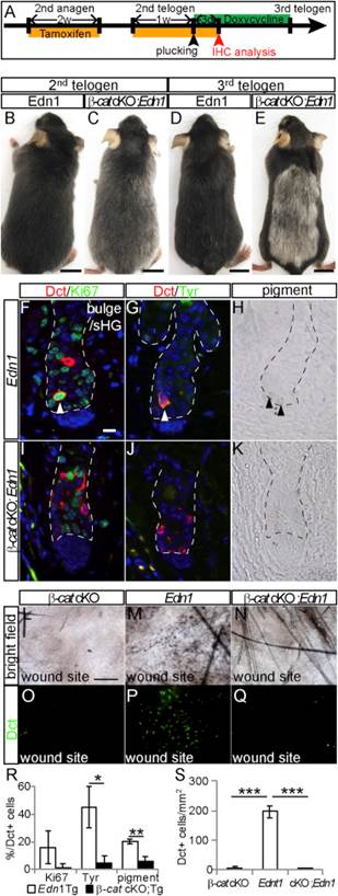
If the Wnt signaling pathway was blocked, stem cell growth and maturation sputtered and stalled and never got going, even when the EdnrB pathway was working properly. These mice had unpigmented fur (see E in figure below).

Loss of beta-catenin Function Suppresses Edn1-Mediated Effects on McSC Proliferation, Differentiation, and Upward Migration (A) Experimental scheme for treatment of Tyr-CreER ; b -catenin fl/fl ; K14-rtTA ; TetO-Edn1-LacZ ( b -cat cKO; Edn1 ) mice and control K14-rtTA ; TetO-Edn1- LacZ ( Edn1 ) mice. (B–E) Gross appearance of Edn1 (B and D) and b -cat cKO; Edn1 mice (C and E) at second (B and C) and third telogen (D and E). (F–K) Immunohistochemistry for indicated markers (F, G, I, and J) and bright- field image (H and K) of bulge/sHG region in skin sections from Edn1 mice (F–H) and b -cat cKO; Edn1 mice (I–K) at anagen II. (L–Q) Bright-field image (L–N) and Dct immunostaining of whole-mount wound site (O–Q) from Tyr-CreER ; b -catenin fl/fl ( b -cat cKO; L and O), Edn1 (M and P), and b -cat cKO; Edn1 mice (N and Q). (R and S) Quantification of the percentage of Dct+ cells positive for Ki67, Tyr, and pigmentation (R) and the number of Dct+ cells in wounded site (S). Dashed lines indicate border between hair follicle and dermis. Arrow- heads indicate double positive cells for indicated markers (F and G) and pigmented cells (H). Data are presented as the mean ± SD.*p<0.05;**p< 0.02; ***p < 0.001. The scale bar represents 1 cm in (B)–(E), 10 m min(F), and 200 m min(L). 1
Loss of beta-catenin Function Suppresses Edn1-Mediated Effects on McSC Proliferation, Differentiation, and Upward Migration
(A) Experimental scheme for treatment of
Tyr-CreER; beta-catenin fl/fl; K14-rtTA; TetO-Edn1-LacZ (beta-cat cKO; Edn1) mice and control K14-rtTA; TetO-Edn1-LacZ (Edn1) mice.
(B–E) Gross appearance of Edn1 (B and D) and
beta-cat cKO; Edn1 mice (C and E) at second (B and C) and third telogen (D and E). (F–K) Immunohistochemistry for indicated markers (F, G, I, and J) and bright-field image (H and K) of bulge/sHG region in skin sections from Edn1 mice (F–H) and beta-cat cKO; Edn1
mice (I–K) at anagen II. (L–Q) Bright-field image (L–N) and Dct immunostaining of whole-mount wound site (O–Q) from
Tyr-CreER; beta-catenin fl/fl (beta-cat cKO; L and O), Edn1 (M and P), and beta-cat cKO; Edn1 mice (N and Q). (R and S) Quantification of the percentage of Dct+ cells positive for Ki67, Tyr, and pigmentation (R) and the number of Dct+ cells in wounded site (S).
Dashed lines indicate border between hair follicle and dermis. Arrow-heads indicate double positive cells for indicated markers (F and G) and pigmented cells (H). Data are presented as the mean ± SD.*p<0.05;**p<
0.02; ***p < 0.001. The scale bar represents 1 cm in (B)–(E), 10 micrometers in (F), and 200 micrometer  in (L).

However, perhaps the most exciting finding for Ito and his colleagues was that Wnt-dependent, EdnrB signaling rescued the defects in melanocyte regeneration caused by loss of the Mc1R receptor. This is precisely the receptor that does not function properly in red-heads, which causes them to have red hair and very light skin that burns easily in the sun. These data suggest that Edn/EdnrB/Wnt signaling in McSCs can be used therapeutically to promote photoprotective-melanocyte regeneration in those patients with increased risk of skin cancers due to their very lightly colored skin.

Melanocyte Stem Cell Modeld

Plant Polyphenol May Help Improve Wound Healing By Activating Mesenchymal Stem Cells


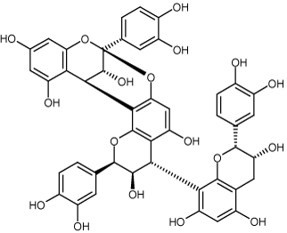
Akito Maeda and his coworkers from Osaka University in Osaka, Japan have discovered that a plant-based polyphenol promotes the migration of mesenchymal stem cells (MSCs) in blood circulation. This same plant polyphenol also causes MSCs to accumulate in damaged tissues and improve wound healing.

This compound, cinnamtannin B-1, might be a candidate drug for stem cell treatments for cutaneous disorders associated with particular diseases and lesions.

Cinnamtannin B-1
Cinnamtannin B-1

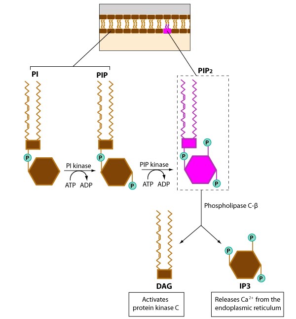
Cinnamtannin B-1, a flavonoid, seems to activate membrane-bound enzymes; specifically the Phosphatidylinositol-3-kinase enzyme, which is an integral enzyme in the phosphoinositol signal transduction pathway, which culminates in the mobilization of intracellular calcium stores and profoundly alters cell behavior and function.

Phosphoinositol pathway signaling

Flow cytometry analysis of mouse blood established that administration of cinnamtannin B-1 increased the release of MSCs from bone marrow. Laboratory experiments with cultured MSCs showed that cinnamtannin B-1 treatment activated MSC migration and recruitment to wounds. This seems to suggest that the enhanced healing caused by cinnamtannin B-1 treatment is due to enhanced MSC migration and homing to damaged tissues.

Imaging analysis of whole animals that had MSCs that expressed the firefly luciferase enzyme showed that cinnamtannin B-1 treatment increased the homing of MSCs to wounds and accelerated healing in a diabetic mouse model.

When Maeda and his colleagues treated MSCs with small molecules that inhibited phosphatidylinositol-3-kinase, those cells no longer responded to cinnamtannin B-1, which confirms the role of the phosphoinositol signal transduction pathway in cinnamtannin B-1 activation of MSCs.

Thus, cinnamtannin B-1 promotes MSC migration in culture and accelerates wound healing in mice. In addition, cinnamtannin B-1-induced migration of MSCs seems to be mediated by specific signaling pathways.

SanBio, Inc Moves Forward With Clinical Stem Cell Trial for Traumatic Brain Injury in Japan


Traumatic brain injuries can result from a variety of causes, ranging from car accidents, falls, occupational hazards, and sports injuries. The cause of traumatic brain injury (TBI) differs from that of ischemic stroke, but many of the clinical manifestations are somewhat similar (motor deficits). Such injuries can cause lifelong motor deficits, and there are currently no approved medicines for the treatment of persistent disability from traumatic brain injury.

SanBio, Inc., has completed the regulatory requirements to conduct a clinical trial using their proprietary SB623 regenerative cell therapy to treat patients who suffer from TBI. The obligatory 30-day review period of clinical trial notification by the Japanese Pharmaceuticals and Medical Devices Agency (PMDA) was completed on March 7, 2016. No safety concerns were voiced, and the trial can proceed.

SanBio’s clinical trial is entitled “Stem cell therapy for traumatic brain injury” or STEMTRA, and it will study the safety and efficacy of SB623 cell therapy in treating patients who suffer from chronic motor impairments following a TBI.

Enrollment in this clinical trial started in the United States in October, 2015. The trial will include clinical sites and patients in Japan and will enroll ~52 patients. The enrollment of Japanese patients is expected to accelerate the overall enrollment of human subjects.

SanBio spokesperson, Damien Bates, the Chief Medical Officer and Head of Medical Research at SanBio, said: “SanBio’s regenerative cell medicine, SB623, has improved outcomes in patients with persistent motor deficits due to ischemic stroke, and our preclinical data suggest that it may also help TBI patients.  This is the first global Phase 2 clinical trial for TBI allogeneic stem cells, and the approval to conduct the trial in Japan, as well as in the United States, brings us one step closer to determining SB623’s efficacy for treatment whose who suffer from the effects of traumatic brain injury.”

SB623 are modified mesenchymal stem cells that transiently express a modified human Notch1 gene that only contains the intracellular domain of the Notch1 protein. This activated gene drives mesenchymal stem cells to form a cell type that habitually supports neural cells and promotes their health, survival, and healing.  When administered into damaged neural tissue, SB623 reverses neural damage. Since SB623 cells are allogeneic (from a donor), a single donor’s cells can be used to treat many patients. In cell culture and animal models, SB623 cells restore function to damaged neurons associated with stroke, traumatic brain injury, retinal diseases, and Parkinson’s disease. SB623 cells function by promoting the body’s natural regenerative process.

SanBio recently completed a US-based Phase 1/2a clinical trial for SB623 in patients with chronic motor impairments six months to five years following an ischemic stroke. The results of this trial demonstrated that SB623 can improve motor function following a stroke. On the strength of these results, SanBio initiated a Phase 2b randomized, double-blind, clinical trial of 156 subjects began enrollment in December 2015.  This trial is entitled ACTIsSIMA (“Allogeneic Cell Therapy for Ischemic Stroke to Improve Motor Abilities”).

Since the therapeutic mechanism of action of SB623 cells and the proposed route of administration are similar in the two trials (the stroke and TBI trials), the results of the TBI trial should be similar to those of the stroke trial.

The Japanese regulatory agencies grant marketing approval for regenerative medicines earlier countries as a result of an amendment to the Pharmaceutical Affairs Law in 2014. This particular amendment defined regenerative medicine products as a new category in addition to conventional drugs and medical devices, and the conditional and term-limited accelerated approval system for regenerative medicine products has started.

Two regenerative medicine products have already gained marketing approval under this new system, and the government-led industrialization of regenerative medicine products has gradually been realized.

SanBio has begun the preparation of clinical trial facilities in Japan and expects the launch of the clinical trial in 2016. the company hopes to market the medicine in Japan by taking advantage of the accelerated approval system.

How Skeletal Stem Cells form the Blueprint of the Face


A new study from the laboratory of University of Southern California (USC) Stem Cell researcher J Gage Crump, who is at the Eli and Edythe Broad CIRM Center for Regenerative Medicine and Stem Cell Research, has identified the key molecular signals that control the critical timing of the development of the vertebrate face.

Previous work has demonstrated that two molecular signals, in particular the JaggedNotch and Endothelin 1 signaling, are integral for shaping the face. Loss of either of these signals results in facial deformities in zebrafish and humans. This illustrates the essential contribution these signaling pathways make to the development of the face.

Lindsey Barske, a researcher in Crump’s laboratory and her colleagues utilized sophisticated genetic, genomic, and imaging tools to study face formation in zebrafish and showed that the Jagged-Notch and Endothelin 1 pathways work in tandem to control when and where the facial stem cells form face-specific cartilage.

In the lower part of the face, the Endothelin 1 signal accelerates cartilage formation early in development, but in the upper face, the Jagged-Notch signal transduction pathway produces signals that prevent stem cells from making cartilage until later in development.

Barske and her colleagues discovered that these timing differences in facial stem cell activity and facial cartilage production play a major role in making the upper and lower cartilage regions of the face.

The earliest blueprint of the facial skeleton is established by intersecting signals that control when stem cells transform cartilage into bone. It also appears that small tweaks to the timing of these events accounts for the different skull shapes observed in vertebrate animals. Also, small, nuanced changes in facial cartilage production and ossification can also account for the diverse array of facial shapes observed in humans.

This work was published in PLOS Genetics 12(4): e1005967. doi:10.1371/journal.pgen.1005967.

Skin Cell to Eye Transplantation Successful


A presentation at the annual meeting of the Association for Research in Vision and Ophthalmology in Seattle, Washington has reported the safe transplantation of stem cells derived from a patient’s skin to the back of the eye in an effort to restore vision. The subject for this research project suffered from advanced wet age-related macular degeneration that did not respond to current standard treatments.

A small skin biopsy from the patient’s arm was collected and reprogrammed into induced pluripotent stem cells (iPSCs). The iPSCs were then differentiated into retinal pigmented epithelial (RPE) cells, which were transplanted into the patient’s eye. The transplanted cells survived without any adverse events for over a year and resulted in slightly, though significantly, improved vision.

iPSCs are adult cells that have been reprogrammed to an embryonic stem cell-like state, which can then be differentiated into any cell type found in the body.

Abstract Title: #3769: Transplantation of Autologous induced Pluripotent Stem Cell-Derived Retinal Pigment Epithelium Cell Sheets for Exudative Age Related Macular Degeneration: A Pilot Clinical Study by Yasuo Kurimoto and others from the laboratory of Masayo Takahashi’s laboratory at the RIKEN Center for Developmental Biology in Kobe, Japan.

Unfortunately, this clinical trial has been suspended because iPSCs made from other patients proved to possess too many genetic abnormalities.  Therefore, Takahashi and her colleagues have decided that allogeneic iPSCs differentiated into RPEs will probably do a better job than the patient’s own skin cell-derived iPSCs.

Positive Results from Phase 2 Study in Spinal Cord Injury


Stem Cells, Inc., has released the six-month results from cohort I of an ongoing Phase 2 clinical trial of human neural stem cells for the treatment of chronic cervical spinal cord injuries. The data displayed significant improvements in muscle strength had occurred in five of the six patients treated. Of these five patients, four of them also showed improved performance on functional tasks that assesses dexterity and fine motor skills. Furthermore, these four patients improved in the level of spinal cord injury according to the classification system provided by the International Standards for Neurological Classification of Spinal Cord Injury or ISNCSCI.

Stem Cells, Inc., expects to release their detailed final 12-month results on this first open-cohort later this quarter.

Chief medical officer, Stephen Huhn, presented these data at the American Spinal Injury Association annual meeting in Philadelphia, on Friday, April 15.  Dr. Huhn also believes that the interim results are very encouraging and reason to be quite hopeful.

“The emerging data continue to be very encouraging,” said Dr. Huhn. “We believe that these types of motor changes will improve the independence and quality of life of patients and are the first demonstration that a cellular therapy has the ability to impact recovery in chronic spinal cord injury. We currently have thirteen sites in the United States and Canada that are actively recruiting patients. We have enrolled and randomized 19 of the 40 total patients in the statistically powered, single-blind, randomized controlled, Cohort II. We are projecting to complete enrollment by the end of September so that we can have final results in 2017.”

The present Phase 2 clinical trial is a multi-center enterprise that includes physicians and scientists at 13 different sites in the united States and Canada. Incidentally, these sites are presently actively recruiting patients.

Stem Cells, Inc., has enrolled and randomized 19 of the 40 total patients in this statistically powered, single-blind, randomized controlled, cohort II.

The Phase 2 study, “Study of Human Central Nervous System (CNS) Stem Cell Transplantation in Cervical Spinal Cord Injury,” will determine the safety and efficacy of transplanting the company’s proprietary human neural stem cells (HuCNS-SC cells) into patients with traumatic injury of the cervical region of the spinal cord.

Cohort I is an open label dose-ranging cohort in six AIS-A or AIS-B subjects. For those of you not familiar with the American Spinal Injury Impairment Scale (ASI A-E scale), here is a summary of the classification scheme:

ASI – A = Complete paralysis; No sensory or motor function is preserved in the sacral segments S4-5.
ASI – B = Sensory Incomplete; Sensory but not motor function is preserved below the neurological level and includes the sacral segments S4-5 (light touch or pin prick at S4-5 or deep anal pressure) AND no motor function is preserved more than three levels below the motor level on either side of the body.
ASI – C = Motor Incomplete; Motor function is preserved below the neurological level**, and more than half of key muscle functions below the neurological level of injury (NLI) have a muscle grade less than 3 (Grades 0-2).
ASI – D = Motor Incomplete; Motor function is preserved below
the neurological level**, and at least half (half or more) of key muscle functions below the NLI have a muscle grade > 3.
ASI – E = Normal; If sensation and motor function as tested with the ISNCSCI are graded as normal in all segments, and the patient had prior deficits, then the AIS grade is E. Someone without an initial SCI does not receive an AIS grade.
Cohort II is a randomized, controlled, single-blinded cohort in forty AIS-B subjects. Cohort III, which will only be conducted at the discretion of the sponsor, is an open-label arm that involves six AIS-C subjects.
The primary efficacy outcome will focus on changes in the upper extremity strength as measured in the hands, arms, and shoulders.  This trial will enroll up to 52 subjects.
StemCells, Inc. has demonstrated the safety and efficacy of their HuCNS-SC cell in preclinical studies in laboratory rodents.  Additional Phase I studies yielded positive human safety data.  Furthermore, completed and ongoing clinical studies in which its proprietary HuCNS-SC cells have been transplanted directly into all three components of the central nervous system: the brain, the spinal cord and the retina of the eye, have further demonstrated the safety of HuCNS SC cells in human patients.
StemCells, Inc. clinicians and scientists believe that HuCNS-SC cells may have broad therapeutic application for many diseases and disorders of the CNS. Because the transplanted HuCNS-SC cells have been shown to engraft and survive long-term, there is the possibility of a durable clinical effect following a single transplantation.
The HuCNS-SC platform technology is a highly purified composition of human neural stem cells (tissue-derived or “adult” stem cells). Manufactured under cGMP standards, the Company’s HuCNS-SC cells are purified, expanded in culture, cryopreserved, and then stored as banks of cells, ready to be made into individual patient doses when needed.

SEPCELL Trial Tests Fat-Derived Stem Cells as a Treatment for Sepsis


The Belgium-based biotechnology company, TiGenix, has launched a clinical trial entitled SEPCELL that uses fat-derived stem cells (called Cx611) to treat severe sepsis secondary to acquired pneumonia (also known as sCAP). SEPCELL is a randomized, double-blind, placebo-controlled, Phase 1b/2a study of sCAP patients who require mechanical ventilation and/or vasopressors.

SEPCELL will, hopefully, enroll 180 patients and will be conducted at approximately 50 centers throughout Europe. Subjects who participate in this trial will be randomly assigned to receive either an investigational product or placebo on days 1 and 3. All patients will be treated with standard care, which usually includes broad-spectrum antibiotics and anti-inflammatory drugs.

The primary endpoint of this clinical trial will examine the number, frequency, and type of adverse reactions during the 90-day period of the trial. The secondary endpoints of the SEPCELL trial include reduction in the duration of mechanical ventilation and/or vasopressors, overall survival, clinical cure of sCAP, and other infection-related endpoints. SEPCELL will also assess the safety and efficacy of the expanded allogeneic adipose stem cells (eASCs) that will be intravenously delivered to some of the patients in this study.

The SEPCELL trial will be managed by TFS International, a company based in Lund, Sweden. TFS has extensive experience in running sepsis trials and hospital-based trials.

Sepsis is a potentially life-threatening complication of infection that occurs when inflammatory molecules (cytokines and chemokines) released into the bloodstream to fight the infection trigger systemic inflammation.  This body-wide inflammation has the ability to trigger a cascade of detrimental changes that damage multiple organ systems and cause them to fail. If sepsis progresses to “septic shock,” blood pressure drops dramatically, which may lead to death. Patients with “severe sepsis” require close monitoring and treatment in a hospital intensive care unit. Drug therapy is likely to include broad-spectrum antibiotics, corticosteroids, vasopressor drugs to increase blood pressure, as well as oxygen and large amounts of intravenous fluids. Supportive therapy may be needed to stabilize breathing and heart function and to replace kidney function. Patients with severe sepsis have a low survival rate so there is a critical need to improve the effectiveness of current therapy. Only a small number of new molecular entities are currently in development for severe sepsis.

Severe sepsis and septic shock significantly affect public health and these event also are leading causes of mortality in intensive care units.

Severe sepsis and septic shock have an incidence of about 3 cases per 1,000, but due to the aging of the population and an increase in drug resistant bacteria.

Cx611 is an intravenously-administered concoction that consists of allogeneic eASCs. These cells are largely mesenchymal stem cells that secrete an impressive array of molecules that suppress the type of immune responses that damage organs during events like septic shock.  eASCs have a higher proliferation rate in culture and faster attachment than bone marrow-based mesenchymal stem cells in cell culture.  ASCs are also less prone to senescence and differentiation.  Their differentiation capacity decreases with expansion time without losing immunomodulatory properties.  These eASCs also have superior inflammation targeting capacities than bone marrow-based mesenchymal stem cells, and are safe, since they do not express ligands for receptors on Natural Killer cells that, and therefore, are unlikely to elicit an immune rejection.

In May 2015, TiGenix completed a Phase 1 sepsis challenge that demonstrated that Cx611 is safe and well tolerated. That trial began in December 2014, and was a placebo-controlled dose-ranging study (3 doses of eASC’s) in which 32 healthy male volunteers were randomized to receive Cx611 or placebo in a ratio of 3:1. Primary endpoints were vital signs and symptoms, laboratory measures and functional assays of innate immunity. All 32 volunteer subjects were recruited and dosed by March 2015. By May, 2015, the phase I trial data essentially demonstrated the safety and tolerability of Cx611.  On the strength of that phase I trial, TiGenix designed a Phase 1b/2a trial in severe sepsis secondary to sCAP in which they expecet to enroll 180 subjects across Europe.

SEPCELL was funded by a €5.4 million grant ($6.14 million) from the European Union.

CardioCell LLC Clincal Trial Tests Ischemia-Resistant Mesenchymal Stem Cells in Heart Failure


The cell therapy company CardioCell LLC has completed enrolling 23 patients for its Phase 2a chronic heart failure trial. These subjects were enrolled at Emory University in Atlanta, GA, MedStar Washington Hospital Center in Washington DC, and three other hospitals.

This study has the ponderous title of “Single-blind, Placebo-controlled, Crossover, Multicenter, Randomized Study to Assess the Safety, Tolerability and Preliminary Efficacy of Single Intravenous Dose of Ischemia-tolerant of Allogeneic Mesenchymal Bone Marrow Cells to Subjects With Heart Failure of Non-ischemic Etiology.”

This clinical trial will examine the safety of CardioCell’s proprietary ischemic-tolerant mesenchymal stem cells in heart failure patients. The trial will also test the ability of these cells to improve the heart function of these safe patients.

Ischemia-resistant mesenchymal stem cells have are extracted from bone marrow and then subjected to harsh cell culture conditions that toughen them up and improves their therapeutic capacities.

Cardiologist Javed Butler said that this clinical trial has been designed to use this novel intervention in a carefully selected group of patients who met rigorous inclusion and exclusion criteria.

This trial will deliver ischemia-tolerant mesenchymal stem cells (itMSCs) by means of intravenous infusion into heart failure patients and then monitor these patients to determine if the itMSC-treated patients show signs of improvement in heat function.

These itMSCs are licensed under the parent company Stemedica and these are allogeneic cells that were isolated from young, healthy donors and grown under hypoxic conditions. Once grown under these harsh culture conditions, the itMSCs increase their ability to home to damaged tissues and engraft into those tissues. itMSCs also secrete increased levels of growth and trophic factors that promote neurogenesis and tissue healing.

Insulin-Secreting Beta Cells from Human Fat


In a study led by Martin Fussenegger of ETH Zurich, stem cells extracted from the fat of a 50-year-old test subject were transformed into mature, insulin-secreting pancreatic beta cells.

Fussenegger and his colleagues isolated stem cells from the fat of a 50-year-old man and used these cells to make induced pluripotent stem cells (iPSCs). These iPSCs were then differentiated into pancreatic progenitor cells and then into insulin-secreting beta cells but means of a “genetic software” approach.

Genetic software refers to the complex synthetic network of genes required to differentiate pancreatic progenitor cells into insulin-secreting beta cells. In particular, three genes, all of which expression transcription factors, Ngn3, Pdx1, and MafA, are particularly crucial for beta cell differentiation.

Fussenegger and his team designed a a protocol that would express within these fat-based stem cells the precise concentration and combination of these transcription factors. This feature is quite important because the concentration of these factors changes during the differentiation process. For example, MafA is not present at the start of beta cells maturation, but appears on day four on the final data of maturation when its concentration rises precipitously. The concentration of Ngn3 rises and then falls and the levels of Pdx1 rise at the beginning and towards the end of maturation.

The Zurich team used ingenious genetic tools to reproduce these vicissitudes of gene expression as precisely as possible. By doing so, they were able to differentiate the iPSC-derived pancreatic progenitor cells into insulin-secreting beta cells.

This work was published in Nature Communications 7, doi:10.1038/ncomms11247.

The fact that Fussenegger’s team was able to use a synthetic gene network to form mature beta cells from adult stem cells is a genuine breakthrough. The genetic network approach also seems to work better than the traditional technique of adding various chemicals and growth factors to cultures cells. “It’s not only really hard to add just the right quantities of these components (growth factors) at just the right time, it’s also inefficient and impossible to scale up,” said Fussenegger.

This new process can successfully transform three out of four fat stem cells into beta cells. Also the beta cells made with this method have the same microscopic appearance of natural beta cells in that they contain internal granules full of insulin. They also secrete insulin in response to increased blood glucose concentrations. Unfortunately the amount of insulin made by these cells is lower than that made by natural beta cells.

Pancreatic islet transplants have been performed in diabetic patients, but such transplantations also require treatment with potent antirejection drugs that have potent side effects.

“With our beta cells, there would likely be no need for this action (administering antitransplantation drugs), since we can make them using endogenous cell material taken from the patient’s own body. This is why our work is of such interest in the treatment of diabetes,” said Fussenegger.

Fussenegger and his group have made these beta cells in the laboratory, but they have yet to transplant them into a diabetic patient. However, the success of this synthetic genetic software technology might also be useful in the reprogramming of adult cells into other types of cells that are useful for therapeutic purposes.

RENEW Trial Shows Stem Cell Mobilization Has Some Potential for Refractory Angina


The RENEW clinical trial has examined the ability of “CD34+” stem cells from bone marrow to alleviate the symptoms of refractory angina.

Angina pectoris is a crushing chest pain that afflicts people when the heart receives too little oxygen to support it for the workload placed upon it. Angina pectoris typically results from the blockage of coronary arteries as a result of atherosclerosis. Treatment of angina pectoris usually includes PCI or percutaneous coronary intervention, which involves the placement of a stent in the narrowed coronary artery, in combination with drug treatments like beta blockers, and/or cardiac nitrate (e.g., nitroglycerine).

Angina pectoris is also classified according to the severity of the disease. The Canadian Cardiovascular Society grading of angina pectoris (which is very similar to the New York Heart Association classification) uses four classes (I-IV) to classify the disease. Patients with Class I angina only experience pain during strenuous or prolonged physical activity. Those with Class II angina have a slight limitation in physical activity and experience pain during vigorous physical activity (climbing several flights of stairs). Class III angina manifests as pain during everyday living activities, such as climbing one flight of stairs. These patients experience moderate limitation of their physical activity. Those with Class IV angina experience pain at rest and are unable to perform any activity without angina, and therefore, suffer from severe limitations on their activity.

Refractory angina pectoris (also known as chronic symptomatic coronary artery disease) stubbornly resists medical therapy and is unamenable to conventional revascularization procedures. Patients with refractory angina pectoris have reproducible lifestyle-limiting symptoms of chest pain, shortness of breath, and easy fatigability.

The results of the RENEW clinical trial were presented at the Society for Cardiovascular Angiography and Interventions 2016 sessions. Even though the trial was prematurely ended for financial reasons, the results that were collected suggest that cell-based therapies might provide relief for suffers of refractory angina pectoris.

RENEW tested the effectiveness of the intravenous infusion of the protein called granulocyte-colony simulating factor (G-CSF), which mobilizes CD34+ stem cells from the bone marrow. Once summoned from the bone marrow, CD34+ stem cells can help establish new blood vessels and increase blood flow throughout the heart. CD34+ stem cells also seem to have some ability to home to sites of damage. Therefore, G-CSF infusions might provide some relief to patients with refractory angina pectoris.

Dr. Timothy D. Henry of the Cedars-Sinai Heart Institute in Los Angles, CA, said: “Clinicians are seeing more RA (refractory angina) patients because people are living longer. Unfortunately, despite better medical care, these people are still confronting ongoing symptoms that affect their daily lives.”

Patients enrolled in the RENEW trial had either class III or IV angina and experiences ~7 chest pain episodes each week. These patients were also not candidates for revascularization (PCI) and their treadmill exercise times were between 3-10 minutes.

112 RA patients were randomly broken into three groups. Group 1 received standard care (28), group 2 received placebo injections (27), and group 3 received treatment with CD34+ cells. The trial was double-blinded and placebo controlled. The original aim was to test 444 RA patients, but financial concerns truncated the study at 112 patients.

All patients were assessed at three, six, 12, and 24 months after treatment by means of exercise tolerance, anginal attacks, and major adverse cardiovascular events (MACEs).

The cell-treated patients increased their exercise times by more than two minutes at three (average 122-second increase), six (average 142-second increase), and twelve (average 124-second increase) months. This is significant, since the other two groups showed no significant increase in their exercise times.

Patients in the cell-treated group also experienced 40 percent fewer anginal attacks at six months relative to the placebo-treated group.

At two years after the treatment, the CD34+-treated group have lower mortality rates (3.7 percent) compared to those who received standard care (7.1 percent) and those who received the placebo (10 percent).

Finally, after two years, the cell-treated group had lower MACE rates (46 percent) than the standard care group (68 percent). The MACE rate for the placebo-treated group was 43 percent.

On the strength of these results, Dr. Henry said, “Cell therapy appears to be a promising approach for these patients who have few options. Our results were consistent with phase 2 results from the ACT34 trial (author’s note: which gave patients infusions of cells and not G-CSF).”

Tom Povsic of the Duke Clinical Research Institute said of the RENEW trial, “It is unfortunate the early termination of this study precludes a full evaluation of the efficacy of this therapy for these patients with very few options.  Studies like RENEW are critical to developing reliable and effective therapies for heart patients, and continued cellular therapies for heart patients, and continued funding is essential to advancing the work that this study began.  We need to find a way to bring these therapies as quickly as safely as possible.”

Dr. Povsic’s words certainly ring true.  Even though the results of the RENEW study are essentially positive, RENEW was planed to be almost three times the size of Douglas Losordo’s earlier, successful ACT34 study.  The results of both the ACT34 and RENEW studies are largely positive.  Perhaps more importantly, both studies have also established that cell-based treatments for RA patients are safe.  However, given the voracity of the FDA for clinical data before it will approve a treatment, even for patients with few current options, it is unlikely that these studies will prove large enough to satisfy the agency.  Until a very large study shows cell-based treatments to be not only safe but efficacious, only then will the mighty turtle known as the FDA approve such treatments for RA patients.

RepliCel Skin Rejuvenation and Tendon Repair Trials With Hair Follicle Stem Cells Underway


RepliCel Life Sciences has enrolled subjects for their skin rejuvenation and tendon repair trial.  The primary goal of these trials is to determine the safety of their cell therapeutic products.

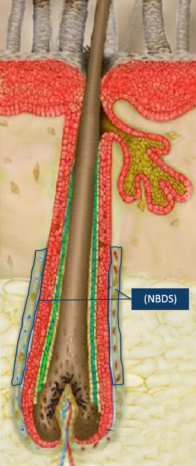
NBDS-Cells-Follicle

In the first trial will test a product called RCS-01, which consists of cells derived from non-bulbar dermal sheath (NBDS) cells, which are taken from the outer regions of hair follicles.  NBDS cells express type 1 collagen, a protein that is steadily degraded in aged skin (hence the formation of wrinkles).  Therefore, RepliCel is confident that RCS-01 injections underneath the skin has the potential to rejuvenate aged or damaged skin.  The trial will examine male and female subjects, between 50-65 years old, and will address the inherent deficit of active fibroblasts required for the production of type 1 collagen, elastin and other critical extracellular dermal matrix components found in youthful skin. The trial will be conducted at the IUF Leibniz-Institut für umweltmedizinische Forschung GmbH in Dusseldorf, Germany.  Originally, RepliCel wished to enrolled 15 men and 15 women, but the large number of female subjects and paucity of men persuaded the company to move forward with the trial despite only enrolling a few men and all the projected women.

The second trial will test the safety and efficacy of RCT-01 in the repair of damaged Achilles tendons.  RCT-01 also consists of NBDS cells and this trial is a phase 1/2 clinical trial that examines the ability of NBDS cells to treat chronic tendinosis caused by acute and chronic tensile overuse.  This trial will take place at the University of British Columbia in Vancouver, BC, and will only treat 10 subjects.  Even though RepliCell wishes to originally test 28 participants, the company shorted the trial in order to have safety data by the end of 2016.

Darrell Panich, RepliCel Vice President of Clinical Affairs, said that the company had a late start on its trials, and therefore truncated the recruitment process in order to have safety data for analysis by the end of 2016.  Despite the small size of these trials (and they are small), the company is hopeful that their safety data will provide the impetus for moving forward with larger phase 2 trials.

Panich said, “We have adjusted our plans for the RCT-01 clinical trial in part because it started later in 2015 and enrolled slower than originally anticipated. While the trial did not meet projected enrollment targets, we are confident the safety and preliminary efficacy data obtained by year-end will provide a signal of the product’s potential to regenerate chronically injured tendon that has failed to respond to other treatments. This will allow our teams to effectively plan larger phase 2 trials in 2017 which are powered to be statistically significant for clinical efficacy (evidence the product works as intended).”

“Future trials involving products from our non-bulbar dermal sheath (NBDS) platform will be designed to investigate the efficacy of these products at different dose levels and treatment frequencies while continuing to collect other data that will be used to support eventual RCS-01 and RCT-01 marketing applications by our commercial partners.”

“The delivery of clinical data when promised is important to management”, said R. Lee Buckler, President & CEO, RepliCel Life Sciences Inc. “We have made critical decisions to keep our commitment to the financial community and we believe the data from these trials will facilitate us closing a licensing and co-development deal on one or both of these products similar to the kind we have in place with Shiseido Company for our RCH-01 product,” he added.

RepliCel is confident that their NBDS fibroblast platform will address numerous indications where impaired tissue healing has been stalled due to a paucity of active fibroblasts, which are required for tissue remodeling and repair.  NBDS fibroblasts, isolated from the hair follicles of healthy individuals, are a rich source of fibroblasts and are unique in their ability to express high levels of type 1 collagen and elastin to push-start the healing process.

RepliCel is also developing products from this same platform to address larger commercial markets in the areas of musculoskeletal and skin-related conditions.

New Gene Therapy Treatment Stops Deadly Brain Cancer in its Tracks


Brain cancers called Diffuse Intrinsic Pontine Gliomas (DIPGs) are often a death sentence. These aggressive, fast-growing, drug-resistant tumors are deadly and they originate from glial cells in the brain.

However a recent report published in the journal Cancer Cell details an experimental gene therapy that stops DIPGs in their tracks. This study included researchers from several different institutions, but was led by scientists at Cincinnati Children’s Hospital Medical Center. The study examined human cancer cells and a mouse model of DIPG.

DIPGs seem to require a gene called Olig2 (which encodes a transcription factor) to grow and survive. The majority of gliomas express the protein encoded by the Olig2 gene and removing this gene halts tumor growth and liquidating Olig2-producing cells inhibits tumor formation. This collaborative team designed a technique scientists found a way to use a gene therapy to shut down Olig2 expression.

“We find that elimination of dividing Olig2-expressing cells blocks initiation and progression of glioma in animal models and further show that Olig2 is the molecular arbiter of genetic adaptability that makes high-grade gliomas aggressive and treatment resistant,” said Qing Richard Lu, PhD, lead investigator and scientific director of the Brain Tumor Center at Cincinnati Children’s. “By finding a way to inhibit Olig2 in tumor forming cells, we were able to change the tumor cells’ makeup and sensitize them to targeted molecular treatment. This suggests a proof of principle for stratified therapy in distinct subtypes of malignant gliomas.”

DIPGs originate from supporting brain cells called oligodendrocytes. Oligodendrocytes make the insulation that surrounds the axons of various nerves in the central nervous system. Olig2 expression appears at the early stages of brain cell development, and is also present in the early-stage dividing and replicating cells in tumors. Olig2 also participates in the transformation of normal oligodendrocyte progenitor cells (OPCs) into cancer cells that divide uncontrollably. Olig2 also facilitates the adaptability of gliomas that helps them evade chemotherapeutic regimens. Indeed, clinically speaking, DIPGs may initially respond to chemotherapeutic agents, but they tend to quickly adapt to these drugs and develop high-levels of resistance to them.

Lu and his colleagues and collaborators eliminated Olig2-positive dividing cells from DIPG tumors that were still in the early stages of tumor formation. Lu and his colleagues used an ingenious technique to remove Oligo2 expression: by genetically engineering a herpes simplex virus-based vector, they delivered a suicide gene (Thymidine kinase) into replicating Olig2-positive cancer cells. Since herpes simplex viruses (HSVs) have the ability to grow in neurons that do not divide a great deal, the HSV-vectors are well suited to this purpose. After infecting the early DIPG cells with the HSV vectors, they administered an anti-herpes drug already in clinical use, ganciclovir (GCV), which kills any cells that have the thymidine kinase gene. The Olig2-deleted tumors were not able to grow.

In follow-up work, Lu and his colleagues observed a fascinating fate for the Olig2- tumors. These cells differentiated into astrocyte-like cells that continued to form tumors, but expressed the epidermal growth factor receptor (EGFR) gene at high levels. EGFR is an effective target for several chemotherapy drugs. In repeated tests in mouse models, Olig2 inhibition consistently transformed the glioma-forming cells into EGFR-expressing astrocyte-like cells. Then these tumors were treated with an EGFR-targeted chemotherapy drug called gefitinib. These treatments stopped the growth of new tumor cells and tumor expansion.

According to Dr. Lu, with additional testing, verification, and, of course, refinement, this experimental therapy that he and his colleagues have designed, could help prevent the recurrence of brain cancer in patients who have undergone initial rounds of successful treatment. Lu also added that these new treatments would probably be used in combination with other existing therapies like radiation, surgery, other chemotherapies and targeted molecular treatments.

Lu and his team will continue their research with other human cell lines and “humanized” mouse models of high-grade glioma. Such mouse models use genetically engineered mice that can grow brain tumors derived from the tumor cells of specific human patients. These tumor cells come from the tumors of patients whose families have donated biopsied tumor samples for research. This allows researchers to test different targeted drugs in their therapeutic protocol that may best match the genetic makeup of tumors from specific individuals.

The entire research team cautions the experimental therapeutic approach they describe will require extensive additional research. Therefore, this type of treatment is years away from possible clinical testing. Having said that, Lu said the data are a significant research breakthrough, since this study identifies a definite weakness in these stubborn cancers that almost always relapse and kill the patients who get them.

ATHENA Clinical Trial Shows that Fat-Based Stem Cell Mixture Improves Heart Patient’s Health, But Does Not Improve Ejection Fraction.


The ATHENA trial is a clinical trial designed to test the ability of a patient’s own “adipose-derived regenerative cells” or ADRCs to improve to improve their heart function. In this trial, heart disease patients received injections of their own ADRCs into their heart muscle. Then these patients were followed and their symptoms, rates of hospitalizations, and heart function were monitored over a period of several months. The initial plans were to examine each patient at one week and at one, three, six, and twelve months after the procedure, and to interview patients via telephone calls from study staff two, three, four, and five years after the procedure.

The ATHENA trial results were presented at the Society for Cardiovascular Angiography and Interventions (SCAI) 2016 Scientific Sessions in Orlando, Fla.

ADRCs are isolated from fat that is collected by means of liposuction. The processing procedure uses a Celution®System (Cytori Therapeutics, San Diego, CA) cell processing unit to separate the mature fat cells and red blood cells (and other unwanted material, i.e., connective tissue and so on) from the other cells. The processed material, or ADRC fraction is an admittedly mixed population of cells that includes some mesenchymal stem cells (a type of adult stem cell), endothelial progenitor cells, leukocytes (white blood cells), endothelial cells (which compose the inner lining of blood vessels), and vascular smooth muscle cells. Several studies in laboratory animals have shown that ADRCs can promote healing of scarred or injured tissue, but the precise exact mechanisms by which ADRCs do this is uncertain. Pre-clinical studies have shown that ADRCs can quell inflammation, stimulate new blood vessel formation, promote cell survival and prevent cell death, and secrete molecules that promote tissue repair and regeneration.

The key advantage of ADRCs come from the fact that fat is the richest source of adult stem and regenerative cells. For example, one gram of fat contains approximately 5,000 stem cells and these cells can be collected and processed in the same day.

The results of the ATHENA trial to data showed that the heart muscle of those who had received injections of their own ADRCs demonstrated symptomatic improvement and a trend towards lower rates of heart failure hospitalizations and angina (chest pain). However, there was no significant improvement in left ventricle ejection fraction (LVEF) or ventricular volumes.

“ADRCs consist of multiple cell types with multiple potential benefits,” said Timothy D. Henry, MD, MSCAI, director, division of cardiology at the Cedars-Sinai Heart Institute and the study’s lead investigator. “Based on the results seen with ADRCs in the PRECISE trial, we designed ATHENA to look at these cells as a possible treatment option for people with refractory chronic myocardial ischemia.”

This phase 2 program consisted of two prospective, randomized double-blind, placebo-controlled, parallel group trials that were called ATHENA and ATHENA II. The patients in this study had an average age of 65 years in both groups. 17 patients received injections of their own ADRCs into their heart muscle and 14 received the placebo. The ejection factions of these patients (the percentage of blood pumped out of the ventricles with each contraction) were between 20-45 percent (normal is around high 40s to low 50s). The ejection fraction or EF can be an early indicator of heart failure if it is 35 percent or below, and the baseline average EF score for both groups was 31.6 percent. The patients were also suffered from angina pectoris, a chest pain that occurs when the heart receives too little oxygen. All patients had blocked coronary arteries but were not candidates for revascularization therapies.

One year after receiving the therapy, the ADRC-treated patients registered improvement in their heart failure classification (57 percent) and angina classification (67 percent) relative to the placebo group (15 percent and 27 percent, respectively). Further, when evaluated with the Minnesota Living with Heart Failure questionnaire, the ADRC-treated patients showed distinct improvements over those who had received the placebo (-21.6 vs. -5.5, p=0.038), and displayed a trend toward fewer heart failure hospitalizations (centrally adjudicated [2/17, 11.7 percent vs. 2/14, 21.4 percent]). However, to emphasize again, there were no between group differences in LVEF or ventricular volume.

The ATHENA trial only examined a small patient population, but the results are potentially promising and consistent with what was seen with PRECISE and might provide the foundation for a large phase 3 trial.

The study, designed to enroll 90 patients, was terminated prematurely due to three neurological events that prolonged trial enrollment, but were not cell related.

The fact that patients feel better and do better with the ADRC treatment is encouraging, but without showing improved objective measures in heart physiology, such as increased ejection fraction, decreased end-diastolic volume and end-systolic volume, such a treatment will have a hard time finding enthusiastic endorsement among cardiologists.

Transdifferentiating Skin Cells into Heart Muscle and Neural Stem Cells With Nothing But Chemicals


A research effort led by Dr. Sheng Ding from the Gladstone Institute and scientists from the Roddenberry Center for Stem Cell Biology and Medicine has successfully transformed skin cells into heart cells and brain cells using little more than a cocktail of chemicals. Previous work that sought to transdifferentiate mature, adult cells into another cell type used gene vectors (such as viruses) that genetically engineered the cells to express new genes at high levels. Because this new protocol uses no genetic engineering techniques, these results are nothing short of unprecedented. This work lays the foundation for, hopefully, being able to regenerate lost or damaged cells with pharmaceutical agents.

In two publications that appeared in the journals Science and Cell Stem Cell, Ding and his collaborators utilized chemical cocktails to drive skin cells to differentiate into organ-specific stem cell-like cells and, then into terminally differentiated heart or brain cells. These results were achieved without genetically engineering cells.

Ding, who was the senior author on both studies, said: “This method brings us closer to being able to generate new cells at the site of injury in patients. Our hope is to one day treat diseases like heart failure or Parkinson’s disease with drugs that help the heart and brain regenerate damaged areas from their own existing tissue cells. This process is much closer to the natural regeneration that happens in animals like newts and salamanders, which has long fascinated us.”

Mature heart muscle cells have very little regenerative ability. Once a patient has suffered a heart attack, the cells that have died are, for the most part, not replaced. Therefore, stem cell scientists have left no stone unturned to find a way to replace dead and dying heart muscle cells. Several clinical trials have transplanted mature adult heart cells or various types of stem cells into the damaged heart. However, such procedures have either not improved heart function or have only modestly improved heart function (with a few exceptions). Typically, transplanted cells do not survive in the hostile environment of the heart after a heart attack and even those cells that do survive fail to properly integrate into the heart. Also, the ability of transplanted cells to differentiate into heart cells is not stellar. Alternatively, Deepak Srivastava, director of cardiovascular and stem cell research at the Gladstone Institute, and his team pioneered a distinctly novel approach in which scar-forming cells in the heart of animals were genetically engineered to differentiate into heart new muscle that greatly improved heart function. Genetic engineering brings its own safety issues to the table and, for these reasons, chemical reprogramming protocols that can do the same thing might provide an easier way to drive heart muscle to regenerate local lesions.

In the Science study, Dr. Nan Cao (a postdoctoral research fellow at Gladstone, and others applied a cocktail of nine chemicals to reprogram human skin cells into beating heart cells. By using a kind of trial-and-error strategy, they discovered the best combination of chemicals to transdifferentiate skin cells into multipotent stem cells. Multipotent stem cells have the ability to differentiate into several distinct cell types from several different types of organs. A second-growth factor/small molecule cocktail drove the multipotent stem cells to differentiate into heart muscle cells.

Perhaps the most surprising result of this protocol is its efficiency. Typically, chemically-induced differentiation is relatively inefficient, but with Ding’s method, over 97% of the cells began beating. These chemically-derived heart muscle cells also responded appropriately to hormones, and they also molecularly resembled heart muscle cells (and not skin cells). Upon transplantation into a mouse heart, these cells developed into healthy-looking heart muscle cells within the heart of the laboratory animal.

“The ultimate goal in treating heart failure is a robust, reliable way for the heart to create new muscle cells,” said Srivastava, co-senior author on the Science paper. “Reprogramming a patient’s own cells could provide the safest and most efficient way to regenerate dying or diseased heart muscle.”

In the second study, published in Cell Stem Cell, which was authored by Gladstone postdoctoral scholar Dr. Mingliang Zhang, PhD, the Gladstone team created neural stem cells from mouse skin cells using a similar approach.

Once again, the chemical cocktail that transdifferentiated skin cells into neural stem cells contained nine different chemicals. Some of the molecules used in the neural stem cell experiment overlapped with those employed in the heart muscle study. Treatment of the skin cells for about ten days with the cocktail transdifferentiated the skins cells into neural-like cells. Virtually all the skin cell-specific genes were shut off and the neural stem cell-specific genes were gradually activated. When these chemical-differentiated cells were transplanted into mice, the cells spontaneously differentiated into neurons, oligodendrocytes, and astrocytes (three basic nerve cells). The neural stem cells were also able to self-replicate, which makes them ideal for treating neurodegenerative diseases or brain injury.

“With their improved safety, these neural stem cells could one day be used for cell replacement therapy in neurodegenerative diseases like Parkinson’s disease and Alzheimer’s disease,” said co-senior author Dr. Yadong Huang, who is a senior investigator at Gladstone. “In the future, we could even imagine treating patients with a drug cocktail that acts on the brain or spinal cord, rejuvenating cells in the brain in real-time.”

Turning Stem Cells in Testes into Testosterone-Producing Cells in a Sustainable Culture System


A research effort led by scientists at Johns Hopkins Bloomberg School of Public Health, in collaboration with researchers from Wenzhou Medical university of China has successfully made testosterone-producing stem cells in culture that can be propagated in the laboratory.

Haolin Chen of the Bloomberg School of Public Health noted that testosterone treatments often produce spikes and troughs in testosterone concentrations that can cause a variety of side effects. Administering testosterone-producing cells might very well prevent these wide variations in testosterone production and decrease the potential side effects. Low testosterone in males has been linked to increased mortality, in addition to depression, decreased cognition and immune function, increase body and reduced muscle mass, and poor healing.

A group of cells called Leydig cells in between the seminiferous tubules in the testes of males typically produce testosterone in response to stimulation by a hormone called luteinizing hormone (LH), which is made by the anterior pituitary. Leydig cells produce testosterone in a rather stable, constant fashion, in contradistinction to the injections that are given to males with low testosterone levels.

Unfortunately, keeping testosterone-producing Leydig cells or Leydig cell progenitors alive in culture has proven rather difficult. To address this problem, Chen and his collaborators started adding combinations of growth factors to the cells to determine if any cocktails of growth factors or nutrients could keep the cells alive. Fortunately, they came upon a combination of platelet-derived growth factor, basic fibroblast growth factor, activin, and a molecule called desert hedgehog that stimulated the proliferation of the Leydig cell precursors. Desert hedgehog and activin in general drove the differentiation of these cells into testosterone-producing Leydig cells.

Further work revealed a cell surface protein called CD90 that earmarked all the stem cells in the testes of rats that could be differentiated into Leydig cells.

Chen thinks that the primary culture-differentiation system that he and his colleagues have devised could serve as a useful model system for stem cells in general, or as a clinically relevant system that could produce testosterone-producing stem cells for males with low testosterone levels.

“Our work could eventually offer a whole new therapy for individuals with low testosterone,” said Chen.

This work was published in the Proceedings of the National Academy of Sciences USA, 2016; 113(10): 2666 DOI:10.1073/pnas.1519395113.

A Faster, Less Expensive Way to Create Heart Tissue for Testing


Researchers from the University of California, San Francisco (UCSF) have designed new stem cell-based procedure that can make three-dimensional heart tissue that can serve as a model system for drug testing and particular diseases. This new technique reduces the number of cells required to make a mini-three-dimensional heart tissue patch. Thus, this procedure can produce a cheaper, more efficient system that is also easier to set up and use.

Bruce Conklin and his colleagues published their results in the internationally acclaimed Proceedings of the National Academy of Sciences USA (DOI:10.1073/pnas.1519395113). This bioengineered microscale heart tissue provides the means for heart researchers to study heart cells in their proper context.

To design their protocol, Conklin and his colleagues used induced pluripotent stem cells (iPSCs), which are made from the mature, adult cells of patients by means of genetic engineering cell culture techniques.  Induced pluripotent stem cells can be differentiated into heart muscle cells, but the cells made iPSCs tend to be rather immature.  Furthermore, experiments with these immature heart muscle cells often requires large quantities of cells that take time and expense to cultivate.

Conklin’s microheart muscles are stretched into highly organized clusters that drive their further differentiation.  After the iPSCs are differentiated into heart muscle cells, they are grown in dog bone-shaped culture dishes that spreads the cells out and forces them to organize properly. This physical arrangement drive their differentiation.  Within a couple of days, the miniheart tissues structurally and functionally resemble heart muscle.  These more mature heart muscles cells provide more realistic information about how a particular experimental drug might affect the heart.  These microscale hearts require up to 1000-fold fewer cells, which allows for more tests, better data, and less hassle all for less expense.

As a demonstration of the maturity of the microscale heart tissue system, Conklin and his group treated their cells with a drug called verapamil.  Verapamil is a member of the “calcium channel blocker” family of drugs.  It inhibits the so-called “L-type” calcium channels, which lowers the delayed rectifier current potassium channel.  The upshot is that heart blood vessels dilate, which send more blood and oxygen to heart muscle, and the activity of the heart muscle is slowed.  However, fetal heart muscle cells are impaired by verapamil, but adult cells, while slowed, are not impaired.  Conklin’s minihearts showed a more adult response to verapamil, which strongly suggests that the cells in this structure are more adult than they are fetal.

The Gladstone Institute researcher, Bruce Conklin, and senior author of this article, said: “The beauty of this technique is that it is very easy and robust, but it still allows you to create three-dimensional miniature tissues that function like normal tissues.  Our research shows that you can create these complex tissues with a simple template that exploits the inherent properties of these cells to self-organize.  We think that the microheart muscle will provide a superior resource for conducting research and developing therapies for heart disease.”