100% Reprogramming Rates


For the first time, stem cell scientists have reprogrammed cultured skin cells into induced pluripotent cells (iPSCs) with near-perfect efficiency.

Even several laboratories have examined protocols to increase the efficiency of cellular reprogramming, a research team at the Weizmann Institute of Science in Rehovot, Israel has managed to increase the conversion rate to almost 100%, ten times the rate normally achieved, by removing a single proteins called Mbd3. This discovery can potentially allow scientists to generate large volumes of stem cells on demand, which would accelerate the development of new treatments.

In 2006, scientists from the laboratory of Shinya Yamanaka showed that mature cells could be reprogrammed to act like embryonic stem cells (ESCs). These reprogrammed adult cells could grow in culture indefinitely and differentiate into any type of cell in the body. However the creation of iPSc lines was notoriously inefficient and labor-intensive. Low cell-conversion rates have slowed the study of the reprogramming process itself. It has also discouraged the development of protocols for producing iPSCs under GMP or “Good Manufacturing Practice” conditions for use in human patients.

However, in a series of experiments that were published in the journal Nature, Weizmann Institute stem-cell researcher Jacob Hanna and his team have reprogrammed cells with nearly 100% efficiency. Moreover, Hanna and his group showed that reprogrammed cells transition to pluripotency on a synchronized schedule.

“This is the first report showing that you can make reprogramming as efficient as anyone was hoping for,” says Konrad Hochedlinger, a stem-cell scientist at Harvard Medical School in Boston, Massachusetts. “It is really surprising that manipulating a single molecule is sufficient to make this switch, and make essentially every single cell pluripotent within a week.”

To make iPSCs from adult cells, scientists typically transfect them with a set of four genes. These genes turn on the cells’ own pluripotency program, which converts them into iPSCs. But even established techniques convert less than 1% of cultured cells. Many cells get stuck in a partially reprogrammed state, and some become pluripotent faster than others, which makes the whole reprogramming process difficult to monitor.

Hanna and his team investigated the potential roadblocks to reprogramming by working with a line of genetically-engineered mouse cells. In these cells, the reprogramming genes were already inserted into the genomes of the cells and could be activated with a small molecule. Such cells normally reprogram at rates below 10%. But when a gene responsible for producing the protein Mbd3 was repressed, reprogramming rates soared to nearly 100%.

Hanna says that the precise timing of embryonic development led him to wonder whether it is possible to “reprogram the reprogramming process.” Cells in an embryo do not remain pluripotent indefinitely, explained Hanna. Usually, Mbd3 represses the pluripotency program as an embryo develops, and mature cells maintain their expression of Mbd3. However, during cellular reprogramming, those proteins expressed from the inserted pluripotency genes induce Mbd3 to repress the cells’ own pluripotency genes.

This hamstrings reprogramming, says Hanna. “It creates a clash, and that’s why the process is random and stochastic. It’s trying to have the gas and brakes on at the same time.” Depleting the cells of Mbd3 allows reprogramming to proceed unhindered.

The team also reprogrammed cells from a human, using a method that does not require inserting extra genes. This technique usually requires daily doses of RNA over more than two weeks. With Mbd3 repressed, only two doses were required.

Stem Cells and LDL Play a Role in Atherosclerosis


Researchers at the University at Buffalo have discovered a new understanding of atherosclerosis in humans that include a key role for stem cells that promote inflammation.

Published in the journal PLOS One, this work extends to humans previous findings in lab animals by researchers at Columbia University that showed that high levels of LDL (“bad”) cholesterol promote atherosclerosis by stimulating production of hematopoietic stem/progenitor cells (HSPC’s).

“Our research opens up a potential new approach to preventing heart attack and stroke, by focusing on interactions between cholesterol and the HSPCs,” says Thomas Cimato, lead author on the PLOS One paper and assistant professor in the Department of Medicine in the UB School of Medicine and Biomedical Sciences.

Cimato noted that the role of stem cells in atherosclerosis could lead to the development of a useful therapy in combination with statins or to a novel therapy that could be used in place of statins for those individuals who cannot tolerate them.

In humans, high total cholesterol recruits stem cells from the bone marrow into the bloodstream. The cytokine IL-17, which has been implicated in many chronic inflammatory diseases, including atherosclerosis, is responsible for the recruitment of HSPCs. IL-17 boosts levels of granulocyte colony stimulating factor (GCSF), which induces the release of stem cells from the bone marrow.

According to Cimato, they observed that statins reduce the levels of HSPCs in the blood but not every subject responded similarly. “We’ve extrapolated to humans what other scientists previously found in mice about the interactions between LDL cholesterol and these HSPCs,” explains Cimato.

The fact that a finding in laboratory animals holds true for humans is noteworthy, adds Cimato. “This is especially true with cholesterol studies,” he says, “because mice used for atherosclerosis studies have very low total cholesterol levels at baseline. We feed them very high fat diets in order to study high cholesterol but it isn’t [sic] easy to interpret what the levels in mice will mean in humans and you don’t know if extrapolating to humans will be valid.”

Cimato added that the LDL concentrations in the blood of mice in their studies is much higher than what is found in patients who come to the hospital with a heart attack or stroke.

“The fact that this connection between stem cells and LDL cholesterol in the blood that was found in mice also turns out to be true in humans is quite remarkable,” he says.

Cimato explains that making the jump from rodents with very high LDL cholesterol to humans required some creative steps, such as the manipulation of the LDL cholesterol levels of subjects through the use of three different kinds of statins.

The study involved monitoring for about a year a dozen people without known coronary artery disease who were on the statins for two-week periods separated by one-month intervals when they were off the drugs.

“We modeled the mechanism of how LDL cholesterol affects stem cell mobilization in humans,” says Cimato.

Cimato and his group found that LDL cholesterol modulates the levels of stem cells that form neutrophils, monocytes and macrophages, the primary cell types involved in the formation of plaque and atherosclerosis.

The next step, he says, is to find out if HSPCs, like LDL cholesterol levels, are connected to cardiovascular events, such as heart attack and stroke.

Biphasic Electrical Stimulation Increases Stem Cell Survival


One of the challenges of stem cell-based therapies is cell survival. Once stem cells are implanted into a foreign site, many of them tend to pack up and die before they can do any good. For this reason, many scientists have examined strategies to improve stem cell survival.

A new technique that improves stem cells survival have been discovered by Yubo Fan and his colleagues at Beihang University School of Biological Science and Medical Engineering. This non-chemical technique, biphasic electrical stimulation (BES) might become important for spinal cord injury patients in the near future.

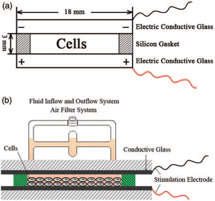
The BES incubation system. (a) Schematic diagram of a longitudinal section of the incubation chamber including: the upper and lower electric conductive glass plates (FTO glass), a closed silicone gasket, the incubation chamber, and a pair of electrode wires; (b) Schematic diagram of a longitudinal section of the entire BES incubation system including the incubation chamber, the fluid inflow-outflow system, the air filter system, a pair of electrode wires, and a fixed cover and base. Conditions of BES: the NPCs were exposed to 12 h of BES at 25mV/mm and 50mV/mm electric field strengths with a pulse-burst pattern and 8ms pulses (20% duty cycle). Cells that were not exposed to BES served as controls. (A color version of this figure is available in the online journal)
The BES incubation system. (a) Schematic diagram of a longitudinal
section of the incubation chamber including: the upper and lower electric  conductive glass plates (FTO glass), a closed silicone gasket, the incubation
chamber, and a pair of electrode wires; (b) Schematic diagram of a longitudinal
section of the entire BES incubation system including the incubation chamber,
the fluid inflow-outflow system, the air filter system, a pair of electrode wires, and
a fixed cover and base. Conditions of BES: the NPCs were exposed to 12 h of
BES at 25mV/mm and 50mV/mm electric field strengths with a pulse-burst
pattern and 8ms pulses (20% duty cycle). Cells that were not exposed to BES
served as controls. 

Spinal cord injury affects approximately 250,000 Americans, with 52% being paraplegic and 47% quadriplegic. There are 11,000 new spinal cord injuries each year and 82% are male.

Stem cell transplantions into the spinal cord to regenerate severed neurons and associated cells provides a potentially powerful treatment. However, once stem cells are implanted into the injured spinal cord, many of them die. Cell death is probably a consequence of several factors such as a local immune response, hypoxia (lack of oxygen), and probably most importantly, limited quantities of growth factors.

Fan said of his work, “We’ve shown for the very first time that BES may provide insight into preventing growth factor deprivation-triggered apoptosis in olfactory bulb precursor cells. These findings suggest that BES may thus be used as a strategy to improve cell survival and prevent cell apoptosis (programmed cell death) in stem cell-based transplantation therapies.”

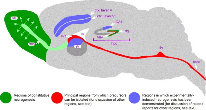
The olfactory bulb is in green in this mouse brain.

Since electrical stimulation dramatically accelerates the speed of axonal regeneration and target innervation and positively modulates the functional recovery of injured nerves, Fan decided to test BES. His results showed that BES upregulated all the sorts of responses in stem cells that you would normally see with growth factors. Thus BES can increase stem cell survival without exogenous chemicals or genetic engineering.

Fan and his team examined the effects of BES on olfactory bulb neural precursor cells and they found that 12 hours of BES exposure protected cells from dying after growth factor deprivation. How did BES do this? Fan and other showed that BES stimulated a growth factor pathway called the PI3K/Akt signaling cascade. BES also increase the output of brain-derived neurotrophic factor.

“What was especially surprising and exciting,” said Fan, “was that a non-chemical procedure can prevent apoptosis in stem cell therapy for spinal cord patients.” Fan continued: “How BES precisely regulates the survival of exogenous stem cells is still unknown but will be an extremely novel area of research on spinal cord injury in the future.”

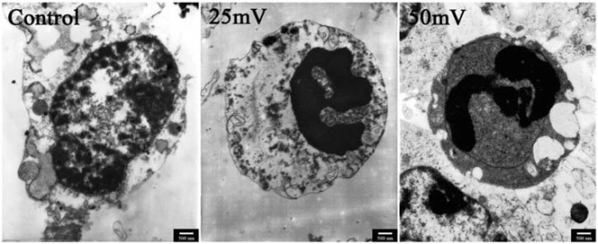
BES alters the ultrastructure of NPCs. The ultrastructural morphological changes of cells were investigated by TEM. In the control group (unstimulated), cells had a necrotic appearance: most cells lost the normal cellular structure with a consequent release of cell contents. In the 25mV/mm and 50mV/mm BES groups, the NPCs showed an apoptotic morphology with nuclear fragmentation and condensation
BES alters the ultrastructure of NPCs. The ultrastructural morphological changes of cells were investigated by TEM. In the control group (unstimulated), cells had a necrotic appearance: most cells lost the normal cellular structure with a consequent release of cell contents. In the 25mV/mm and 50mV/mm BES groups, the NPCs showed an apoptotic morphology with nuclear fragmentation and condensation

BES can improve the survival of neural precursor cells and will provide the survival of neural precursor cells and will provide the basis or future studies that could lead to novel therapies for patients with spinal cord injury.

Cardiac Muscle Repair with Molecular Beacons


Pure heart muscle cells that are ready for transplantation. This is one of the Holy Grails of regenerative medicine. Of course when working with pluripotent stem cell lines, isolating nothing but beating heart muscle cells is rather difficult. A new technique makes the isolation of pure cultures of beating heart muscle cells that much easier.

Researchers at Emory and Georgia Tech have developed a method that utilizes molecules called “molecular beacons” to isolate heart muscle cells from pluripotent stem cells. Molecular beacons fluoresce when they come into contact with cells that express certain genes. In this case, the beacons target cells that express heart-specific myosin.

Physicians can use these purified cardiac muscle cells to heal damaged areas of the heart in patient that have suffered a heart attack or are suffering heart failure. This molecular beacon technique might also have applications in other fields of regenerative medicine as well.

“Often, we want to generate a particular cell population from stem cells for introduction into patients,” said Young-sup Yoon, professor of medicine and director for stem cell biology at Emory University School of Medicine. “But the desired cells often lack a readily accessible surface marker, or that marker is not specific enough, as is the case for cardiac muscle cells. This technique could allow us to purify almost any type of cell.”

Gang Bao pioneered he use of molecular beacons and was a co-author of this publication. Yoon and is colleagues and collaborators grew mouse and human embryonic stem cells and induced pluripotent stem cells and differentiated them into heart muscle cells (cardiomyocytes). They then used molecular beacons to label only those cells that expressed messenger RNAs with just the right sequences. These molecular beacons hybridized with the mRNAs and fluoresced. Bao and others then used flow cytometry to sort the fluorescent cells from the non- fluorescent cells. The fluorescent cells have differentiated into heart muscle cells and were isolated from all the other cells.

These purified heart muscle cells could engraft into the heart of a mouse that had suffered a heart attack and they improved heart function and formed no tumors. This proof-of-principle experiment shows that this technique is feasible.

“In previous experiments with injected bare cells, investigators at Emory and elsewhere found that a large proportion of the cells are washed away. We need to engineer the cells into compatible biomaterials to enhance engraftment and retention,” said Yoon,

Spiking Stem Cells to Generate Myelin


Regenerating damaged nerve tissue represents a unique challenge for regenerative medicine. Nevertheless, some experiments have shown that it is possible to regenerate the myelin sheath that surrounds particular nerves.

Myelin is a fatty, insulating sheath that surrounds particular nerves and accelerates the transmission of nerve impulses. The myelin sheath also helps neurons survive, and the myelin sheath is attacked and removed in multiple sclerosis, a genetic disease called Charcot-Marie-Tooth disease, and spinal cord injuries. Being able to regenerate the myelin sheath is an essential goal of regenerative medicine.

Fortunately, a new study from a team of UC Davis (my alma mater) scientists have brought this goal one step closer. Wenbig Deng, principal investigator of this study and associate professor of biochemistry and molecular medicine, said, “Our findings represent an important conceptual advance in stem cell research. We have bioengineered the first generation of myelin-producing cells with superior regenerative capacity.”

The brain contains two main cell types; neurons and glial cells. Neurons make and transmit nerve impulses whereas glial cells support, nourish and protect neurons. One particular subtype of glial cells, oligodendrocytes, make the myelin sheath that surrounds the axons of many neurons. Deng and his group developed a novel protocol to induce embryonic stem cells (ESCs) to differentiate into oligodendrocyte precursor cells or OPCs. Even though other researchers have made oligodenrocytes from ESCs, Deng’s method results in purer populations of OPCs than any other available method.

Making OPCs from ESCs is one thing, but can these laboratory OPCs do everything native can do? When Deng and his team tested the electrophysiological properties of their laboratory-made OPCs, they discovered that their cells lacked an important component; they did not express sodium channels. When the lab-made OPCs were genetically engineered to express sodium channels, they generated the characteristic electrical spikes that are common to native OPCs. According to Deng, this is the first time anyone has made OPCs in the laboratory with spiking properties. Is this significant?

Deng and his colleagues compared the spiking OPCs to non-spiking OPCs in the laboratory. Not only did the spiking OPCs communicate with neurons, but they also did a better job of maturing into oligodentrocytes.

Transplantation of these two OPC populations into the spinal cord and brains of mice that are genetically unable to produce myelin also showed differences. Both types of OPCs were able to mature into oligodendrocytes and produce myelin sheaths, but only the spiking OPCs had the ability to produce longer and thicker myelin sheaths.

Said Deng, “We actually developed ‘super cells’ with an even greater capacity to spike than natural cells. This appears to give them an edge for maturing into oligodendrocytes and producing better myelin.

Human neural tissue has a poor capacity to regenerate and even though OPCs are present, they do not regenerate tissue effectively when disease or injury damages the myelin sheath. Deng believes that replacing glial cells with the enhanced spiking OPCs to treat injuries and diseases has the potential to be a better strategy than replacing neurons, since neurons are so problematic to work with in the laboratory. Instead providing the proper structure and environment for neurons to live might be the best approach to regenerate healthy neural tissue. Deng also said that many diverse conditions that have not been traditionally considered to be myelin-based diseases (schizophrenia, epilepsy, and amyotrophic lateral sclerosis) are actually now recognized to involve defective myelin.

On that one, I think Deng is dreaming. ALS is caused by the death of motor neurons due to mechanisms that are intrinsic to the neurons themselves. Giving them all the myelin in the world in not going to help them. Also, OPCs made from ESCs will be rejected out of hand by the immune system if they are used to regenerate myelin in the peripheral nervous system. The only hope is to keep them in the central nervous system, but even there, any immune response in the brain will be fatal to the OPCs. This needs to be tested with iPSCs before it can be considered for clinical purposes.

The Use of Synthetic Messenger RNAs Augment Heart Regeneration and Healing After a Heart Attack


A collaborative effect between researchers at Harvard University and Karolinska Institutet has shown that the application of particular factors to the heart after a heart attack can heal the heart and induce the production of new heart muscle.

Kenneth Chien, who has a dual appointment at the medical university Karolinska Institutet and Harvard University, led this research teams said this about this work: “This is the beginning of using the heart as a factory to produce growth factors for specific families of cardiovascular stem cells, and suggests that it may be possible to generate new heart parts without delivering any new cells to the heart itself.”

This study builds upon previous work by Chien and his colleagues in which the growth factor VEGFA, which is known to activate the growth of endothelial cells in the adult heart (endothelial cells line blood vessels), also serves as a switch that converts heart stem cells away from making heart muscle to forming coronary vessels in the fetal heart.

To drive the expression of VEGFA in the heart, Chien and others made synthetic messenger RNAs that encoded VEGFA and injected them into the heart cells. Injections of these synthetic VEGFA messenger RNAs produced a short burst of VEGFA.

Chien induced a heart attack in mice and then administered the synthetic VEGFA messenger RNAs to some mice and buffer to others 48 hours after the heart attacks. Chien and his crew was sure to inject the synthetic VEGFA mRNAs into the regions of the heart known to harbor the resident cardiac stem cell populations.

Not only did the VEGFA-mRNA-injected mice survive better than the other mice, but their hearts had smaller heart scars, and had clear signs of the growth of new heart muscle that had been made by the resident cardiac stem cell populations. One pulse of VEGFA had long-term benefits and those cells that would have normally made the heart scar ended up making heart muscle instead as a result of one pulse of VEGFA.

Chien said of this experiment, “This moves us very close to clinical studies to regenerate cardiovascular tissue with a single chemical agent without the need for injecting any additional cells into the heart.”

At the same time, Chien also noted that this technology is in the early stages of development. Even though these mice had their chests cracked open and their hearts injected, for human patients, the challenge is to adapt heart catheter technologies to the delivery of synthetic messenger RNAs. Also, to demonstrate the safety and efficacy of this technology to humans, Chien and others will need to repeat these experiments in larger animals that serve as a better model system for the human heart than rodents. Chien’s laboratory is presently in the process of doing that.

To adapt catheter technology to deliver these reagents, Chien had co-founded a company called Moderna Therapeutics to research this problem and develop the proper platform technology for clinical use. Chien is also collaborating with the biotechnology company AstraZeneca to help expedite moving the synthetic RNA technology into a clinical setting.

Forming Induced Pluripotent Stem Cells Inside a Living Organism


A team from the Spanish National Cancer Research Centre (CNIO) has become the first research team to convert adult cells that are still within a living organism into cells that show characteristics of embryonic stem cells.

The CNIO researchers also say that these embryonic stem cells, which were obtained directly from inside an organism, have a broader capacity for differentiation than those obtained by means of an in vitro culture system. Specifically, they have the characteristics of totipotent cells, a primitive state never before obtained in a laboratory, according to the CNIO team.

Manuel Serrano, Ph.D., director of CNIO’s Molecular Oncology Program and head of the Tumor Suppression Laboratory, led this study. It was supported by Manuel Manzanares, Ph.D., and his team from the Spanish National Cardiovascular Research Centre.

The CNIO researchers say their work extends that of Nobel Prize winner Shinya Yamanaka, M.D., Ph.D., one step forward. Yamanaka opened a new horizon in regenerative medicine when, in 2006, he demonstrated that stem cells could be created from adult cells by using a cocktail of genes. But while Yamanaka induced his cells in culture in the lab (in vitro), the CNIO team created theirs directly in mice (in vivo). Generating these cells within an organism brings this technology even closer to regenerative medicine, they say.

In a study published online Sept. 11 in the journal Nature, the CNIO research team details how it used genetic manipulation techniques to create mice in which Dr. Yamanaka’s four genes could be activated at will. When these genes were activated, they observed that the adult cells were able to de-differentiate into embryonic stem cells in multiple tissues and organs.

María Abad, Ph.D., lead author of the article and a researcher in Dr. Serrano’s group, said, “This change of direction in development has never been observed in nature. We have demonstrated that we can also obtain embryonic stem cells in adult organisms and not only in the laboratory.”

Dr. Serrano added, “We can now start to think about methods for inducing regeneration locally and in a transitory manner for a particular damaged tissue.” Stem cells obtained in mice also show totipotent characteristics never generated in a laboratory. Totipotent cells can form all the cell types in a body, including the placental cells. Embryonic cells within the first couple of cell divisions after fertilization are the only cells that are totipotent.

The researchers reported that they were also able to induce the formation of pseudo-embryonic structures in the thoracic and abdominal cavities of the mice. These pseudo-embryos displayed the three layers typical of embryos (ectoderm, mesoderm, and endoderm), and extra-embryonic structures such as the vitelline membrane, which surrounds the egg, and even signs of blood cell formation, which first appears in the primary embryonic vesicle (otherwise known as the “yolk sac”).

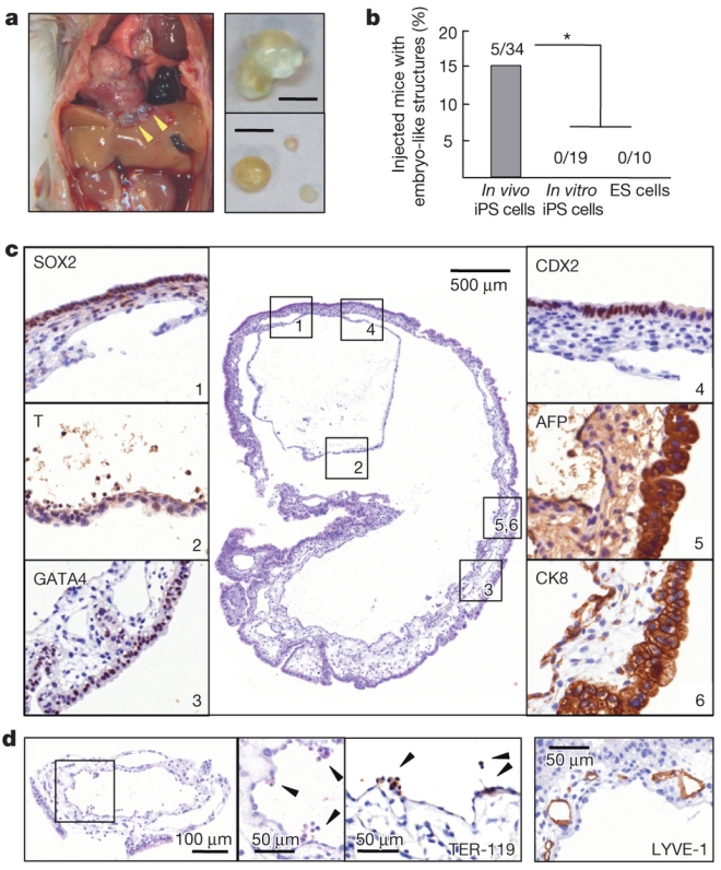
“This data tell us that our stem cells are much more versatile than Dr. Yamanaka’s in vitro inducted pluripotent stem cells, whose potency generates the different layers of the embryo but never tissues that sustain the development of a new embryo, like the placenta,” the CNIO researcher said.  Below is a figure from their paper.  The pictures look pretty convincing.

a, Cysts in the abdominal cavity of a reprogrammable mouse. b, Frequency of embryo-like structures after intraperitoneal injection of in vivo iPS cells (3 clones), in vitro iPS cells (2 clones) and ES cells (JM8.F6). Fisher’s exact test: *P < 0.05. c, Cyst generated by intraperitoneal injection. Left panels, germ layer markers: SOX2 (ectoderm), T/BRACHYURY (mesoderm) and GATA4 (endoderm). Right panels, extraembryonic markers: CDX2 (trophectoderm), and AFP and CK8, both specific for visceral endoderm of the yolk sac. d, Cyst generated by intraperitoneal injection presenting TER-119+ nucleated erythrocytes and LYVE-1+ endothelial cells in structures resembling yolk sac blood islands.

The researchers emphasize that any possible therapeutic applications of their work are still distant, but they believe that it could mean a change of direction for stem cell research, regenerative medicine and tissue engineering.

“Our stem cells also survive outside of mice in a culture, so we can also manipulate them in a laboratory,” said Dr. Abad. “The next step is studying if these new stem cells are capable of efficiently generating different tissues such as that of the pancreas, liver or kidney.”

This paper is very interesting, but I find it rather unlikely that their approach will take regenerative medicine by storm.  Engineering mice to express these four genes in an inducible manner caused the formation of unusual tumors throughout the mice.  Maybe they can be coaxed to differentiate into kidney or heart muscle or whatever, but learning how to get them to do that will take a fair amount of in vitro work.  This is interesting, but I doubt that it will change the field overnight.

Do Stem Cells from Bone Outdo Those from the Heart in Regenerating Cardiac Tissue?


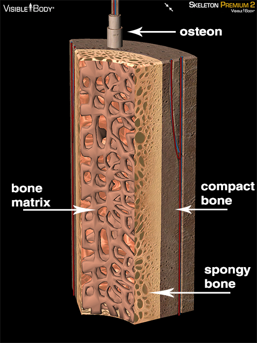
Scientists at Tulane University in New Orleans, La. (US) have completed a study that suggests that stem cells derived from cortical, or compact bone do a better job of regenerating heart tissue than do the heart’s own stem cells.

The study, led by Steven R. Houser, Ph.D., FAHA, director of Tulane’s School of Medicine’s Cardiovascular Research Center (CVRC), could potentially lead to an “off the rack” source of stem cells for regenerating cardiac tissue following a heart attack.

Cortical bone stem cells (CBSCs) are considered some of the most pluripotent cells in the adult body. These cells are naïve and ready to differentiate into just about any cell type. However, even though CBSCs and similar pluripotent stem cells retain the ability to develop into any cell type required by the body, they have the potential to wander off course and land in unintended tissues. Cardiac stem cells, on the other hand, are more likely to stay in their resident tissue.

Bone cross-section

To determine how CBSCs might behave in the heart, Houser’s team, led by Temple graduate student Jason Duran, collected the cells from mouse tibias (shin bones), expanded them in the lab and then injected them into back the mice after they had undergone a heart attack.

The cells triggered the growth of new blood vessels in the injured tissue and six weeks after injection had differentiated into heart muscle cells. While generally smaller than native heart cells, the new cells had the same functional capabilities and overall improved survival and heart function.

Similar improvements were not observed in mice treated with cardiac stem cells, nor did those cells show evidence of differentiation.

“What we did generates as many questions as it does answers,” Dr. Houser said. “Cell therapy attempts to repopulate the heart with new heart cells. But which cells should be used, and when they should be put into the heart are among many unanswered questions.”

The next step will be to test the cells in larger animal models. The current study was published in the Aug. 16 issue of Circulation Research.

Transformation of Non-Beating Human Cells into Heart Muscle Cells Lays Foundation for Regenerating Damaged Hearts


After a heart attack, the cells within the damaged part of the heart stop beating and become ensconced in scar tissue. Not only does this region not beat, it does not conduct the signal to beat either and that can not only lead to a slow, sluggish heartbeat, it can also cause irregular heart rates or arrhythmias.

Now, however, scientists have demonstrated that this damage to the heart muscle need not be permanent. Instead there is a way to transform those cells that form the human scar tissue into cells that closely resemble beating heart cells.

Last year, researchers from the laboratory of Deepak Srivastava, MD, the director of Cardiovascular and Stem Cell Research at the Gladstone Institute, transformed scar-forming heart cells (fibroblasts) into beating heart-muscle cells in live mice. Now they report doing the same to human cells in a culture dishes.

“Fibroblasts make up about 50 percent of all cells in the heart and therefore represent a vast pool of cells that could one day be harnessed and reprogrammed to create new muscle,” said Dr. Srivastava, who is also a professor at the University of California, San Francisco. “Our findings here serve as a proof of concept that human fibroblasts can be reprogrammed successfully into beating heart cells.”

In 2012, Srivastava and his team reported that fibroblasts could be reprogrammed into beating heart cells by injecting just three genes (collectively known as GMT, which is short for Gata4, Mef2c, and Tbx5), into the hearts of live mice that had been damaged by a heart attack (Qian L, et al., Nature. 2012 31;485(7400):593-8). From this work, they reasonably concluded that the same three genes could have the same effect on human cells.

“When we injected GMT into each of the three types of human fibroblasts (fetal heart cells, embryonic stem cells and neonatal skin cells) nothing happened—they never transformed—so we went back to the drawing board to look for additional genes that would help initiate the transformation,” said Gladstone staff scientist Ji-dong Fu, Ph.D., the study’s lead author. “We narrowed our search to just 16 potential genes, which we then screened alongside GMT, in the hopes that we could find the right combination.”

The research team began by injecting all candidate genes into the human fibroblasts. They then systematically removed each one to see which were necessary for reprogramming and which were dispensable. In the end, they found that injecting a cocktail of five genes—the 3-gene GMT mix plus the genes ESRRG and MESP1—were sufficient to reprogram the fibroblasts into heart-like cells. They then found that with the addition of two more genes, called MYOCD and ZFPM2, the transformation was even more complete.

To help things along, the team used a growth factor known as Transforming Growth Factor-Beta (TGF-Beta) to induce a signaling pathway during the early stages of reprogramming that further improved reprogramming success rates.

“While almost all the cells in our study exhibited at least a partial transformation, about 20 percent of them were capable of transmitting electrical signals—a key feature of beating heart cells,” said Dr. Fu. “Clearly, there are some yet-to-be-determined barriers preventing a more complete transformation for many of the cells. For example, success rates might be improved by transforming the fibroblasts within living hearts rather than in a dish—something we also observed during our initial experiments in mice.”

The immediate next steps are to test the five-gene cocktail in hearts of larger mammals. Eventually, the team hopes that a combination of small, drug-like molecules could be developed to replace the cocktail, which would offer a safer and easier method of delivery.

This latest study was published online August 22 in Stem Cell Reports.

First Patient Treated in Study that Tests Stem Cell-Gene Combo to Repair Heart Damage


The first patient has been treated in a groundbreaking medical trial in Ottawa, Canada, that uses a combination of stem cells and genes to repair tissue damaged by a heart attack. The first test subject is a woman who suffered a severe heart attack in July and was treated by the research team at the Ottawa Hospital Research Institute (OHRI). Her heart had stopped beating before she was resuscitated, which caused major damage to her cardiac muscle.

The therapy involves injecting a patient’s own stem cells into their heart to help fix damaged areas. However, the OHRI team, led by cardiologist Duncan Stewart, M.D., took the technique one step further by combining the stem cell treatment with gene therapy.

“Stem cells are stimulating the repair. That’s what they’re there to do,” Dr. Stewart said in an interview. “But what we’ve learned is that the regenerative activity of the stem cells in these patients with heart disease is very low, compared to younger, healthy patients.”

Stewart and his colleagues will supply the stem cells with extra copies of a particular gene in an attempt to restore some of that regenerative capacity. The gene in question encodes an enzyme called endothelial nitric oxide synthase (eNOS). Nitric oxide is a small, gaseous molecule that is made from the amino acid arginine by the enzyme nitric oxide synthase. Nitric oxide or NO signals to smooth muscle cells that surround blood vessels to relax, which causes blood vessels to dilate and this increases blood flow. In the damaged heart, NO also helps build up new blood vessels, which increase healing of the cardiac muscle. Steward added, “That, we think, is the key element. We really think it’s the genetically enhanced cells that will provide the advantage.”

Nitric oxide synthesis

The study will eventually involve 100 patients who have suffered severe heart attacks in Ottawa, Toronto and Montreal.

Benefits of stem cells in treating MS declines with donor’s age


MS is a neurodegenerative disease characterized by inflammation and scar-like lesions throughout the central nervous system (CNS). There is no cure and no treatment eases the severe forms of MS. But previous studies on animals have shown that transplantation of mesenchymal stem cells (MSCs) holds promise as a therapy for all forms of MS (see Bai L, et al., Glia 2009 Aug 15;57(11):1192-203). The MSCs migrate to areas of damage, release trophic (cell growth) factors and exert protective effects on nerves and regulatory effects to inhibit T cell proliferation.

Several clinical trials examining the ability of fat-derived MSCs to treat MS patients have been conducted. Unfortunately, most of these studies are rather small and the results are all over the place. One study treated ten patients with MSCs injected intrathecally (just under the meninges that cover the brain and spinal cord) and the results were mixed; 6/10 improved, 3 stayed the same and one deteriorated. Another study treated ten patients with intravenous fat-derived MSCs and the patients showed symptomatic improvement, but when MRIs of the brain were examined, no improvements could be documented. A third study treated 15 people with intrathecal injections and IV administrations of MSCs, and some stabilized. A fourth study only examined 3 patients treated with a mixture of their own fat-derived MSCs and fat-derived MSCs from another person. In all three cases, their MRIs and symptoms improved. A fifth study used umbilical cord MSCs administered intravenously and the patient showed substantial improvement (for review see Tyndall, Pediatric Research 71(4):433-438).

These results are somewhat encouraging, but also somewhat underwhelming and clinical trials go. Why did some work and other not work as well? In order to understand why, researchers must understand the biologic changes and therapeutic effects of older donor stem cells. A new study appearing in the journal STEM CELLS Translational Medicine is the first to demonstrate that adipose-derived MSCs donated by older people are less effective than cells from their younger counterparts.

Fortunately, all the available MS-related clinical trials have confirmed the safety of autologous MSC therapy. As to the efficacy of these cells, however, it is unclear if MSCs derived from older donors have the same therapeutic potential as those from younger ones.

“Aging is known to have a negative impact on the regenerative capacity of most tissues, and human MSCs are susceptible to biologic aging including changes in differentiation potential, proliferation ability and gene expression. These age-related differences may affect the ability of older donor cells to migrate extensively, provide trophic support, persist long-term and promote repair mechanisms,” said Bruce Bunnell, Ph.D., of Tulane University’s Center for Stem Cell Research and Regenerative Medicine. He served as lead author of the study, conducted by a team composed of his colleagues at Tulane.

In their study, Bunnell and his colleagues induced an MS-like disease in laboratory mice called chronic experimental autoimmune encephalomyelitis (EAE). Then they treated them before disease onset with human adipose-derived MSCs derived from younger (less than 35 years) or older (over age 60) donors. The results corroborated previous studies that suggested that older donors are less effective than their younger counterparts.

“We found that, in vitro, the stem cells from the older donors failed to ameliorate the neurodegeneration associated with EAE. Mice treated with older donor cells had increased inflammation of the central nervous system, demyelination leading to an impairment in movement, cognition and other functions dependent on nerves, and a proliferation of splenocytes [white blood cells in the spleen], compared to the mice receiving cells from younger donors,” Dr. Bunnell noted.

In fact, the proliferation of T cells (immune cells that attack the myelin sheath in MS patients) in these mice indicated that older MSCs might actually stimulate the proliferation of the T cells, while younger stem cells inhibit T cell proliferation. T cells are a type of white blood cell in the body’s immune system that help fight off disease and harmful substances. When they attack our own tissues, they can cause diseases like MS.

As such, Dr. Bunnell said, “A decrease in T cell proliferation would result in a decreased number of T cells available to attack the CNS in the mice, which directly supports the results showing that the CNS damage and inflammation is less severe in the young MSC-treated mice than in the old MSC-treated mice.”

“This study in an animal model of MS is the first to demonstrate that fat-derived stem cells from older human donors have less therapeutic effectiveness than cells from young donors,” said Anthony Atala, M.D., editor of STEM CELLS Translational Medicine and director of the Wake Forest Institute for Regenerative Medicine. “The results point to a potential need to evaluate cell therapy protocols for late-onset multiple sclerosis patients.”

Stem Cell-Conventional Treatment Combo Offers New Hope in Fighting Deadly Brain Cancer


A new type of treatment that combines neural stem cells with conventional cancer fighting therapies shows promise in animal studies for the most common and deadliest form of adult brain cancer — glioblastoma multiforme (GBM). The details are revealed in a groundbreaking study led by Maciej Lesniak, M.D., that appeared in the journal STEM CELLS Translational Medicine.

“In this work, we describe a highly innovative gene therapy approach, which is being developed along with the NIH and the FDA. Specifically, our group has developed an allogeneic neural stem cell line that is a carrier for a virus that can selectively infect and break down cancer cells,” explained Dr. Lesniak, the University of Chicago’s director of neurosurgical oncology and neuro-oncology research at the Brain Tumor Center.

The stem cell line used is a neural stem cell line called HB1.F3 NSC. The US Food and Drug Administration has recently approved this cell line for use in a phase I human clinical trial.

Glioblastoma multiforme remains fatal despite intensive treatment with surgery, radiation and chemotherapy. Cancer-killing viruses have been used in clinical trials to treat those tumors that resist treatment with other therapies and infiltrate throughout the brain. Unfortunately, according to Lesniak, this therapy was subject to some “major drawbacks.”

“When you inject a virus into a tumor alone (without a carrier, like NSC), the virus stays at the site of the injection, and does not spread. Moreover, our immune system clears it. By using NSCs, we can achieve a widespread distribution of the virus throughout the tumor mass, since the NSC travel. Also, they act like a stealth fighter, hiding the virus from the immune system.” Lesniak and his co-workers used NSCs loaded with a novel oncolytic adenovirus. This virus selectively targets glioblastoma multiforme in combination with chemo-radiotherapy. Using this strategy, Lesniak’s team was able to overcome the limitations associated with anticancer viral therapies.

Using mice that had glioblastoma multiforme, the research team showed that their neural stem cell line, which is derived from human fetal cells, significantly increased the median survival time of the mice beyond conventional treatments alone. The addition of chemo-radiotherapy further enhanced the benefits of this novel stem cell-based gene therapy approach.

“Our study argues in favor of using stem cells for delivery of oncolytic viruses along with multimodal chemo-radiotherapy for the treatment of patients with glioblastoma multiforme, and this is something that we believe warrants further clinical investigation,” Dr. Lesniak concluded.

Lesniak’s team is completing final FDA-directed studies. He expects to start a human clinical trial, in which a novel oncolytic virus will be delivered via NSCs to patients with newly diagnosed glioblastoma multiforme, early in 2014.

Treatment of glioblastoma multiforme depends on novel therapies,” said Anthony Atala, M.D., Editor of STEM CELLS Translational Medicine and director of the Wake Forest Institute for Regenerative Medicine. “This study establishes that a combination of conventional and gene therapies may be most effective and suggests a protocol for a future clinical investigation.”

Producing blood cells from stem cells could yield a purer, safer cell therapy


The journal Stem Cells Translational Medicine has published a new protocol for reprogramming induced pluripotent stem cells (iPSCs) into mature blood cells. This protocol uses only a small amount of the patient’s own blood and a readily available cell type. This novel method skips the generally accepted process of mixing iPSCs with either mouse or human stromal cells. Therefore, is ensures that no outside viruses or exogenous DNA contaminates the reprogrammed cells. Such a protocol could lead to a purer, safer therapeutic grade of stem cells for use in regenerative medicine.

The potential for the field of regenerative medicine has been greatly advanced by the discovery of iPSCs. These cells allow for the production of patient-specific iPSCs from the individual for potential autologous treatment, or treatment that uses the patient’s own cells. Such a strategy avoids the possibility of rejection and numerous other harmful side effects.

CD34+ cells are found in bone marrow and are involved with the production of new red and white blood cells. However, collecting enough CD34+ cells from a patient to produce enough blood for therapeutic purposes usually requires a large volume of blood from the patient. However, a new study outlined But scientists found a way around this, as outlined by Yuet Wai Kan, M.D., FRS, and Lin Ye, Ph.D. from the Department of Medicine and Institute for Human Genetic, University of California-San Francisco has devised a way around this impasse.

“We used Sendai viral vectors to generate iPSCs efficiently from adult mobilized CD34+ and peripheral blood mononuclear cells (MNCs),” Dr. Kan explained. “Sendai virus is an RNA virus that carries no risk of altering the host genome, so is considered an efficient solution for generating safe iPSC.”

“Just 2 milliliters of blood yielded iPS cells from which hematopoietic stem and progenitor cells could be generated. These cells could contain up to 40 percent CD34+ cells, of which approximately 25 percent were the type of precursors that could be differentiated into mature blood cells. These interesting findings reveal a protocol for the generation iPSCs using a readily available cell type,” Dr. Ye added. “We also found that MNCs can be efficiently reprogrammed into iPSCs as readily as CD34+ cells. Furthermore, these MNCs derived iPSCs can be terminally differentiated into mature blood cells.”

“This method, which uses only a small blood sample, may represent an option for generating iPSCs that maintains their genomic integrity,” said Anthony Atala, MD, Editor of STEM CELLS Translational Medicine and director of the Wake Forest Institute for Regenerative Medicine. “The fact that these cells were differentiated into mature blood cells suggests their use in blood diseases.”

Stem Cell Therapy Repairs Brain Damage Hours After Stroke Occurs


According to the Center for Disease Control, stroke is a leading cause of death in the United States. Fortunately stroke has been the subject of significant research efforts, but unfortunately, developing treatments that ensure complete recovery for stroke patients is extremely challenging. The challenge increase when more than a few hours have passed between onset of the stroke and administration of treatment.

Thus a new study released in STEM CELLS Translational Medicine has generated more than a little excitement. This study indicates that indicates that endothelial precursor cells (EPCs), which are found in the bone marrow, umbilical cord blood, and rarely in peripheral blood, can make a significant difference for these patients’ recovery. The contribution of EPCs even extends to the later stages of stroke. In animal studies, EPC implantation into the brain after a stroke minimized the initial brain injury and helped repair the stroke damage.

“Previous studies indicated that stem/progenitor cells derived from human umbilical cord blood (hUCB) improved functional recovery in stroke models,” noted Branislava Janic, Ph.D., a member of Henry Ford Health System’s Cellular and Molecular Imaging Laboratory in Detroit and lead author of the study. “We wanted to examine the effect of hUCB-derived AC133+ endothelial progenitor cells (EPCs) on stroke development and resolution in rats.”

Dr. Janic and his team injected EPCs into the brains of rats that had suffered strokes. When they later examined the animals using MRI, they found that the transplanted EPCs had selectively migrated to the injured area, stopped the tissue damage from spreading, initiated regeneration, and affected the time course for stroke resolution. The lesion size in the brain was significantly decreased at a dose of 10 million cells, if the cells were given as early as seven days after the onset of the stroke.

“This led us to conclude that cord blood-derived EPCs can significantly contribute to developing more effective treatments that allow broader time period for intervention, minimize the initial brain injury and help repair the damage in later post-stroke phases,” Dr. Janic said.

“The early signs of stroke are often unrecognized, and many patients cannot take advantage of clot-busting treatments within the required few hours after stroke onset,” said Anthony Atala, M.D., editor of STEM CELLS Translational Medicine and director of the Wake Forest Institute for Regenerative Medicine. “In this animal study, a combination of stem cells shows promise for healing stroke damage when administered 24 hours after the stroke.”

Overexpression of a Potassium Channel in Heart Muscle Cells Made From Embryonic Stem Cells Decreases Their Arrhythmia Risk


Embryonic stem cells have the capacity to differentiate into every cell in the adult body. One cell type into which embryonic stem cells (ESCs) can be differentiated rather efficiently is cardiomyocytes, which is a fancy term for heart muscle cells. The protocol for making heart muscle cells from ESCs is well worked out, and the conversion is rather efficient and the purification schemes that have been developed are also rather effective (for example, see Cao N, et al., Highly efficient induction and long-term maintenance of multipotent cardiovascular progenitors from human pluripotent stem cells under defined conditions. Cell Res. 2013 Sep;23(9):1119-32. doi: 10.1038/cr.2013.102 and Mummery CL et al., Differentiation of human embryonic stem cells and induced pluripotent stem cells to cardiomyocytes: a methods overview. Circ Res. 2012 Jul 20;111(3):344-58).

Using these cells in a clinical setting has two large challenges. The first is that embryonic stem cell derivatives are rejected by the immune system of the recipient, thus setting up the patient for a graft versus host response to the implanted tissue, thus making the patient even sicker than when they started. The second problem is that heart muscle cells made from ESCs are immature and cause the heart to beat abnormally fast thus causing “tachyarrythmias” and died within the first two weeks after the transplant (see Liao SY, et al., Heart Rhythm 2010 7:1852-1859).

Both of these problems are large problems, but the laboratory of Ronald Li at the University of Hong Kong at used a genetic engineering trick to make heart muscle cells from mouse embryonic stem cells to seemingly fix this problem.

Li and his colleagues engineered mouse ESCs with a gene for a potassium rectifier channel that could be induced with drugs. Then they differentiated these genetically ESCs into heart muscle cells. This potassium rectifier channel (Kir2.1) is not present in immature heart muscle cells and putting it into these cells might cause them to beat at a slower rate.

These engineered ESC-derived heart muscle cells were tested for their electrophysiological properties first. Without the drug that induces KIR2.1, the heart muscle cells showed very abnormal electrical properties. However, once the drug was added, their electrical properties looked much more normal.

Then they induced heart attacks in laboratory animals and implanted their engineered ESC-derived heart muscle cells 1 hour after the heart attacks were induced. Animals not given the drug to induce the expression of Kir2.1 faired very poorly and had episodes of tachyarrythmia (really fast heart beat) and over half of them died by 5 weeks after the implantation. Essentially the implanted animals did worse than those animals that had had a heart attack that were not treated. However, those animals that were given the drug that induces the expression of Kir2.1 in heart muscle cells did much better. The survival rate of these animals was higher than the untreated animals after about 7 weeks after the procedure. Survival rates increased by only a little, but the increase was significant. Also, the animals that died did not die of tachyarrythmias. In fact the rate of tachyarrythmias in the animals given the inducing drug (which was doxycycline by the way) had significantly lower levels of tachyarrythmia than the other two groups.

Other heart functions were also significantly affected. The ejection fraction in the animals that ha received the Kir2.1-expression heart muscle cells was 10-20% higher than the control animals. Also the density of blood vessels was substantially higher in both sets of animals treated with ESC-derived heart muscle cells. The echocardiogram of the hearts implanted with the Kir2.1-expressing heart muscle cells was altogether more normal than that of the others.

This paper is a significant contribution to the use of ESC-derived cells to treat heart patients. The induction of heart arrhythmias by ESC-derived heart muscle cells is a documented risk of their use. Li and his colleagues have effectively eliminated that risk in this paper by forcing the expression of a potassium rectifier channel in the ESC-derived heart muscle cells. Also, because these cells were completely differentiated and did not have any interloping pluripotent cells in their culture, tumor formation was not observed.

There are a few caveats I would like to point out. First of all, the increase in survival rate above the control is not that impressive. The improvement in heart function parameters is certainly encouraging, but because the survival rates are not that higher than the control mice that received no treatment, it appears that these benefits were only conferred to those mice who survived in the first place.

Secondly, even though the heart attacks were induced in the ventricles of the heart, Li and his colleagues injected a mixture of heart muscle cells that included atrial, ventricular, nodal and heart fibroblasts. This provides an opportunity for beat mismatches and a “substrate for ventricular tachycardia” as Li puts it. In the future, the transplantation of just ventricular heart muscle cells would be cleaner experiment. Since these mice were not observed long enough to observe potential arrythmias that might have arisen from the presence of a mixed population in the ventricle.

Finally, in adapting this to humans might be difficult, since the hearts of mice beat so much faster than those of humans. It is possible that even if human cardiomyocytes were engineered with Kir2.1-type channels, that arrythmias might still be a potential problem.

Despite all that, Li’s publication is a large step forward.

Treating Crohn’s Disease Fistulas with Fat Stem Cells


All of us have probably heard of Crohn’s disease or have probably known someone with Crohn’s disease. While the severity of this disease varies from patient to patient, some people with Crohn’s disease simply cannot get a break.

Crohn’s disease is one of a group of diseases known as IBDs or “Inflammatory Bowel Diseases.” IBDs include Crohn;s disease, which can affect either the small or large intestine and rarely the esophagus and mouth, ulcerative colitis, which is restricted to the large intestine, and other rarer types of IBDs known that include Collagenous colitis, Lymphocytic colitis, Ischaemic colitis, Diversion colitis, Behçet’s disease, and Indeterminate colitis.

Crohn’s disease (CD) involves the patient’s immune system attacking the tissues of the gastrointestinal tract, which leads to chronic inflammation within the bowel. While the exact mechanism by which this disease works is still not completely understood and robustly debated, Crohn’s disease was originally thought to be an autoimmune disease in which the immune system recognizes some kind of surface protein in the gastrointestinal tract as foreign and then attacks it. However, genetic studies of CD, linked with clinical and immunological studies have shown that this is not the case. Instead, CD seems to be due to a poor innate immunity so that the bowel has an accumulation of intestinal contents that breach the lining of the gastrointestinal tract, resulting in chronic inflammation. A seminal paper by Daniel Marks and others in the Lancet in 2006 provided hard evidence that this is the case. When Marks and others tested the white blood cells from CD patients and their ability to react to foreign invaders, those cells were sluggish and relatively ineffective. Therefore, Crohn’s seems to be an overactivity of the acquired immunity to make up for poor innate immunity.

Given all that, one of the biggest, most painful consequences of CD are anal fistulas. If those sound painful it’s because they are. A fistula is a connection between to linings in your body that should not normally be connected. In CD patients, the anus and the attached rectum get kicked about by excessive inflammation and tears occur. These tears heal, but the healing can cause connections between linings that previously did not exist. Therefore fecal material not comes out of the body in more than one place. Sounds disgusting? It gets worse. Those areas that leak feces are not subject to extensive pus formation and they must be fixed surgically. But how do you fix something that is constantly inflamed? It’s an ongoing problem in medicine.

Enter stem cells to the rescue, maybe. In Spain, a multicenter clinical study has just been published that shows that fat-derived mesenchymal stem cells might provide a better way to treat these fistulas in CD patients. Mesenchymal stem cells have the ability to suppress inflammation, and for that reason, they are excellent candidates to accelerate healing in cases such as these.

Galindo and his group took 24 CD patients who had at least one draining fistula (yes, some have more than one) and gave them 20 million fat-derived mesenchymal stem cells. These cells were extracted from someone else, which is an important fact, since liposuction procedures on these patients might have added to their already surfeit of inflammation.

For this treatment, the cells were administered directly on the lesion, which is almost certainly important. If the closing of the fistula was incomplete after 12 weeks, then the patients were given another dose of 40 million fat-derived mesenchymal stem cells right on the lesion. All these patients were followed until week 24 after the initial stem cell administration.

The results were very hopeful. There were no major adverse effects six months after the stem cell treatment. This is a result seen over and over with mesenchymal stem cells – they are pretty safe when administered properly. Secondly, full analysis the data showed that at week 24 69.2% of the patients showed a reduction in the number of draining fistulas. Even more remarkably, 56.3% of the patients achieved complete closure of the treated fistula. That is just over half. Also, 30% of the cases showed complete closure of all existing fistulas. These results are exciting when you consider the criteria they used for complete closure: absence of draining pus through its former opening. complete “re-epithelization” of the tissue, which means that the lining of the tissue is healed, looks normal and is properly attached to the proper neighbors, and magnetic resonance image (MRI) scans of the region must look normal. For these patients, the MRI “Score of Severity,” which is a measure of the structural abnormality of the anal region, showed statistically significant reductions at week 12 with a marked reduction at week 24. Folks that’s good news.

Galindo interprets his results cautiously and notes that this is a small study, which is true. He also states that the goal of this study was to ascertain the safety of this technique, and when it comes to safety, this technique is certainly safe. When it comes to efficacy, another larger study is required that specifically examined the efficacy of this technique. Galindo is, of course, quite correct, but this is certainly a very exciting result, and hopefully these cells will get further chances to “strut their therapeutic stuff.”

See de la Portilla F, et al Expanded allogeneic adipose-derived stem cells (eASCs) for the treatment of complex perianal fistula in Crohn’s disease: results from a multicenter phase I/IIa clinical trial.  Int J Colorectal Dis. 2013 Mar;28(3):313-23. doi: 10.1007/s00384-012-1581-9. Epub 2012 Sep 29.

Reducing the Heart Scar After a Heart Attack


After a heart attack, inflammation in the heart kills off heart muscle cells and fibroblasts in the heart make a protein called collagen, which forms a heart scar. The heart scar does not contract and does not conduct electrochemical signals. The scar will contract over time, but its presence can lead to abnormal heart rhythms, also known as arrhythmias. Arrythmias can be fatal, since they can cause a heart attack. To prevent a heart attack, physicians will treat heart attack patients with a group of drugs called beta-blockers that slow down the heart rate and protect the heart from the deleterious effects of norepinephrine (secreted by the sympathetic nerve inputs to the heart). An alternative treatment is digoxin or digitalis, which is a chemical found in foxglove. Digitalis inhibits ion pumps in heart muscle cells and slows the heart and the force of its contractions. Digitalis, however, interacts with a whole shoe box fill of drugs, has a very long half-life, and is hard to dose. Therefore it is not the first choice.

Given all this, helping the heart to make a smaller heart scar is a better strategy for treating a heart after a heart attack. To accomplish this, you need to inhibit the heart fibroblasts that make the heart scar in the first place. Secondly, you must move something into the place of the dead cells. Otherwise, the heart could burst or scar tissue will move into the area anyway.

To that end, Yigang Wang and his colleagues at the University of Cincinnati Medical Center in Ohio have published an ingenious paper in which they tried two different strategies to reduce the size of the heart scar, which concomitantly increased the colonization of the heart by induced pluripotent stem cells engineered to express a sodium-calcium exchange pump.

Previously, Wang and his colleagues used a patch to heal the heart after a heart attack. The patch consisted of endothelial cells, which make blood vessels, induced pluripotent stem cells engineered to make a sodium-calcium exchange pump called NCX1, and embryonic fibroblasts. This so-called tri-cell patch makes new blood vessels, establishes new heart muscle, and the foundational matrix molecules to form a platform for beating heart muscle.

In order to get these cells to spread throughout the injured heart, Wang and others used a reagent that specifically inhibits heart fibroblasts. They used a small non-coding RNA molecule. A group of microRNAs called miR-29 family are downregulated after a heart attack. As it turns out, these microRNAs inhibit a group of genes that involved in collagen deposition. Therefore, by overexpressing miR-29 microRNAs, they could prevent collagen deposition and reduce scar formation.

The experimental design in this paper is rather complex. Therefore, I will go through it slowly. First, they tried to overexpress miR-29 microRNAs in cultured heart fibroblasts and sure enough, they inhibited collagen synthesis. Cells overexpressing miR-29 made less than a third of the collagen of their normal counterparts. When they placed these fibroblasts into the heart and induced heart attacks, again, they made significantly less collagen when they were expressing miR-29.

Then they used their miR-29 RNAs by injecting them directly into the heart before inducing a heart attack, and then after the heart attack, they applied the tri-patch. Their results were significant. The scar size was smaller (almost one-third the size of the controls), and the density of blood vessels was much higher in the tri-patched hearts treated with miR-29. The induced pluripotent stem cells differentiated into heart muscle cells and spread throughout the heart. Heart function measures also consistently went up too.  The echiocardiograph before more normal, the ejection fraction went up, the % shortening of the heart muscle fibers was increased, and the relaxation phase of the heart (diastole) also was not so puffy (see graphs and figures below).

(A): M-mode echocardiograph data in three groups. (B): Quantification analysis for heart function. Quantitative data for LVDd (B-1), LVDs (B-2), EF (B-3), and FS (B-4) 4 weeks after Tri-P implantation. *p,0.05 vs. Ctrl+MI+Tri-P group; {p,0.05 vs. Anti-29b+MI+Tri-P group. LVDd, left ventricular enddiastolic diameters; LVDs, left ventricular end-systolic diameters; EF, ejection fraction index; FS, fractional shortening. All values expressed as mean 6 SEM. n = 6 for each group. (C): Two-D mode echocardiograph data in three groups, analyzed by long-axis and short-axis views. *p,0.05 vs. Ctrl+MI+Tri-P group; {p,0.05 vs. miR-29b+MI+Tri-P group. Ctrl, control mimic pretreatd rat with Tri-cell patch graft; miR-29b, miR- 29b mimic pretreated rat with Tri-cell patch graft; Anti-29b, miR-29b inhibitor pretreated rat with Tri-cell patch graft. White dotted lines indicate endocardium and epicardium.
(A): M-mode echocardiograph data in three groups. (B): Quantification analysis for heart function. Quantitative data for LVDd (B-1), LVDs (B-2), EF (B-3), and FS (B-4) 4 weeks after Tri-P implantation. *p,0.05 vs. Ctrl+MI+Tri-P group; {p,0.05 vs. Anti-29b+MI+Tri-P group. LVDd, left ventricular enddiastolic diameters; LVDs, left ventricular end-systolic diameters; EF, ejection fraction index; FS, fractional shortening. All values expressed as mean 6 SEM. n = 6 for each group. (C): Two-D mode echocardiograph data in three groups, analyzed by long-axis and short-axis views. *p,0.05 vs. Ctrl+MI+Tri-P group; {p,0.05 vs. miR-29b+MI+Tri-P group. Ctrl, control mimic pretreatd rat with Tri-cell patch graft; miR-29b, miR-29b mimic pretreated rat with Tri-cell patch graft; Anti-29b, miR-29b inhibitor pretreated rat with Tri-cell patch graft. White dotted lines indicate endocardium and epicardium.

There is a cautionary note to this study. Inhibiting collagen formation after a heart attack could create soft fragile regions of the heart that are subject to rupture should the vascular systolic pressure increase. While that threat was not observed in this study, human hearts, which are much larger, would be much more susceptible to such a mishap. Therefore, while this study is interesting and suggest a strategy in humans, it requires more testing and refinement before anyone can even think about applying it to humans.

Drug Corrects Brain Abnormalities in Mice With Down Syndrome


Down syndrome (DS) results when human babies have three copies of chromosome 21 rather than the normal two copies. However, three copies of pieces of chromosome 21 can also cause DS, and the region of chromosome 21 called the “Down Syndrome Critical Region” can also cause the symptoms of DS. The Down Syndrome Critical Region is located 21q21–21q22.3. Within this region are several genes, that, when present in three copies, seem to be responsible for the symptoms of DS. These genes are APP or amyloid beta4 precursor protein, SOD1 or Superoxide dismutase, DYRK or Tyrosine Phosphorylation-Regulated Kinase 1A, IFNAR or Interferon, Alpha, Beta, and Omega, Receptor, DSCR1 or the Down Syndrome Critical Region Gene 1 (some sort of signaling protein), COL6A1 or Collagen, type I, alpha 1, ETS2 or Avian Erythroblastosis Virus E26 Oncogene Homolog 2, and CRYAz or alpha crystalline (a protein that makes the lens of the eye).

All of these genes have been studied in laboratory animals, and the overproduction of each one of them can produce some of the symptoms of DS. For example, APP overproduction in mice leads to the death of neurons in the brain and inadequate transport of growth factors in the brain (see A.Salehi et al., Neuron, July 6, 2006; and S.G. Dorsey et al., Neuron, July 6, 2006). Also, the overexpression of CRYA1 seems to cause the increased propensity of DS patients to suffer from cataracts. Likewise, overexpression of ETS2 leads to the head and facial abnormalities in mice that are normally seen in human DS patients (Sumarsono SH, et al. (1996). Nature 379 (6565): 534–537).

People can also have only portions of the DS Critical Region triplicated and this leads to graded types of DS that only have some but not all of the symptoms of DS.

Why all this introduction to DS? It is among the most frequent genetic causes of intellectual disability. Therefore, finding a way to improve the cognitive abilities of DS patients is a major goal. T

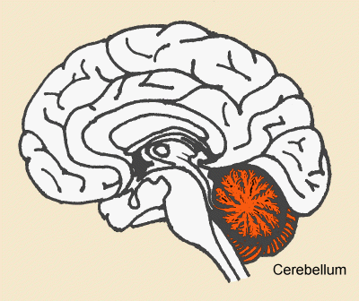
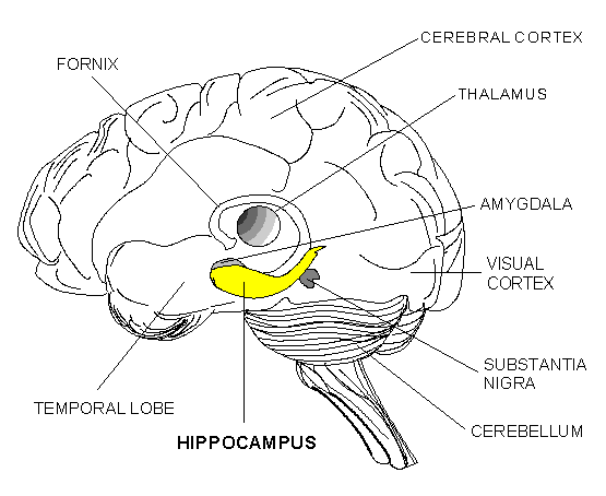
There is a mouse strain called Ts65Dn mice that recapitulates some major brain structural and behavioral symptoms of DS and these include reduced size and cellularity of the cerebellum and learning deficits associated with the hippocampus.

Roger Reeves at Johns Hopkins University has used a drug that activates the hedgehog signaling pathway to reverse the brain deficits of Ts65Dn mice. Yes you read that right.

A single treatment given the newborn mice of the Sonic hedgehog pathway agonist SAG 1.1 (SAG) results in normal cerebellar morphology in adults.

cerebellum

But wait, there’s more. SAG treatment at birth also improved the hippocampal structure and function. The hippocampus is involved in learning and memory.

hippocampus

SAG treatment resulted in behavioral improvements and normalized performance in a test called the “Morris water maze task for learning and memory. The Morris water maze test essentially takes a mouse from a platform in shallow water and then moves the mouse through the maze and then leave it there. The mouse has to remember how they got there and retrace their steps to get back to the platform before they get too tired from all that swimming. Normally Ts65Dn mice do very poorly at this test. However, after treating newborn Ts65Dn mice with SAG, they improved their ability to find their way back.

SAG treatment also produced other effects in the brain. For example, the ratios of different types of receptors in the brain associated with memory are skewed in Ts65Dn mice, but after treatment with SAG, these ratios became far more normal. Also, the physiology of learning and memory was also more normal in the brains of SAG-treated Ts65Dn mice.

These results are extremely exciting. They confirm an important role for the hedgehog pathway in cerebellar development. Also, they suggest that the development of the cerebellum (a small lobe at the back of the brain involved in coordination and fine motor skills, direct influences the development of the hippocampus. These results also suggest that it might be possible to provide a viable therapeutic intervention to improve cognitive function for DS patients.

This excitement must be tempered. This is an animal model and not a perfect animal model. Also, it is unclear if such a compound will work in humans. Much more work must be done, but this is a fascinating start.

BMP-2 Release By Synthetic Coacervates Improves Bone Making Ability of Muscle Stem Cells


Johnny Huard and his co-workers from the McGowan Institute for Regenerative Medicine at the University of Pittsburgh have isolated a slowly-adherent stem cell population from skeletal muscle called muscle-derived stem cells or MDSCs (see Deasy et al Blood Cells Mol Dis 2001 27: 924-933). These stem cells can form bone and cartilage tissue in culture when induced properly, but more importantly when MDSCs are engineered to express the growth factor Bone Morphogen Protein-2 (BMP-2), they make better bone and do a better job of healing bone lesions than other engineered muscle-derived cells (Gates et al., J Am Acad Orthop Surg 2008 16: 68-76).

In most experiments, MDSCs are infected with genetically engineered viruses to deliver the BMP-2 genes, but the use of viruses is not preferred if such a technique is to come to the clinic. Viruses elicit and immune response and can also introduce mutations into stem cells. Therefore a new way to introduce BMP-2 into stem cells is preferable.

To that end, Huard and his colleagues devised an ingenious technique to feed BMP-2 to implanted MDSCs without using viruses. They utilized a particle composed of heparin (a component of blood vessels) and a synthetic molecule called poly(ethylene arginylaspartate diglyceride), which is mercifully abbreviated PEAD. The PEAD-heparin delivery system formed a so-called “coacervate,” which is a tiny spherical droplet that is held together by internal forces and composed of organic molecules. These PEAD-heparin coacervates could be loaded with BMP-2 protein and they released slowly and steadily to provide the proper stimulus to the MDSCs to form bone.

When tested in culture dishes, the BMP-2-loaded coacervates more than tripled the amount of bone made by the MDSCs, but when they were implanted in living rodents the presence of the BMP-2-loaded coacervates quadrupled the amount of bone made by the MDSCs.

This technique provides a way to continuously deliver BMP-2 to MDSCs without using viral vectors to infect them. These carriers do inhibit the growth or function of the MDSCs and activate their production of bone.

This paper used a “heterotropic bone formation assay” which is to say that cells were injected into the middle of muscle and they formed ectopic bone. The real test is to see if these cells can repair actual bone lesions with this system.