Cardiospheres Aid Heart Healing by Secreting Endoglin and Inhibiting TGF-beta signaling


Eduardo Marbán from Cedars-Sinai Heart Institute in Los Angeles and his team have invested a great deal of work into the development of cardiospheres, which are self-assembling heart-derived stem cells that grow as little balls of cells in culture. Several preclinical experiments and a few clinical trials have established the effectiveness of cardiospheres are treatments for the heart after a heart attack. However, Marbán and his co-workers have also worked very hard at determining why cardiospheres heal a damaged heart.
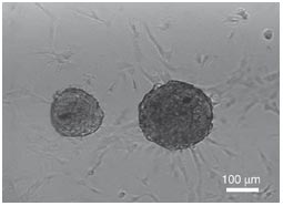
Cardiospheres
Cardiospheres in culture


To that end, Marbán and others have returned to their mouse model to do very detailed experiments with their cardiospheres and define exactly why these cells help heal the heart. To date, it is clear that cardiospheres increase the density of blood vessels in the heart tissue, decrease scar deposition, and prevent heart remodeling (the enlargement of the heart after a heart attack to compensate for the increase load placed on smaller amount of heart tissue). Marbán and others wanted to know precisely how cardiospheres managed these feats.

It has been known for some time that scar formation in the heart is largely a consequence of the activation of the TGF-beta signaling pathway (see NG Frangogiannis, Circulation Research 110: 159-173). Inhibition of this pathway can prevent the scar-making cells (fibroblasts) from migrating to the site of damage, dividing, and depositing the protein collagen, which is the main component of heart scars.

An abundant literature on heart scars show that the heart scar plays an important short-term role, but that in the long-run, it prevents the heart from resuming full function because it does not communicate with the rest of the heart muscle cells and does not contract. Therefore, helping the heart get through the first month after the heart attack without a scar is a crucial time.

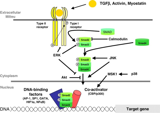
In a recent paper published by Marbán and his team, cardiospheres were tested in culture and in the heart of mice that had suffered a heart attack. Marbán thought that since the cardiospheres were attenuating scar formation, they must be inhibiting TGF-beta signaling. TGF-beta proteins are secreted by cells and they bind to a receptor complex that then activate intracellular proteins called “SMADs.” These activated SMAD proteins enter the nucleus and activate the transcription of target genes.

TGF-beta signaling

In his co-culture experiments, Marbán and others used normal human fibroblasts from the lower layers of human skin and cultured them with and without cardiospheres. The co-culturing experiments showed that without cardiospheres, the dermal fibroblasts made lots of collagen and activated their internal SMAD proteins. When these human dermal fibroblasts were incubated with cardiospheres, their SMAD proteins were largely inactivated and they made very little collagen.

Such a result is not surprising, but how are the cardiospheres doing this? As it turns out, there is an inhibitor of the TGF-beta receptor complex called “endoglin” that can also be secreted known as sE. When Marbán and others examined their cardiospheres, they were secreting a fair amount of sE.

Thus, the production of sE could definitely prevent dermal fibroblasts from activating their SMADs and making collagen, but what if these sE molecules were inactivated? Marbán and others made antibodies against sE and then used them to inactivate the sE made in culture by the cardiospheres. Under such conditions, the cardiospheres no longer were able to prevent SMAD activation in dermal fibroblasts and the fibroblasts made lots of collagen, even in the presence of cardiospheres.

This is all fine and good, but it is in cell culture, and cell culture experiments must always be confirmed by experiments in a living creature. Therefore, Marbán and his colleagues used cardiospheres to treat mice that had suffered a heart attack. As observed before, the cardiosphere-treated mice showed increases in ejection fraction and fractional shortening, and decreases in end-diastolic and end-systolic volume. The cardiosphere-treated animals also had much less scar tissue after one month and greater blood vessel density. Furthermore, the cardiosphere-treated mice did not show the maladaptive enlargement of the heart muscle cells seen in post-heart attack patients. When the heart tissue was assayed one month after treatment, it was clear that the cardiosphere-treated heart tissue showed increased sE expression and much less TGF-beta signaling. The downstream targets of SMAD activation were much less, and SMADs also showed less activation. Expression of the TGF-beta receptors was also decreased.

This paper shows that endoglin expression plays a key role in preserving and healing the heart after a heart attack. Would it be possible to give soluble endoglin to heart attack patients? This remains to be seen.

One caveat with this paper is that human dermal fibroblasts are similar but different from heart fibroblasts. While it is reasonable to suppose that these two cell types react in a similar way to cardiospheres, such a supposition must be rigorously confirmed experimentally.
Healing Corneal Blindness with Stem Cells from Extracted Teeth


Scientists at the University of Pittsburgh have found a new way to treat corneal blindness, which affects millions of people around the world.

James Funderburgh and his colleagues at the University of Pittsburgh School of Medicine showed that stem cells isolated from the dental pulp of extracted wisdom teeth can be differentiated into specialized cells that can maintain the health and integrity of corneas and rid them of the scars caused by illness or injury that compromise the ability to see clearly.  These cells can be safely injected into the corneas of mice.

According to Funderburgh, who is a professor of ophthalmology, this new approach can replace damaged corneal eye tissue with tissue made from the patient’s own cells rather than cells from a donor. Such a procedure circumvents the problems of immunological rejection that dog the reconstruction of corneal tissue with cells from donors. Furthermore, donor corneas are in short supply in certain parts of the world (e.g., Africa and Asia).

“Our work is promising because using the patient’s own cells for treatment could help avoid these problems,” said Dr. Funderburgh, who is the senior author of a new paper describing the research, in a written statement.

A post-doctoral research fellow in Dr. Funderburgh’s laboratory, Dr. Fatima Syed-Picard, took cells from the pulp of extracted wisdom teeth and chemically processed them to differentiate them into specialized corneal cells. Then Syed-Picard and others injected these “keratocytes” into the corneas of healthy mice. Once in the eyes of laboratory mice, the tooth pulp-derived cells integrated with the existing tissue with no sign of rejection even after several weeks.

Could such a treatment work in human patients? “We are thinking that in the future people may ‘bank’ their extracted wisdom teeth or the cells from those teeth,” Funderburgh told The Huffington Post in an email. “For someone who did not do that it is possible to extract dental pulp with a root canal procedure, but this is still hypothetical. In the worst-case scenario, someone might consider having a tooth extracted to provide cells for this procedure.”

Last year more than 70,000 corneal transplants were performed in the U.S., According to Kevin Corcoran, president and CEO of the Washington, D.C.-based Eye Bank Association of America (EBAA), there were more than 70,000 corneal transplants performed in the US alone.

“There’s a lot of exciting research being done in the area of [corneal] transplant, and EBAA is interested in any outcome that can help restore sight to the blind or visually impaired,” said Corcoran, who was unfamiliar with the Pitt research.

Dr. Syed-Picard stressed that this research is still in the formative stages and the results are preliminary, and added that it would probably take a few years before human testing could begin. The next step, she said, would be to conduct a similar set of experiments in rabbits.

European Knee Meniscus Injury Pilot Trial to Evaluate Cytori Cell Therapy Begins


Cytori Therapeutics is a cell therapy company that is in the process of developing cell therapies from a patient’s own fat tissue that can potentially treat a variety of medical conditions. To date, the preclinical studies and clinical trials suggest that their Cytori Cell Therapy can improve blood flow, modulate the immune system, and facilitating wound repair.

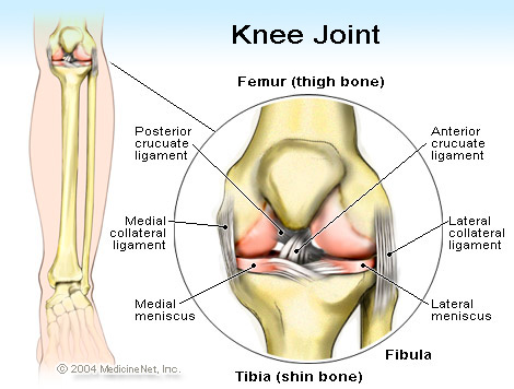
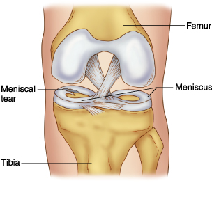
Recently, Cytori has announced that it has enrolled its first patients in an ambitious clinical trial that will test their stem cell product in patients undergoing surgery for traumatic injuries to the meniscus of the knee.  The meniscus is a wedge of cartilage on either side of the knee joint that acts a a shock absorber between the femur and the head of the tibia.

meniscus

Ramon Cugat, who is the Co-Director of the Orthopedic Institute, Hospital Quiron Barcelona, Spain, is the principal investigator for this trial. Dr. Cugat serves as an orthopedic surgeon at Hospital Quiron Barcelona. This trial will test the ability of Cytori Cell Therapy to heal the meniscus and is being conducted in parallel with a similar trial that is testing the Cytori Cell Therapy as a treatment for anterior cruciate ligament (ACL) repairs. The patients treated with Cytori Cell Therapy for ACL repairs are still being evaluated, but to date, no safety related concerns have emerged and the patients seem to have improved. These preliminary results were presented at the Barcelona Knee Symposium in November 2014.

knee_joint

“Dr. Cugat is a leading expert in treating traumatic knee injuries in elite athletes,” said Dr. Marc H. Hedrick, President and CEO of Cytori Therapeutics. “These trials are important to Cytori because, at minimal cost to us, they provide additional clinical evidence that our therapy can be safely used in treating a multitude of knee conditions.”

The meniscus trial is a two-center, phase I study that will assess the safety and efficacy of Cytori’s ECCM-50 adipose-derived regenerative cell therapy in meniscus repair. In this trial, up to 60 patients who have had meniscus surgery to repair the meniscus will receive injection of the cells directly into the meniscus. Each patient will be evaluated by several clinical read outs that assess the recovery of the patient after meniscus surgery. As in the case of the ACL repair study, the goal of this trial is to determine if Cytori Cell Therapy can be safely delivered to the meniscus and whether efficacy can be observed.

“Tears to the meniscus are problematic injuries for active individuals, particularly athletes. Based on the early results from a recent series of 20 patients treated for complete anterior cruciate ligament injuries, we are eager to evaluate whether augmentation surgery with Cytori Cell Therapy will lead to quicker and more complete healing,” said Dr. Cugat.

Injected patients will fill out a patient questionnaire that assesses knee pain, function and activity, This questionnaire is called the Knee Injury and Osteoarthritis Outcome Score (KOOS), but patients will also be physically examined to ascertain the extent of their knee function and the degree of their movement, with or without pain. Patients will be given a visual analogue score to assess knee pain, and knee function will be assessed by the Lysholm Knee Scoring Scale, Tegner Activity Scale, and the Lower Extremity Functional Scale. Each patient will also have their knees examined by Magnetic Resonance Imaging (MRI) in order to examine the structural integrity of their meniscus. These assessments will be taken before and 60, 90, 180 and 365 days after surgery and the MRIs will be done before and at 90, 180 and 365 days after surgery.

The preliminary results of the ACL study showed that the Cytori strategy was feasible and did not result in any significant safety issues above that seen with a standard small volume liposuction. All the injected patients recovered without any complications. The results of the ACL trial were compared to a historical control group of patients who had the same surgical procedure by the same surgical team but without other interventions. Overall, the patient’s recovery from pain and their ability to return to daily activities was accelerated as a result of the therapeutic enriched bone-patellar tendon-bone transplant. Both the patient questionnaires and serial MRI scans of the knees following cell therapy were consistent with accelerated healing of the graft. Presently, Dr. Cugat and his coworkers are obtaining one year follow-up information on the treated patients and they will report their data in a peer-reviewed journal in the future.

ACL and meniscus tears are among the most common sports-related knee injuries and unfortunately, these two injuries often are sustained simultaneously. According to the American Academy of Orthopedic Surgeons, ACL injuries have an annual incidence of more than 200,000 cases with nearly half undergoing surgical reconstruction. Further, an estimated 850,000 patients undergo surgical procedures to address meniscus injuries each year.

Preclinical Study Results Pave the Way for Newly Opened Clinical Trial of Immune Cells Engineered to Attack Protein Found on Tumors in 30 Percent of Patients with Glioblastoma


Scientists from the University of Pennsylvania have engineered immune cells to seek out and attack a type of deadly brain cancer. In an important preclinical study, these souped-up immune cells were shown to be both safe and effective at controlling tumor growth in mice treated with these modified cells. This work is the result of collaboration between a team from the Perelman School of Medicine at the University of Pennsylvania and the Novartis Institutes for BioMedical Research. These results will hopefully be the impetus for a newly opened clinical trial for glioblastoma patients at Penn.

Marcela Maus, assistant professor of Hematology/Oncology at the Penn Abramson Cancer Center, said: “A series of trials that began in 2010 have found that engineered T cells have an effect in treating some blood cancers, but expanding this approach into solid tumors has posed challenges. A challenging aspect of applying engineered T cell technology is finding the best targets that are found on tumors but not normal tissues. This is the key to making this kind of T cell therapy both effective and safe.”

This new preclinical study, which was conducted with Hideho Okada and his colleagues at the University of Pittsburgh, makes use of T cells engineered to express a chimeric antigen receptor (CAR) that specifically binds to a mutant epidermal growth factor receptor protein called EGFRvIII. EGFRvIII is found on the cell surfaces of approximately 30 percent of glioblastoma tumors. Over 22,000 Americans are diagnosed with glioblastoma each year, and those patients whose glioblastomas express the EGFRvIII mutation tend to be more aggressive and are less likely to respond favorably to standard therapies and more likely to recur after treatment.

“Patients with this type of brain cancer have a very poor prognosis. Many survive less than 18 months following their diagnosis,” said M. Sean Grady, who is the Charles Harrison Frazier Professor and chair of the department of Neurosurgery. “We’ve brought together experts in an array of fields to develop an innovative personalized immunotherapy for certain brain cancers.”

This new trial is being led by Donald O’Rourke, associate professor of Neurosurgery, who heads an interdisciplinary collaboration of neurosurgeons, neuro-oncologists, neuropathologists, immunologists, and transfusion medicine experts.

In order to bring this experiment to fruition, Maus and her colleagues had to characterize the EGFRvIII CAR T cell. They had to develop and tested multiple antibodies that bind to cells expressing EGFRvIII on their surface. The single-chain variable fragments or scFvs that recognized the mutant EGFRvIII protein were then extensively tested in order to confirm that they do not also bind to those normal, EGFR proteins that are widely expressed on cells in the human body.

Maus and her group then generated a panel of humanized scFvs and tested their specificity and function in CAR modified T cells. The humanized scFvs have distinct amino acid sequences that more closely resemble human antibodies. From this huge panel of humanized scFvs, they selected one scFv to explore further based on its binding selectivity for EGFRvIII over normal non-mutated EGFR. They also evaluated the EGFRvIII CAR T cells by testing them against normal EGFR-expressing skin cells in mice grafted with human skin. This test showed that the engineered EGFRvIII CAR T cells did not attack cells with normal EGFR, at least under these conditions.

In order to test the selected scFv for its anti-cancer efficacy, Maus and others used human tumor cells that expressed EGFRvIII and showed that the EGFRvIII CAR T cells could multiply and secrete cytokines in response such to tumor cells. When used inside living animals, it was clear that the EGFRvIII CAR T cells ably controlled tumor growth in several mouse models of glioblastoma. The tumors were measured with magnetic resonance imaging (MRI) and the EGFRvIII CAR T cells caused tumor shrinkage, and were also effective with used in combination with the anticancer drug temozolomide, which is normally used to treat patients with glioblastoma.

On the strength of these preclinical successes, this team designed a phase 1 clinical study of CAR T cells transduced with humanized scFv directed to EGFRvIII for both newly diagnosed and recurrent glioblastoma patients who carry the EGFRvIII mutation. “There are unique aspects about the immune system that we’re now able to utilize to study a completely new type of therapy,” said O’Rourke.

For these glioblastoma patients, their T cells were removed by means of apheresis (a process similar to dialysis), and then the T cells were genetically engineered using a viral vector that programs them to find EGFRvIII-expressing cancer cells. The patient’s own engineered cells are infused back into their body, and when the T cells find the EGFRvIII-expressing cells, a signaling domain built into the CAR promotes proliferation of these “hunter” T-cells. This procedure is distinct from other T cell-based therapies that also target some healthy cells, since EGFRvIII seems to only be found on tumor tissue, which the study’s leaders hope will minimize side effects.

This new phase I clinical trial will enroll 12 adult patients whose tumors express EGFRvIII, in two groups: One arm of 6 patients whose cancers have returned after receiving other therapies, and one arm of 6 patients who are newly diagnosed with the disease and still have 1 cm or more of tumor tissue remaining after undergoing surgery to remove it.

The clinical trial is sponsored by the biotech company Novartis. In 2012, the University of Pennsylvania and Novartis announced an exclusive global research and licensing agreement to further study and commercialize novel cellular immunotherapies using CAR technologies. This STM study is the first pre-clinical paper developed within the Penn-Novartis alliance, with Penn and Novartis scientists working collaboratively. Ongoing clinical trials evaluating a different type of Penn-developed CAR therapy known as CTL019 have yielded promising results among some patients with certain blood cancers. In July 2014, the FDA granted CTL019 its Breakthrough Therapy designation for the treatment of relapsed and refractory acute lymphoblastic leukemia in both children and adults.

Long-term Tumorgenicity of Induced Pluripotent Stem Cells


A paper from the Okano laboratory has shown that implantation of neural stem cells made from induced pluripotent stem cells can still form tumors ever after a long period of time.

This paper is an important contribution to the safety issues surrounding induced pluripotent stem cells (iPSCs). As noted in previous posts, iPSCs are made from adult cells by means of genetic engineering and cell culture techniques. In short, by introducing four different genes into adult cells and then culturing them in a special culture medium, a fraction of these cells will de-differentiate into cells that resemble embryonic stem cells in many ways, but are not exactly like them.

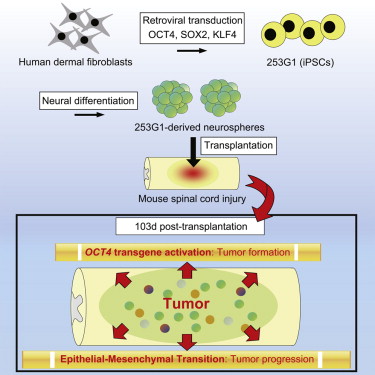
The Okano laboratory made iPSCs using viruses that integrate into the genome of the host cell, which is not the safest option. However, because in the four-gene cocktail that is normally used to reprogram these cells (Oct-4, Klf-4, Sox2, and c-Myc), the c-Myc gene is often thought to be the main cause of tumor formation. Okano and his collaborators made their iPSCs without the c-Myc gene, but only used the three-gene cocktail of Oct-4, Klf-4, and Sox2. Such a cocktail is much less efficient that the four-gene cocktail, but it supposed to make iPSCs that are altogether safer.

These iPSCs were differentiated into neural stem cells that grew as tiny spheres of cells, and these “neurospheres” were transplanted into the spinal cords of mice that had suffered a spinal cord injury. The implanted cells differentiated into neurons and glial cells and restored some neural function to these mice. However, the mice were observed for a long period of time after the implantations to assess the long-term safety of these implanted cells.

After 105 days, the implanted mice began to show deterioration of their neural function and their spinal cords showed tumors. It is clear that the Oct-4 gene that was used in the reprogramming procedure was the reason for the tumor transformation.

Graphical Abstract 20141213

This experiment, once again, calls into question the safety of any method for iPSC generation that leaves the transfected genes in the reprogrammed cells. I reported in a previous post that skin cells made from iPSCs that had their transgenes left in them were good at causing tumors and not as good as forming skin cells, but iPSCs without their reprogramming transgenes were safer and more effective tools for regenerative medicine.  This experiment also shows that c-Myc is not the only concern with iPSCs.  Any of the transgenes used for reprogramming can cause problems, and they must be removed if iPSCs are going to produce safe, differentiated cells.  Finally, this experiment pretty much shows that the use of retrovirus tools to introduce genes into cells for the sake of reprogramming is a bad idea if those cells are going to be used for regenerative medicine.  Non-integrating tools are much safer and preferable in these cases.

The Okano paper appeared in Stem Cell Reports.

Human Stem Cells Repair Radiation Damage in Rat Brains


Radiation is a powerful treatment for brain cancer, but this potentially life-saving treatment comes with a heavy cost, which is permanent damage to the brain.

Preclinical work at Memorial Sloan Kettering Cancer Center has shown that human stem cells can be used to make cells that repair radiation-induced damage in the brain.

When rats were treated with radiation and then given cocktails to the human stem cells, they regained the cognitive and motor functions that were lost after brain irradiation.

In the brain, stem cells called OPCs or oligodendrocyte progenitor cells mature into oligodendrocytes that produce the protective myelin coating that surrounds axons in the central nervous system. During radiation treatment, OPCs die off and are depleted. Because OPCs help shield and repair the myelin sheath throughout the life of the organism, depletion of them threatens the integrity of the myelin sheath, which threatens the proper transmission of neural impulses throughout the brain.

A research project led by neurosurgeon Viviane Tabar and her research associate Jinghua Piao wanted to use stem cells to replace these lost OPCs. They used human embryonic stem cells and human induced pluripotent stem cells to make cultured OPCs.

In the next phase of the experiment, Taba, Piao and their coworkers treated rats whose brains had been irradiated with their cultured OPCs. After injection of the stem cell-derived OPCs, brain repair was evident and the rats regained their cognitive and motor function that they had previously lost as a consequence of radiation exposure.

The treatment appeared quite safe since none of the animals developed any tumors or aberrant growths.

The ability to repair radiation damage could mean that the quality of cancer survivors could be greatly improved and it could also expand the therapeutic window of radiation, according to Tabor.

“This will have to be proven further, but if we can repair the brain effectively, we could be bolder with our radiation dosing, within limits,” said Tabor.

Such a treatment scheme could also be very important in children, for whom physicians must use lower doses of radiation to limit brain damage.

Infertility Treatment with Stem Cells is Unlikely


Because several laboratories have managed to differentiate embryonic stem cells into cells that look very much like human eggs and sperm, many have predicted that infertility will be treated with stem cell treatments (see Volarevic V, et al., Biomed Res Int. 2014;2014:507234). However, new work from the University of Gothenburg and Karolinska Institute has cast doubt on this hope.

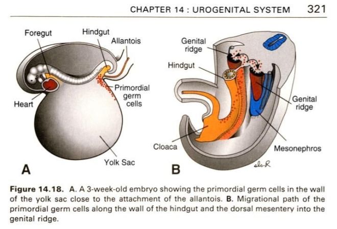
At about 24 days of life, large, spherical sex cells are recognizable among the endodermal cells of the umbilical vesicle close to the allantois. These cells are the primordial germ cells and they are the progenitor cells of the sperm in men and eggs in women. As the embryo folds during about the late 4th week, the dorsal portion of the umbilical vesicle is incorporated into the embryo. This incorporation of the umbilical vesicle occur concurrently with the migration of these primordial germ cells along the dorsal mesentery of the hindgut to the gonadal ridges. During the 6th week of life, the primordial germ cells enter the underlying mesenchyme and are incorporated into the gonadal cords. Primordial germ cell migration is mainly regulated by three genes: Stella, Fragilis, and BMP-4.

PGC migration

These primordial germ cells divide as they migrate, and by five months of gestation, embryonic ovaries contain about six to seven million oogonia. Most of these oogonia experience cell death before birth, but the remaining oogonia begin meiosis toward the end of gestation. At this time, the oogonia are called primary oocytes. Meiosis is arrested in prophase of the first meiotic division, and this is the same stage at which spermatogenesis in the male is blocked. Primary oocytes decrease in number throughout a woman’s life. The ovaries of a newborn girl contain about two million primary oocytes and these are all the gametes she will ever have. Each primary oocyte is contained within a hollow ball of cells called the ovarian follicle. By the time a woman reaches puberty, that number of primary oocytes has been reduced to 400,000. Only about 400 of these cells will ovulate during a woman’s reproductive years. The rest will die by means of programmed cell death. Once all the primary oocytes are gone, ovulation stops and the woman undergoes menopause.

Kui Liu from the University of Gothenburg said: “Ever since 2004, the studies on stem cell research and infertility have been surrounded by hype. There has been a great amount of media interest in this, and the message has been that the treatment of infertility with stem cells is about to happen. However, many researchers, including my research group, have tried to replicate these studies and not succeeded. This creates uncertainty about whether it is all possible to create new eggs with the help of stem cells.”

In collaboration with Outi-Hovatta’s laboratory at the Karolinska Institute and Jan-Åke Gustafsson’ research team at the university of Houston in the US, Lui’s research team carried out experiments on mice that failed to demonstrate that functional gametes could be formed from pluripotent stem cells. Essentially, the only gametes that could that the female mice had were the ones they were born with.

In Liu’s opinion, fertility clinics should place their attention on using the eggs that women still have in order to treat infertility.

Stem Cells Derived From Amniotic Tissues Have Immunosuppressive Properties


Ever since they were first isolated, amnion-based stem cells have been considered promising candidates for cell therapies because of their ease of access, plasticity, and absence of ethical issues in their derivation and use. However, a Japanese research team has discovered that stem cells derived from human female amnion also have the ability to suppress the inappropriate activation of the immune system and that there are straight-forward ways to enhance their immunosuppressive potential.

The amniotic membrane is a three-layered structure that surrounds the baby and suspends it in amniotic fluid. Amniotic fluid acts as a protective shock-absorber, a lubricant and an important physiological player in the life of the embryo and fetus. Because the fetus is a privileged entity that escapes attack from the mother’s immune system, researchers have been very interested in determining the immunological properties of the amnion cells.

“The human amniotic membrane contains both epithelial cells and mesenchymal cells,” said study co-author Dr. Toshio Nikaido, Department of Regenerative Medicine, Graduate School of Medicine and Pharmaceutical Sciences at the University of Toyama. “Both kinds of cells have proliferation and differentiation characteristics, making the amniotic membrane a promising and attractive source for amnion-derived cells for transplantation in regenerative medicine. It is clear that these cells have promise, although the mechanism of their immune modulation remains to be elucidated.”

In this study by Nikaido and his coworkers, amnion-derived cells inhibited natural killer cell activity and induced white blood cell activation. Nikaido reported that he and his colleagues saw the amnion-derived cells increase production of a molecule called interleukin-10 (IL-10).

“We consider that IL-10 was involved in the function of amnion-derived cells toward NK cells,” explained Dr. Nikaido. “The immunomodulation of amnion-derived cells is a complicated procedure involving many factors, among which IL-10 and prostaglandin E2 (PGE2) play important roles.”

Molecules called “prostaglandins,” such as PGE2, mediate inflammation, smooth muscle activity, blood flow, and many other physiological process. In particular, PGE2 exerts important effects during labor and stimulates osteoblasts (bone-making cells) to release factors that stimulate bone resorption by osteoclasts. PGE2 also suppresses T cell receptor signaling and may play a role in the resolution of inflammation.

When Nikaido and others used antibodies against PGE2 and IL-10, they removed the immunosuppressive effects of the amnion-derived cells on natural killer cells. These data imply that these two factors contribute to the immunosuppressive abilities of amnion-derived cells.

“Soluble factors IL-10 and PGE2 produced by amnion-derived cells may suppress allogenic, or ‘other’ related immune responses,” concluded Dr. Nikaido. “Our findings support the hypothesis that these cells have potential therapeutic use. However, further study is needed to identify the detailed mechanisms responsible for their immodulatory effects. Amnion-derived cells must be transplanted into mouse models for further in vivo analysis of their immunosuppressive activity or anti-inflammatory effects.”

Given the levels of autoimmune diseases on the developed world, these results could be good news for patients who suffer from diseases like Crohn’s disease, systemic lupus erythematosus, or rheumatoid arthritis. While more work is needed, amnion-based cells certainly show promise as immunosuppressive agents.

The study will be published in a future issue of Cell Transplantation.

Induced Pluripotent Stem Cells Differentiated into Intestinal Cells


Even the liver is the main organize when it comes to the metabolization of drugs, the small intestine also plays an important role in all aspects of drug metabolism. Unfortunately, no laboratory system exists at present that serves as a standardized system for evaluating the way drugs interact with the small intestine.

A new study by Tamihide Matsubara and his colleagues from Nagoya City University in Japan has sought to alleviate this problem. Matsubara and his coworkers used human induced pluripotent stem (iPS) cells to produce functional human intestinal enterocytes and showed that they faithfully recapitulated the drug metabolism of normal, human intestinal enterocytes.

To make intestinal enterocytes from iPS cells, Matsubara and others treated these cells with chemicals called activin A and fibroblast growth factor 2 to drive the cells to become intestinal-like stem cells. These cultured intestinal-like stem cells them differentiated into enterocytes when grown in a culture medium that contained epidermal growth factor and other small-molecule compounds.

The differentiated cells expressed intestinal marker genes and drug transporters. For example, they expressed sucrase-isomaltase, an intestine-specific marker, and enterocyte drug-metabolizing enzymes such as CYP1/2, CYP2C9, CYP2C19, CYP2D6, CYP3A4/5, UGT, and SULT. Inhibitor studies showed that the intestinal oligopeptide transporter SLC15A1/PEPT1 was inhibited by the pain reliever ibuprofen, just like in naturally-occurring enterocytes. Also, active forms of vitamin D increased the expression of the enzymes CYP3A4 and CYP3A4/5, which is also observed in naturally-occurring human enterocytes.

These results show that Matsubara and his colleagues have successfully generated enterocyte-like cells that have the same drug metabolizing capacities as naturally-occurring enterocytes. These cells would be very useful for developing novel evaluation systems to predict individual human intestinal drug metabolism.

Compact Spinal Implants to Help Spinal Cord Injured Patients Walk


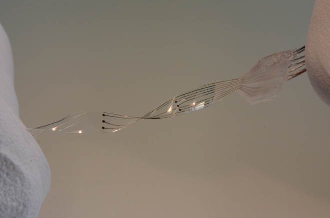
An interdisciplinary research team at the Ecole Polytechnique Fédérale de Lausanne (EPFL) in Switzerland led by Dr. Grégoire Courtine and Dr. Stéphanie P. Lacour has recently lifted the curtain on their flexible spinal implant called the electronic dura (or e-Dura). According to Courtine and Lacour, this implant greatly improves spinal injury rehabilitation in spinal cord injured rats.

In a paper published by this team earlier in the journal Science this month, the EPFL team showed that, because of its flexibility, this next-generation e-Dura implant lasts longer (up to two months) and causes much less damage than traditional implants.

These latest results are an extension of earlier research in 2012 in which Courtine and Lacour published breath-taking results that showed spinal cord-injured rats that should have been paralyzed had regained the ability to walk, run, and even climb stairs.

Spinal cord injury results in loss of control over the part of the body below the point of injury. Courtine and his coworkers were able to reactivate the spinal cord in rats with a specific combination of drugs plus electrical stimulation to simulate the excitatory input from the brain. The drugs they used, monoamine agonists, bind to receptors and activate them in the same way that such neurotransmitters would in healthy subjects. When the spinal cord was exposed to these drugs plus mild electrical stimulation, the activated nerve cells in the spinal column produced movement in the paralyzed animals.

Spinal Cord Implant to help the Cripples walk

This movement, however, was largely involuntary, since the brain was not able to communicate with the area below the injured spinal cord. However, over time, as the animals trained and repeatedly walked in their harnesses (which kept them safe from falling); they became more confident in their ability to walk again. In fact, the EPFL team noticed a fourfold growth of new nerves in the spinal cord. This new nerve growth eventually restored communication between the brain and the injured area of the spine.

Courtine, whose eyes sparkle as he passionately talks about his research, is eager to take these findings to the clinic to see if they can help human beings who have suffered a catastrophic spinal cord injury. In preparation for clinical trials, however, they came up against another problem and that was the need for a long-term spinal implant that could deliver the chemical and electrical stimulation needed to initiate spinal cord healing. Courtine hopes that his e-Dura can satisfy this need.

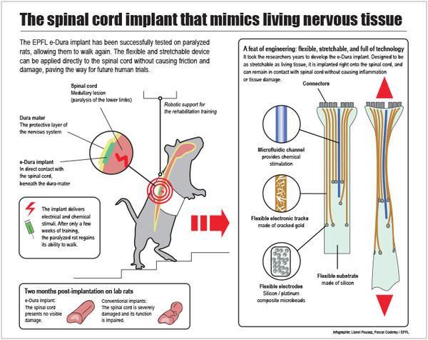
e-Dura

The e-Dura meets two important criteria for spinal implants: durability and biocompatibility. In order to reduce the number of surgical procedures an injured patient must undergo, an implantable device needs to last a long time. It also has to be biocompatible and flexible. Early generation implants caused inflammation and the formation of scar tissue, which usually offset any positive results the implant provided.

When tested on laboratory animals, Courtine’s laboratory applied the e-Dura implant beneath the protective dura mater, directly on the spinal cord. Thankfully, the implant did not cause any adverse effects and lasted long enough for the paralyzed animals to complete their rehabilitation. Functionally, the implant also performed very well.

The e-Dura unit contains very small microfluidic channels that are embedded on a flexible silicon substrate. The device delivers precise amounts of drugs directly to the nerve cells in the spinal cord. Cracked gold conducting wires and electrodes that are made of a composite material that consists of silicon and platinum send electrical signals to the injured spinal cord. Electronic circuitry in the implant also provides the opportunity for the EPFL team to monitor the electrical messages sent back and forth to the brain as the new nerves are activated.

Indeed, this research is wonderfully exciting, but it is unclear how well it will work in humans. First of all, humans will probably need a different cocktail of drugs or a distinct electrical stimulation pattern to stimulate the spinal cord to heal itself. As with much clinical research at the beginning stages, there many unanswered questions to date. Advanced clinical trials will hopefully uncover some of these idiosyncrasies that characterize the injured human spinal cord and such answers are an integral part of providing a protocol that applies uses technology to human patients.

While exoskeleton technology also continues to approach consumer markets, it would be better to return to people their natural ability to walk. However you slice it, spinal cord injury patients may have more options in the coming years.

New Technology Reprograms Skin Fibroblasts


Fibroblasts are one of the main components of connective tissue, which is the main reason scientists typically exploit them for experiments. A collaborative team of scientists from the University of Pennsylvania, Boston University, and the New Jersey Institute of Technology have invented a way to reprogram fibroblasts without going through a pluripotent stage.

The senior author of this study, Xiaowei Xu, associate professor of pathology and laboratory medicine at the University of Pennsylvania School of Medicine, said, “Through direct reprogramming, we do not have to go through the pluripotent stem cell stage, but directly convert fibroblasts to melanocytes . So these cells do not have tumorigenicity” (the ability to cause tumors).

Melanocytes are found in the skin and they are responsible for the pigment in our skin. They are in the uppermost layer of the skin, known as the epidermis, and produce melanin, a brown pigment that helps screen against the harmful effects of UV light.

Turning a fibroblast into a melanocytes might seem trivial for a stem cell scientist; just reprogram the fibroblast into an induced pluripotent stem cells and then differentiate it into a melanocytes. However, this procedure utilized direct reprogramming, in which the fibroblast was converted into a melanocytes without traversing through the pluripotent stage. The difficultly with direct reprogramming is finding the right cocktail of genes and/or growth factors that will accomplish the deed.

Xu and his colleagues began their search by examining the genes that are specific to melanocytes. They found 10 different transcription factors that are important for melanocytes development. Next they screened these ten genes for their ability to convert a fibroblast into a melanocytes. They found that of the ten melanocytes-specific genes, three of them, Sox10, MITF, and PAX3 could do the job effectively. They called this gene combination “SMP3.”

When Xu and others tested SMP3 on mouse embryonic fibroblasts, they quickly expressed melanocytes-specific genes. When Xu’s group used SMP3 on human fetal dermal cells, once again, the cells rapidly differentiated into melanocytes. Xu and his team referred to these cells as hi-Mel, which is short for human, induced melanocytes.

When hi-Mel were grown in culture they produced melanin a plenty. When they were implanted into the skin of pigmentless mice, once again they rose to the challenge and made a great deal of pigment. Thus hi-Mel express the same genes as melanocytes and they behave for all intents and purposes as melanocytes.

Xu and his colleagues think that their procedure might be able to treat human patients with a condition called vitiligo in which the skin has patches that are devoid of pigment.

Another potential use of this technology is a way to effectively study melanoma, one of the most dangerous skin cancers known to human medicine. My good friend and SAU colleague died over a year ago from melanoma and having better ways to treat this monster would have been marvelous for Charlie, and his family, who miss him dearly. By generating melanocytes from the fibroblasts of melanoma patients, they can “screen not only to find why these patients easily develop melanoma, but possibly use their cells to screen for small compounds that can prevent melanoma from happening.”.

Also, because so the body contains so many fibroblasts in the first place, this reprogramming technique is well-suited for other cell-based treatments.

Teaching Old Cells New Tricks


The laboratory of Helen Blau at Stanford University has devised a technique to lengthen the sequences that cap the ends of chromosomes in skin cells. This treatment enlivens the cells and makes them behave as though they were younger.

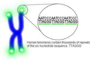
In order to properly protect linear chromosomes from loosing DNA at their ends, chromosomes have a special set of sequences called “telomeres” at their ends. Telomeres consists of short sequences that are repeated many times. A special enzyme called the telomerase replicates the telomeres and maintains them. As we age, telomerase activity wanes and the telomeres shorten. This threatens the genetic integrity of the chromosomes, since a loss of genes from the ends of chromosomes can be deleterious for cells. In young humans, the telomeres may be 8,000 to 10,000 bases long. When the telomeres shorten to a particular length, growth stops and the cells become quiescent.

Human telomeres

Embryonic stem cells have long telomeres at the ends of their chromosomes and they also have robust telomerase activity. Adult stem cells have varied telomerase activity and telomere length, but it seems that the length of the telomeres and the activity of the telomerase correlates with the vitality of the stem cell population and its capacity to heal (see H. Saeed and M. Iqtedar (2013). J. Biosci. 38, 641–649). As we age our stem cell quality decreases as their telomeres shorten.

Blau and her colleagues used a modified type of RNA to lengthen the telomeres of large numbers of cells. According to Blau: “Now we have found a way to lengthen human telomeres by as much as 1,000 nucleotides, turning back the internal clock in these cells by the equivalent of many years of human life. This greatly increases the number of cells available for studies such as drug testing or disease modeling.”

In these experiments, Blau and her coworkers used chemically modified messenger RNA molecules that code for TERT, which is the protein component of the telomerase. The expression of these messenger RNAs in human cells greatly increased the levels of telomerase activity.

This technique devised by Blau and her team have distinct advantages of previously described protocols. First, this technique boosts telomerase activity temporarily. The modified messenger RNA sticks around for several hours and is translated into TERT protein, but this protein only lasts for about 48 hours, after which its activity dissipates. After the telomerase have lengthened the telomeres, they will shorten again after each cell division as before.

“This new approach paves the way toward preventing or treating diseases of aging,” said Blau. “There are also highly debilitating genetic diseases associated with telomere shortening that could benefit from such a potential treatment.”

Blau and her team are testing their technique in other cell types besides skin cells.

Cardiosphere-Derived Injections Improve Heart Function in Children with Hypoplastic Left Heart Syndrome


Hypoplastic Left Heart Syndrome or HLHS accounts for 2 to 3 percent of all congenital heart disease. It shows a prevalence rate of two to three cases per 10,000 live births in the United States. HLHS is the most common form of functional single ventricle heart disease. The National Inpatient Sample database has estimated that there were an estimated 16,781 cases of HLHS among neonates born between 1988 and 2005 in the United States. More males have HLHS than females with the male to female ratio being about 1.5:1. Despite its low incidence relative to other congenital cardiac disorders, HLHS, if left untreated, is responsible for 25 to 40 percent of all neonatal cardiac deaths.

In HLHS patients, the left ventricle (the main pumping chamber of the heart), aorta, and related components are underdeveloped.

Children born with HLHS typically require surgery within a few days of birth and additional long-term treatment is required to address issues associated with right ventricular-dependent circulation.

Results from a clinical trial conducted by researchers at Okayama University and Okayama University Hospital show that children who suffer from HLHS seem to benefit from injections of cardiosphere-derived cells (CDCs).

Apparently in children, cardiac progenitor cells that can differentiate into several different heart-specific cell types are more abundant and self-renewing in children than adults.

The research group, led by Hidemasa Oh, monitored the heart function of seven patients who had received injections of cells and a control group of seven patient who had not received any such injections. They concluded that, “Our prospective controlled study, the first pediatric phase I clinical trial of stem cell therapy for heart disease to our knowledge, suggests that intracoronary infusion of autologous cardiac progenitor cells is a feasible and safe approach to treat children with HLHS.”

The cardiac progenitor cells used in this study came directly from the hearts of the patients. When these heart-specific progenitor cells are isolated and grown in cell culture, they form tiny balls of cells called “cardiospheres.” These patient-derived cardiosphere-derived cells (CDCs) were administered to the experimental subjects in this study after they were confirmed to contain a normal number of chromosomes and express a host of heart-specific genes. The transcoronary administration of the CDCs did not produce any adverse effects.

The heart functions monitored by the research group included the right ventricular ejection fraction or RVEF, end-systolic volume (ESV), which is the volume of blood within the ventricle at the maximum point of contraction, and the end-diastolic volume or EDV, which is the volume of blood at the maximum filling point, stroke volume, and cardiac output. Additionally, the levels of brain natriuretic peptide or BNP (a direct measure of heart failure) were also monitored. BNP is made by the ventricles of the heart in response to excessive stretching of the heart muscle.

Because of the rarity of this disease, this study was necessarily small. This study was also a non-randomized study. Therefore, this study is more of an evaluation of the safety of this procedure rather than its efficacy. However, the improvement in the RVEP in the stem cell-treated patients compared to the non-treated group 18 months after CDC administration provides possible evidence of the efficacy of this treatment.

Clearly more work is needed, but we will know more as the data rolls in.

Human Placenta-Derived Multipotent Cells Modulate Cardiac Injury in Large and Small Animal Models


Placental-derived multipotent cells or PDMCs have been isolated from human term placental tissues. PDMCs have the ability to differentiate into neurons, bone, fat, and liver. Can cells like these help heal a damaged heart?

Men-Luh Yen and his colleagues from the National Taiwan University Hospital, Taipei, Taiwan, have recently published a large study of PDMCs that have examined the characteristics of these cells in culture and in small and large animals.

In culture, when PDMCs are grown with mouse heart muscle cells for eight days that differentiate into cells that look a lot like heart muscle cells.  These cells express the heart-specific gene alpha-sarcomeric actinin.  This is not evidence that PDMCs can differentiate into heart muscle cells, but it is evidence that they differentiate into heart muscle-like cells.  It is possible that these cells might be able to completely differentiate into heart muscle cells with the right signals.

When the culture medium from PDMCs are used to grow human umbilical vein endothelial cells, the human umbilical vein endothelial cells formed blood vessel-like tubes.  This indicates that PDMCs secrete a host of growth factors that induce the formation of blood vessels.  When Yen and his group examined the genes expressed by cultured PDMCs, they discovered that they expressed several growth factors known to induce blood vessel formation, such as hepatocyte growth factor (HGF), interleukin-8 (IL-8), and growth-regulated oncogene (GRO).  When these growth factors were given to cultured umbilical vein endothelial cells, they formed blood vessel-like tubes.  Thus HGF, GRO and IL-6 promote the formation of blood vessels.

When PDMCs were used to treat the heart of mice that had suffered a heart attack.  This part of the paper is less satisfying because many of their mice died as a result of this procedure (5 or 18).  However, the PDMS-treated mice did show a steady improvement in their ejection fractions (percentage of blood volume ejected from the heart) compared to mice that were only injected with culture medium.  These PDMC-injected mice also had extensive capillary beds in their heart tissue, suggesting that the increased heart function was due to the induction of new blood vessels.  In all honesty, this section of the paper should have had better controls and more animals should have been tested.  A sham group should have been included with an untreated group as well.

To extend their experiments in living animals, Yen’s group used a similar experimental strategy in Lanyu minipigs.  Here again, a lack of proper controls and large numbers of dead animals (5 of 17) diminish the clarity of the data.  The PDMC-treated minipigs showed a significant increase in ejection fraction (53.8 plus or minus 4.4 percent in the PDMC-treated minipigs vs. 39.2 plus or minus 2.3 percent in the culture medium-treated minipigs).  Also the blood vessel density in the hearts of the PDMC-treated pigs was over three times that of the other group.  Cell death studies showed that the hearts of the PDMC-treated minipigs that half that of the non-stem cell-treated minipigs.  This shows that PDMCs secrete molecules that promote cell survival.

Finally, Yen and others present what they think is evidence that the injected PDMCs in the hearts of the minipigs differentiated into heart muscle cells.  First of all, implanted PDMCs were observed eight weeks after they were injected.  There is little reason to suppose that these cells would have survived if they were not tightly associated with resident heart cells.  Secondly, these PDMCs expressed two heart-specific genes:  cardiac troponin T (cTNT), which is important for heart muscle contraction, and connexin 43, which is integral for forming gap junctions between heart muscle cells.  Gap junctions allow heart muscle cells to stay electrically connected with one another and allow them to contract as a single unit and these cells were expressing connexin 43 and were apparently integrated into the heart muscle.

I must say that I do not find this convincing, since the fusion of heart muscle cells and injected stem cells can account for such data.  Before I would believe that PDMCs can transdifferentiate into heart muscle cells, I would need to see compelling evidence that the connexin 43, cTNT, and human HLA-expressing cells also do not express minipig-specific genes.  Secondly, I would need to see PDMCs express the genes for the calcium-handling system that is unique to heart muscle cells.  The lack of express of these proteins is the single best reason to doubt that mesenchymal stem cells can transdifferentiate into heart muscle cells.  There is evidence that mesenchymal stem cells that stimulate endogenous heart stem cells to make new heart muscle, but little good evidence that mesenchymal stem cells can form mature, functional heart muscle cells.

All in all, the Yen paper shows some interesting data, even if some of it is not top quality.  Clear PDMCs are interesting cells that have a potential future in regenerative medicine.