Brain Regeneration Promoting Compound to be Tested in Alzheimer’s Clinical Trial


A research team at the University of Southern California (USC) will be initiating a Phase 1 clinical trial to test the effectiveness of their compound “Allo,” which promotes brain cell regeneration, in Alzheimer’s patients.

This new trial is one of four that are investigating new therapeutic targets in Alzheimer’s disease. These trials will also incorporate novel approaches to participant identification and selection.

These trials were reported at the Alzheimer’s Association International Conference in Boston. According to Roberta Brinton of USC, again and Alzheimer’s disease (AD) are characterized by a decline in the ability of the body to self-renew and repair (and this includes the brain). However, the capacity for regeneration is retained, albeit at a decreased level.

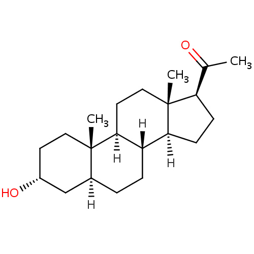
Allopregnanolone (3α-hydroxy-5α-pregnan-20-one), or Allo for short, is a neurosteroid that naturally occurs in the brain. Small quantities of it can also be found in the bloodstream. Previous studies have shown that Allo can improve cognitive function in older laboratory animals and in animal models of AD (see Chen S, Wang JM, Irwin RW, Yao J, Liu L, et al. (2011) Allopregnanolone Promotes Regeneration and Reduces β-Amyloid Burden in a Preclinical Model of Alzheimer’s Disease. PLoS ONE 6(8): e2429).

Allopregnanolone
Allopregnanolone

Robert Diaz Brinton, Professor of Pharmacology and Pharmaceutical Sciences, Biomedical Engineering and Neurology at USC, reported the design of her clinical study at the Alzheimer’s Association International Conference. In this trial, participants diagnosed with mild cognitive impairment due to Alzheimer’s disease and mild Alzheimer’s disease will receive doses of Allo, administered once-per-week to establish a safe dose that is well tolerated.

Since Allo is already naturally synthesized in the brain, and reaches high levels during the third trimester of pregnancy, Brinton and her colleagues were able to circumvent the first few stages of safety testing. The secondary goals of this clinical trial include assessing potential short-term effects of Allo dosing on cognition and MRI indicators of AD. Such data will inform a Phase 2 proof of concept trial with MRI-based biomarkers of regeneration efficacy.

“Allopregnanolone is a well-characterized agent with a very promising track record of promoting neural stem cells generation and restoring cognitive function in animal models of Alzheimer’s,” said Brinton. “We consider Allopregnanolone a first class regenerative therapeutic for mild cognitive impairment and Alzheimer’s. Our hope is that, through further research, we will add Allo to the roster of Alzheimer’s treatments.”

One of the critical issues to consider in clinical trials such as this is the ongoing and relentlessly progressive burden of brain death caused AD. It is not sufficient to only generate new neurons and promote the survival of those neurons. It is also necessary to reduce the ongoing burden of the pathology of AD in order for treatments to accrue long-term benefits.

Brinton commented that “we were very encouraged to discover that Allo reduced the burden of Alzheimer’s pathology. Out findings are very exciting as they show that Allo increases the energy capacity of the brain. This is important because the generation of new neurons, new synaptic circuits and synaptic transmission all require substantial energy.”

Induced Pluripotent Stem Cells Replace Liver Function in Mice


Liver transplants save lives and in the United States there is a shortage of livers for transplantation. Between July 1, 2008 and June 30, 2011, well over 14,601 adult donor livers were recovered and transplanted. Of these livers that were transplanted, many other patients died from liver failure. If there was a way to restore liver function in patients with liver failure without dependence on a liver from a liver donor, then we might be able to extend their lives.

A paper from the laboratory of Hossein Baharvand at the University of Science and Culture in Tehran, Iran provides a step towards doing just that. In this paper, Baharvand and his colleagues used human induced pluripotent stem cells to make hepatocyte-like cells or HLCs. Hepatocyte is a fancy word for a liver cell. These HLCs were then transplanted into the spleen of mice that have damaged livers, and they rescued liver function in these mice.

The liver is a vital organ. It processes molecules absorbed by the digestive system, processes foreign chemicals to make them more easily excreted. It also produces bile, which helps dispose of fat-soluble waste and solubilize fats for degradation in the small intestine during digestion. It also produces blood plasma proteins, cholesterol and special proteins to cholesterol and fat transport, converts excess glucose into glycogen for storage, regulates blood levels of amino acids (the building blocks of proteins), processes used hemoglobin to recycle its iron content, converts poisonous ammonia to urea, regulates blood clotting, and helps the body resist infections by producing immune factors and removing bacteria from the bloodstream. Thus without a functioning liver, you are in deep weeds.

Induced pluripotent stem cells or iPSCs are made from adult cells that have been genetically engineered to de-differentiate into embryonic-like stem cells. They can be grown in culture to large numbers, and can also be differentiated into, potentially, any cell type in the adult body.

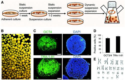
In this paper, Baharvand and his colleagues grew human iPSCs in “matrigel,” and then grew them in suspension. Matrigel is gooey and the cells stick to it and grow, and they were grown in matrigel culture for 1 week. After one week, the cells were grown in liquid suspension for 1-2 weeks. The cells have better access to soluble growth factors in liquid culture and tend to grow faster. After this they were grown in a stirred culture (known as a spinner).  This expanded the cells into large numbers for further use.

 Expansion and characterization of human induced pluripotent stem cells (hiPSCs) in a dynamic suspension culture. (A) Schematic representation of the suspension culture and expansion of human pluripotent stem cells (hPSCs) from adherent to stirred bioreactor. The cells were transferred to bacterial dishes to adapt to three-dimensional (3D) environment and then transferred into the dynamic phase, stirred flask. (B) Morphology of a tested hiPSC line (hiPSC1) after passage in a stirred suspension bioreactor as monitored by dark-field microscopy. (C) Immunostaining of cross sections of spheroids for OCT4 and TRA-1-81. Scale bar: 50 μm. (D) Flow cytometry analysis and (E) normal karyotype of suspended cells in the bioreactor.
Expansion and characterization of human induced pluripotent stem cells (hiPSCs) in a dynamic suspension culture. (A) Schematic representation of the suspension culture and expansion of human pluripotent stem cells (hPSCs) from adherent to stirred bioreactor. The cells were transferred to bacterial dishes to adapt to three-dimensional (3D) environment and then transferred into the dynamic phase, stirred flask. (B) Morphology of a tested hiPSC line (hiPSC1) after passage in a stirred suspension bioreactor as monitored by dark-field microscopy. (C) Immunostaining of cross sections of spheroids for OCT4 and TRA-1-81. Scale bar: 50 μm. (D) Flow cytometry analysis and (E) normal karyotype of suspended cells in the bioreactor.

Getting cells to grow in liquid suspension tends to be a bit of an art form, but these iPSCs grew rather well. Also, the iPSCs were differentiated into definitive endoderm, which is the first step in bringing cells to the liver cell stage. The drug Rapamycin and activin (50 ng / L for those who are interested) were used to bring the growing iPSCs to the definitive endoderm.  The cells expressed all kinds of endoderm-specific genes.  Endoderm is the embryonic germ layer from which the digestive system and its accessory organs forms.

 Induction of hiPSCs into definitive endoderm. (A) Diagrammatic representation of the experimental groups for endoderm induction of hiPSCs in the bacterial dish static suspension, which include rapamycin (Rapa) “priming” and activin A “inducing” phases, and positive control groups of hiPSCs cultured in the absence of Rapa in suspension or adherent cultures in the presence of activin A. (B) Gene expression analysis of hiPSCs induced into endodermal cells. Quantitative reverse transcriptase–polymerase chain reaction (qRT-PCR) showed no significant differences in SOX17 and FOXA2 [definitive endoderm (DE) markers], and BRA (mesoendoderm marker) in all groups. SOX7 (visceral endoderm marker) was expressed at a low level. Influence of the refreshment strategy of the induction medium on DE formation in suspension cultures by qRT-PCR (C), immunostaining (D), and flow cytometry (E). We compared single (SR), double (DR), and triple (TR) refreshment of induction medium per 24 h for 4 days after Rapa administration in the static suspension of hiPSCs. Scale bar: 100 μm. The target gene expression level in qRT-PCR was normalized to GAPDH and calibrated with (presented relative to) hiPSCs. Data are presented as mean±SD. Statistical analysis as determined by one-way ANOVA with Tukey's post hoc test, n=3 for B, C, and E. *P<0.05, **P<0.01.
Induction of hiPSCs into definitive endoderm. (A) Diagrammatic representation of the experimental groups for endoderm induction of hiPSCs in the bacterial dish static suspension, which include rapamycin (Rapa) “priming” and activin A “inducing” phases, and positive control groups of hiPSCs cultured in the absence of Rapa in suspension or adherent cultures in the presence of activin A. (B) Gene expression analysis of hiPSCs induced into endodermal cells. Quantitative reverse transcriptase–polymerase chain reaction (qRT-PCR) showed no significant differences in SOX17 and FOXA2 [definitive endoderm (DE) markers], and BRA (mesoendoderm marker) in all groups. SOX7 (visceral endoderm marker) was expressed at a low level. Influence of the refreshment strategy of the induction medium on DE formation in suspension cultures by qRT-PCR (C), immunostaining (D), and flow cytometry (E). We compared single (SR), double (DR), and triple (TR) refreshment of induction medium per 24 h for 4 days after Rapa administration in the static suspension of hiPSCs. Scale bar: 100 μm. The target gene expression level in qRT-PCR was normalized to GAPDH and calibrated with (presented relative to) hiPSCs. Data are presented as mean±SD. Statistical analysis as determined by one-way ANOVA with Tukey’s post hoc test, n=3 for B, C, and E. *P<0.05, **P<0.01.
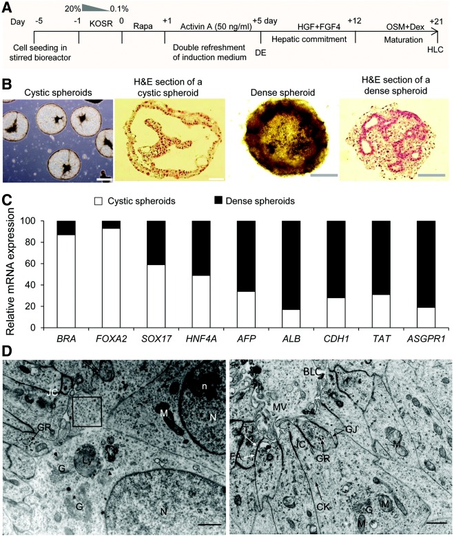
After the cells went through this culture protocol, they were grown in a stirred liquid culture called a “spinner.” The culture system contain a cocktail of growth factors that differentiated the definitive endoderm cells into HLCs.  The cells formed little spheres that expressed a host of liver-specific genes.

Differentiation of hepatocyte-like cells (HLCs) from hiPSCs in the stirred bioreactor. (A) Stepwise protocol for differentiation of hiPSCs into HLCs. (B) The morphology and cross section of spheroids at day 21. The spheroids in this step were observed as cystic and dense spheroids. Hematoxylin and eosin (H&E) staining of spheroid cross sections indicated cystic and dense epithelioid appearances in the transplant and dense spheroids, respectively. Scale bar: 100 μm. (C) Comparative relative mRNA expression in cystic and dense spheroids normalized to GAPDH and calibrated to undifferentiated hiPSCs. Transplant and dense spheroids expressed more early and late hepatic lineage markers, respectively. Data are presented as mean. n=3. (D) Transmission electron microscopy (TEM) of differentiated cells in dense spheroids at day 21. Nucleus (N), nucleoli (n), mitochondria (M), Golgi apparatus (G), lysosomes (Ly), rough endoplasmic reticuli (arrowhead), glycogen granules (GR), intermediate filaments (CK), tight junctions (TJ), gap junctions (GJ), fascia adherens (FA), junctional complex (JC), microvilli (MV), and bile-like canaliculus (BLC). Scale bar: 1 μm.

From the figure above, we can see that these HLCs, not only express liver-specific genes, but when they are examined in the electron microscope they look, for all intents and purposes, like liver cells.  Functional tests of these spheres of HLCs showed that they 1) took up low-density lipoprotein; 2) produced albumin (a major blood plasma protein); 3) expressed cytochrome P450s, which are the major enzymes used to process drugs; 4) produced urea from amino acids, just like real liver cells; 5) accumulated glycogen; 6) and made liver proteins (HNF4a, ALB, etc).

So it looks like liver, quacks like liver, but can it replace liver?  These HLCs were transplanted into the spleen of mice whose livers had been treated with carbon tetrachloride.  Carbon tetrachloride tends to make mincemeat of the liver, and these mice are in trouble, since their livers are toast.  Transplantation of the iPSC-derived HLCs into the spleens of these mice increased their survival rate and decreased the blood levels of liver enzymes that are usually present when there is liver damage.

This paper is significant because the procedure used provides an example of a “scalable” protocol for making large quantities of iPSCs, and their mass differentiation into definitive endoderm and then liver cells,  Because this can potentially provide enough cells to replace a nonfunctional liver, it represents a major step forward in regenerative medicine.


Molecular Signature Distinguished Old Stem Cells from New Stem Cells


Eukaryotic organisms include every living thing with the exception of bacteria, Bacteria are known as prokaryotes, and they do not have an organized nucleus. Eukaryotic cells, on the other hand, have an organized nucleus in which that houses the chromosomes, which are linear molecules of DNA.

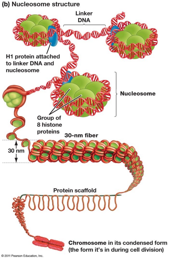
DNA is the molecule that stores genetic information. The chromosomes of eukaryotic organisms are sometimes rather long. How then does the cell manage to store all that DNA in such a small compartment such as the nucleus? The answer is that DNA in eukaryotic cells is wound into a tight configuration known as chromatin.

Chromatin consists of DNA molecules that are spooled around a cylindrical structure made of histone proteins. There are four so-called “core histones” that compose the cylinders and the DNA winds around these histone cores. Then a non-core histone called H1 pulls the histone cylinders with their DNA wound about them together to form higher-order structures. The histone cylinders wound about with DNA are called “nucleosomes” or “core particles.” The assembled clusters to nucleosomes are called “30 nanometer solenoids.”

Chromatin1

You might think that DNA all wound into chromatin would be difficult to access and transcribe.  If you think that, then you are correct.  How then does the cell access DNA wound into chromatin? It modifies the histones so that the grip the histones have on the DNA is loosened.  Since histones are positively charged and DNA is negatively charged (lots of phosphate), the two molecules bind to each other rather tightly.  However, If histones are decorated with acetate groups, they become less positively charged and bind to DNA less tightly.  This opens up the chromatin for gene expression.  However, if histones are decorated with methyl groups (CH3), then proteins bind the histones and cinch the DNA even more tightly so that nothing is expressed.  This is known as the “histone code,” since geneticists can use the chemical modifications of histones to make highly educated guesses about if genes will be expressed and the levels at which they will be expressed.

A research team at Stanford University in Palo Alto, CA, led my Thomas Rando, professor of neurological sciences and chief of the Veterans Affairs Palo Alto Health Care System’s Neurology service, has identified characteristic differences in histone modifications between stem cells from the muscles of young mice and old mice.  Rando’s team also identified histone signatures characteristic of sleeping or quiescent and active stem cells in the muscles of young mice.

Rando said, “We’ve been trying to understand both how the different states a cell finds itself in can be defined by the markings on the histones surrounding its DNA, and to find an objective way to define the ‘age’ of a cell.”

All the cells of our body share the same genes, but these cells can be remarkably different in their function, structure, shape, and metabolism.  Only a fraction of a cell’s genes are actually turned one and are actively making proteins.  A muscle cells produced muscle-specific proteins and a liver cell makes liver cell specific proteins.  Rando’s team has generated data that suggests that these same kinds of on/off differences may distinguish old stem cells from young stem cells.

First a little background in necessary.  In 2005, Rando and others published a study that demonstrated that stem cells in several tissues from older mice, including muscle, seemed to act younger after continued exposure to the blood of a younger mouse.  The capacity of these stem cells in older mice to divide, differentiate, and repopulate tissues declines with advancing age.  However, after these stem cells from older mice were exposed to younger mouse’s blood, their ability to proliferate and repair tissues resembled those of their stem-cell counterparts in younger animals (see Conboy IM et al., Nature. 2005 433(7027):760-4).

Rando and his group asked the next question: “What is happening inside these cells that make them act as though they are younger?”  The first place Rando and others decided to look was the chemical modifications of their histones.  The cell population they examined was muscle satellite cells, which are relatively easy to isolate and grow in culture.  Normally, muscle satellite cells sit within skeletal muscles and do well little.  However, once the muscle is damaged, muscle satellite cells wake up, swing into action, and divide and fuse with damaged muscle fibers to repair them.

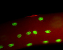
Muscle Satellite Cells in green

In mice that are old, histones in muscle satellite cells are a mixture of signals that tell expression to stop and signals that tell gene expression to go.  However, in satellite cells from younger mice, the histones are largely a collection of go signals with only few stop signals.  According to Rando, “Satellite cells can sit around for practically a lifetime in a quiescent state, not doing much of anything.  But they’re ready to transform to an activated state as soon as they get the word that the tissue needs repair.  So you might think that satellite cells would be already programmed in a way that commits them solely to the ‘mature muscle cell’ state.”  Thus you would expect those genes specific for other tissues like skin, brain or fat would be marked with stop signals.

Instead quiescent satellite cells taken from the younger mice contained histones with a mixture of stop and go signals in those genes ordinarily reserved for other tissues.  This was similar to what was observed in mature muscle-specific genes.  Satellite cells from older mice were pockmarked with stop signals interspersed with go signals.

Are these changes typical of those that occur in other types of stem cells in other tissues?  That is presently unknown.  Also, what is the signal in the blood from the younger mice that causes the satellite cells function as though they are young?”  Rando said, “We don’t have the answers yet.  But now that we know what kinds of these changes occur as these cells age, we can ask which of these changes reverse themselves when an old cell goes back to becoming a young cell.”

Rando’s group is presently examining if the signatures they have identified in satellite cells generalize to other kinds of adult stem cells as well.

Human Embryonic Stem Cell Communication Network Discovered


Cells use a variety of mechanisms to talk to each other. These signaling pathways are called “signal transduction” pathways, and they vary extensively from one cell type to another.

Therefore, it should be no surprise that human embryonic stem cells signal to each other. The precise signal transduction pathway that human embryonic stem cells use to communicate with each other is the subject of a research project from a laboratory in Singapore.

Human embryonic stem cells or hESCs can differentiate into any adult cell type. The factors that keep hESCs in their pluripotent state are of interest to stem cell scientists because they might allow them to better direct the differentiation of hESCs or even grow them in culture better.

Cell-to-cell communication is vitally important to multicellular organisms. The coordinated development of tissues in the embryo that culminate in the formation of specific organs requires that cells receive signals and respond accordingly. If there are errors in these signals, the cells will respond differently and the embryo will either be grossly abnormal, or the cell might divide uncontrollably to make a tumor.

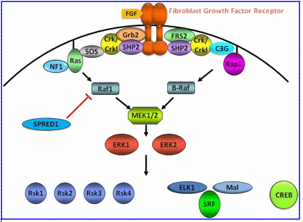
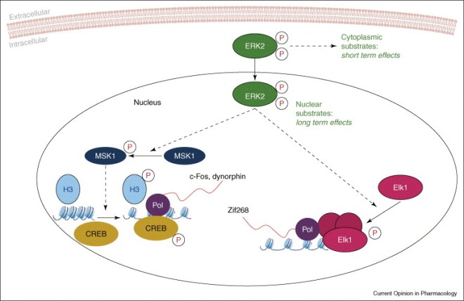
Human ESCs communicate by means of a signal transduction pathway known as the extracellular regulated kinase or ERK pathway.  The ERK signal transduction pathway begins with the binding of a growth factor receptor by a growth factor.  These growth factors are almost always bound to the extracellular matrix, which is the goo that surrounds cells and provides a structure in which the cells live.  The binding of the receptor causes the receptor to pair with another copy of itself, and that activates the bits of the receptor found inside the cell (tyrosine kinase domain for the interested).  The activated receptor attaches phosphates to itself, which causes particular proteins to find and bind the receptor, which recruits particular proteins to the cell membrane.  One of the recruited proteins is a protein kinase called RAF.  RAF attaches phosphate groups to the protein kinase MEK, and MEK attaches phosphate groups to the protein kinase ERK.  Once ERK has a phosphate attached to it, it can move into the nucleus and regulate transcription factors involved in the control of gene expression.  Thus a phenomenon that began at the cell membrane culminates in a change in gene expression.

ERK_pathway

Stem cell scientists a A*STAR’s Genomic Institute of Singapore and the Max Planck Institute of Molecular Genetics (MPIMG) in Berlin, Germany studied how genetic information is accessed in hESCs. To do this they mapped the kinase interactions across the entire human genome (kinases are enzymes that attach phosphate groups to other molecules) and discovered that ERK2, a protein that belongs to the ERK signal transduction pathway targets important sites such as non-coding genes, and histones, cell cycle, metabolism, and stem cell-specific genes.

The ERK signaling pathway involves an additional protein called ELK1 that interacts with ERK2. However, this research team discovered that ELK1 has a second, totally opposite function. At genomic sites not targeted by ERK signaling, ELK1 silences genetic information, which keeps the cell in its undifferentiated state.

ELK1 Interaction with ERK2

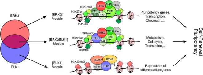
The authors propose a model that integrates this bi-directional control to keep the cell in the stem cell state, in which genes necessary for differentiation are repressed by ELK1 that is not associated with ERK2, and cell-cycle, translation and other pluripotency genes are activated by ELK1 in association with ERK2 or ERK2 plus other transcription factors.

Model of the Transcriptional Regulatory Network of ERK2 Signaling in hESCsTranscription factors such as ELK1 link ERK2 to sequence-specific regulation of gene expression. ERK2 and ELK1 colocalization defines three distinct modules that target different sets of genes. In this model, combinatorial binding of ERK2 and ELK1 with transcription factors, chromatin regulators, and the basal transcriptional machinery integrates external signaling into the cell-type-specific regulatory network. In hESCs, ERK2 and ELK1 participate in the regulation of pluripotency and self-renewal pathways, whereas differentiation genes are repressed.

First author Jonathan Göke from Stem Cell and Developmental Biology at the GIS said, “The ERK signaling pathway has been known for many years, but this is the first time we are able to see the full spectrum of the response in the genome of stem cells. We have found many biological processes that are associated with this signaling pathway, but we also found new and unexpected patterns such as this dual-mode of ELK1. It will be interesting to see how this communication network changes in other cells, tissues, or in disease.”

A co-author of this study, Martin Vingron said, “A remarkable feature of this study is, how information was extracted by computational means from the data.”

Professor Ng Huck Hui, managing author of this paper, added, “This is an important study because it describes the cell’s signaling network and its integration into the general regulatory network. Understanding the biology of embryonic stem cells is a first step to understanding the capabilities and caveats of stem cells in future medical applications.”

Both Copies of the Nanog Gene Are Expressed in Embryonic Stem Cells


Commonly held ideas are sometimes held because there is a great of evidence to substantiate them. However, other times, an idea is commonly held because simply because it has been repeated over and over and over even though the evidence for it is poor. Thus, when new evidence come to light showing the commonly held believe to be untrue, it becomes incumbent on us to readjust what we think.

When it comes to embryonic stem cells and the genes that keep them pluripotent, the transcription Nanog plays a very critical role in the self-renewal of embryonic stem cells and there is a great deal of evidence for this assertion. However, the expression of the gene that encodes Nanog was thought to follow the same mode of expression as some of the other pluripotency promoting genes. Namely, that only one of the copies of the Nanog gene were thought to be expressed in embryonic stem cells. This turns out to be probably false.

First a little background. In 2007, Ian Chambers and others published a paper in the journal Nature that examined the expression and function of Nanog in embryonic stem cells. Chambers and others found that Nanog expression levels in individual embryonic stem cells from a culture derived from a single cell varied wildly.  The figure from the Chambers et al paper is shown below.

Immunofluorescence of TNG cells for Oct4 and Nanog. Individual signals from 4,6-diamidino-2-phenylindole (DAPI), GFP, anti-Oct4 and anti-Nanog are shown on the left alongside a combined view of GFP with the stainings from anti-Oct4 and anti-Nanog.
Immunofluorescence of TNG cells for Oct4
and Nanog. Individual signals from 4,6-diamidino-2-phenylindole (DAPI),
GFP, anti-Oct4 and anti-Nanog are shown on the left alongside a combined
view of GFP with the stainings from anti-Oct4 and anti-Nanog.

The reason for this fluctuation in Nanog levels was uncertain, but Chambers and others showed that Nanog could be deleted from mouse embryonic stem cells without affecting their ability to contribute to various sundry embryonic tissues during mouse development, even though they do not make functional gametes (eggs and sperm).  In fact, mouse embryonic stem cells can self-renew under particular conditions without a functional copy of the Nanog gene even though they are prone to differentiation.  From this, Chambers and others concluded that Nanog stabilized rather than promoted pluripotency of embryonic stem cells by “resisting or reversing alternative gene expression states.”

Fast forward to 2012 and another Nature paper by Yusuke Miyanari and Maria-Elena Torres-Padilla from the IGBMC in Strasbourg, France, which showed that before mouse embryos implanted into the uterus, only one copy of the Nanog gene was expressed, but after implantation, both copies of the Nanog gene was expressed.  Miyanari and Torres-Padilla also made mouse embryonic stem cells that had copies of the Nanog gene labeled with different glowing proteins.  This ingenious experiment showed confirmed that Nanog levels were variable, but also showed that only one copy of the Nanog gene was expressed in growing embryonic stem cells in culture.

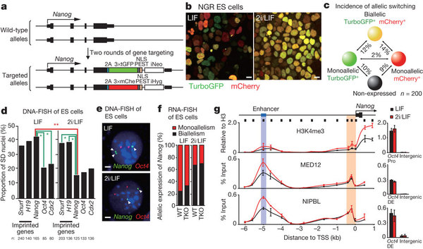
a, Schematic of the Nanog knock-in reporter NGR. A PEST motif in the carboxy terminus of the fluorescent proteins allows monitoring of dynamic Nanog expression. iHyg, internal ribosome entry site (IRES) hygromycin; iNeo, IRES neomycin; mChe, mCherry; NLS, nuclear localization signal; tGFP, TurboGFP. b, Representative image of NGR ES cells cultured with LIF or 2i/LIF. Scale bar, 10 µm. c, The incidence of allelic switching of Nanog expression in ES cells. Cells were classified into four groups: monoallelic (TurboGFP-positive, green), monoallelic (mCherry-positive, red), biallelic (TurboGFP- and mCherry-positive, yellow) and no expression (black). The proportion of cells undergoing a transition between these four groups during a single cell cycle is indicated. Overall, 47% of cells showed a colour change in this period. n, number of cells analysed. d, The asymmetric replication of Nanog in ES cells cultured with LIF changes to symmetric replication upon treatment with 2i. The cell nuclei were classified as single/double (SD), single/single (SS) and double/double (DD) according to DNA-FISH signals5. n, number of nuclei analysed. *, P < 4 × 10−7; **, P < 1.4 × 10−3 (Fisher’s exact test). e, Representative image of DNA-FISH for Nanog (arrowheads) and Oct4 in ES cells cultured with LIF or 2i/LIF. Scale bar, 2 µm. f, Nanog allelic expression is unaffected in the absence of DNA methyltransferase activity. Quantification of RNA-FISH for Nanog in wild-type (WT) ES cells and ES cells lacking all three DNA methyltransferases (TKO) cultured with LIF or 2i/LIF. g, ChIP for H3K4me3, MED12 or NIPBL along the Nanog locus (black line, top) in ES cells cultured with LIF or 2i/LIF. The position of the ChIP amplicons is depicted by the thick boxes below the line, the TSS by an arrow, the first exon by the black box on the line, and the distal enhancer by the blue box on the line. The Oct4 promoter region (Oct4 Pro) and distal enhancer (Oct4 DE) were positive controls (right)20. The mean ± s.d. of three independent biological replicates is shown.

Now if we fast forward one more year and a paper from the journal Cell Stem Cell and a letter to the same edition of this journal, we have an article by Dina Faddah and others from the laboratory of Rudolf Jaenisch at the Whitehead Institute (MIT, Cambridge, MA), and a supporting letter from Adam Filipczyk and others from Germany and Switzerland.  In this article, the authors also double-labeled mouse embryonic stem cells and examined multiple cells and showed that BOTH copies of Nanog were expressed, and that the range of variability of Nanog expression was approximately the same as other pluripotency genes.

Filipczyk and others used a similar approach to examine the expression of Nanog in mouse embryonic stem cells and they came to the same conclusions as those of Faddah and others.

What is the reason for the differences in findings?  Faddah and others did an important experiment to answer this question.  Some of the cells that with labeled copies of the Nanog gene disrupted the production of a functional Nanog protein.  The constructs used in the papers by Faddah and others and by Filipczyk and others did not disrupt Nanog protein production.  When Faddah and others tested these other constructs that disrupted Nanog protein production to determine is the amount of glowing protein tracked with the amount of Nanog protein produced, it was clear that the amount of Nanog protein made by the cells did not reflect the amount of glowing protein produced.  According to Dina Faddah, “The way the reported was inserted into the DNA seems to disrupt the regulation of the alleles so that when the reported said Nanog isn’t being expressed, it actually is.”

Jaenisch sees this as an instructional tale for all stem cell scientists.  He noted: “Clearly, the conclusions for this particular gene need to be reconsidered.  And it raises the question for other genes.  For some genes, there might be similar issues.  For other genes, they might be more resistant to this type of disturbances caused by a reporter.”

Bottom line – read the materials and methods part of the paper carefully because the way these experiments are done can determine if the results are trustworthy.

MSNBC Host Says That Life Begins Whenever You Feel Like It Does


I lived in Great Britain for three years for my first postdoctoral research fellowship at Sussex University. To be completely honest, I never got into the whole royal family thing, but the birth of George Alexander to Prince William and Kate Middleton is certainly an event to celebrate. George has little chance of ever ascending to the throne, but he is certainly a bundle of joy to his parents and to the British people.

Therefore, I find it rebarbative that media kill joys have used the joyous birth of William and Kate’s baby to be an opportunity to talk about abortion. In addition to this, one particular pro-choice news correspondent, Melissa Harris-Perry decided to wax philosophically about the nature of the unborn.

After noting the worldwide excitement that has surrounded Kate Middleton’s pregnancy and birth, MSNBC host Melissa Harris-Perry compared the buzz surrounding the British royal birth to Texas abortion politics, and then offered her own answer to the question “when does life begin:”

“When does life begin? I submit the answer depends an awful lot on the feeling of the parents. A powerful feeling – but not science,”

News correspondents say stupid things, but this has to rank as one of the most brain-dead things I have ever heard. Let’s not forget who said it, since Melissa Harris-Perry, is the news anchor who wore tampon earrings and received Planned Parenthood’s Maggie Award.

Once the egg in the fallopian tube of the mother fuses with a sperm cell from the father, the egg undergoes a complex sequences of biochemical and cellular events that culminate in the fusion of the genetic material of the mother with that of the father. This marks the end of the process known as fertilization and the beginning of the embryonic stages of development. The embryo has begun the journey of human development, growth, and maturation that will not stop until the individual dies. The embryo is genetically distinct from the mother and the father, and is a human being, albeit, a very young human being. The embryo is not a plant, an alligator, or some facsimile or something else, it is human, but a young human. That is not a feeling, but a scientific fact.

Can we kill the embryo just because it is very young? Reflection leads me to say no, no, a thousand times no. Do we value two-year old children more than one-year old children? Do we value six-year old children more than four-year old children? Age is irrelevant to the moral worth of an individual.

But, you say, the embryo is underdeveloped relative to a new-born baby. Does the extent of development determine moral worth? Again, a one-month old baby is more developed than a two-week old baby. Does that make the one-year old baby more valuable? No. Are teenagers who are more physically developed more morally valuable than eight-year old children? No. Therefore, the extent of development is not a good measure of a human being’s moral worth.

Ms. Harris-Perry seems to thing that feelings or perhaps she means how deeply a mother wants her baby is the factor that determines if he or she should continue to live. Again I say no. This would justify genocide. The dictators of North Korea can simply say that killing their own people is due to the fact that they did not want them anymore. They had those kind of feelings you know. How about Hitler and the Third Reich and their slaughter of six million Jews and many millions of  others? Hitler and his officers killed them because they did not feel that Jews and others were worthy of life. In fact, Harris-Perry’s ethic can justify any heinous, insidious acts simply on the basis of feelings.

This is, as I have said, brain-dead and she should be called out for it. The unborn human beings are still human beings regardless of how we feel about them. That is a fact of genetics and embryology regardless of your feelings about it. If MSNBC has news correspondents that say things that are this stupid, then maybe they deserve to have such low ratings.

New Pluripotent Stem Cell Production Protein Identified


Large scale production of stem cells requires an intimate knowledge of the genetic networks that convert adult cells into induced pluripotent stem cells (iPSCs). The original protocol established by Shinya Yamanaka and his colleagues used four genes all clustered on a retrovirus vector, but there are safer, more technically subtle ways to make iPSCs.

Because iPSCs are made from a patient’s own cells, they are less likely to be rejected by the patient’s immune system. They also show tremendous developmental flexibility, they can potentially be differentiated into any adult cell type in the body.  The problem with iPSCs comes from the difficulty of making large quantities of them in a reasonable amount of time.  However, a new research publication from scientists at the University of Toronto, the University for Sick Children and Mount Sinai Hospital, in collaboration with colleagues from the United States and Portugal, identifies specific proteins that play central roles in controlling pluripotency that may mean a potential breakthrough in producing iPSCs.

Researchers discovered these proteins by using something called the “splicing code.”  Benjamin Blencowe discovered the splicing code a few years ago.  “The mechanisms that control embryonic stem cell pluripotency have remained a mystery for some time.  However, what Dr. Blencowe and the research team found is that the proteins identified by our splicing code can activate or deactivate stem cell pluripotency,” said Brendan J. Frey, from the University of Toronto Departments of Electrical Engineering and Medicine, who published with Benjamin Blencowe the paper that deciphered this splicing code (see Nature 2010 465: 53-59).  While a complete recipe for producing iPSCs may not be available yet, it is beginning to look more likely, according to Frey.

In this paper, Blencowe and his collaborators identified two proteins known as muscleblind-like RNA binding proteins, or MBNL1 and MBNL2.  These proteins are conserved and direct negative regulators of a large program of cassette exon alternative splicing events that are differentially regulated between embryonic stem cells and other cell types.

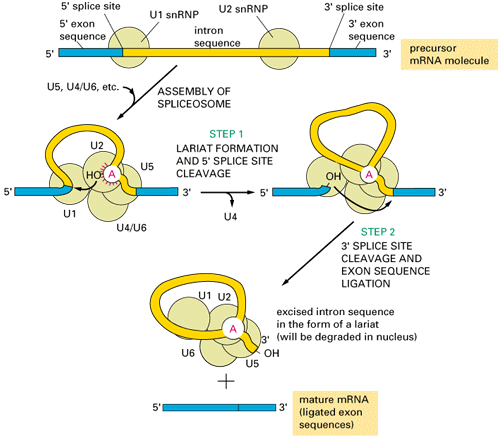
RNA splicing occurs in plant, animal, fungal, and protist cells (only very, very rarely in bacteria), and involves the removal of segments of primary RNA transcripts.  When RNA molecules are transcribed in eukaryotic cells, they are engaged by cellular machinery called the RNA spliceosome.  The RNA spliceosome removes segments known as “introns” and the excised introns are degraded and the remaining RNA segments, which are known as “exons, are ligated together to form a mature messenger RNA.

mRNA splicing

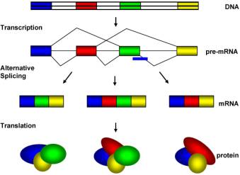
Some introns are removed from primary RNA transcripts by all cells, but others are removed in some cells but not others.  This phenomenon is known “alternative splicing” and it is responsible for the differential regulation of particular genes.

alternative_splicing

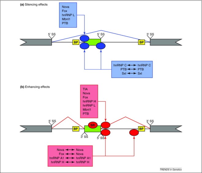
Alternative splicing is mediated by sequences called splicing enhancers and splicing silencers that are six to either nucleotides long and bind proteins that either induce or repress alternative splicing in those cells that express the proteins that bind these splicing enhancers or silencers.

Alternative RNA splicing mechanism

MBNL is one of these proteins that bind to RNA splicing silencers.  If the quantity of MBNL proteins in differentiated cells is decreased, then these cells switch to an embryonic stem cell-like alternative splicing pattern for approximately half of their genes.  Conversely, overexpression of MBNL proteins in ES cells promotes differentiated-cell-like alternative splicing patterns.  Among the MBNL-regulated events is an ES-cell-specific alternative splicing switch in a protein-coding gene called the forkhead family transcription factor FOXP1.  FOXP1 controls pluripotency, and consistent with a central and negative regulatory role for MBNL proteins in pluripotency, knockdown of MBNL significantly enhances the expression of key pluripotency genes and the formation of induced pluripotent stem cells during somatic cell reprogramming.

Thus MBNL proteins should be one of the main targets for the mass production of iPSCs.

Breast Cancer Clinical Trial Targets Cancer Stem Cells


Even though my previous posts about cancer stem cells have generated very little interest, understanding cancer as a stem cell-based disease has profound implications for how we treat cancer. If the vast majority of the cells in a tumor are slow-growing and not dangerous but only a small minority of the cells are rapidly growing and providing the growth the most of the tumor, then treatments that shave off large numbers of cells might shrink the tumor, but not solve the problem, because the cancer stem cells that are supplying the tumor are still there. However, if the treatment attacks the cancer stem cells specifically, then the tumor’s cell supply is cut off and the tumor will wither and die.

In the case of breast cancer, the tumors return after treatment and spread to other parts of the body because radiation and current chemotherapy treatments do not kill the cancer stem cells.

This premise constitutes the foundation of a clinical trial operating from the University of Michigan Comprehensive Cancer Center and two other sites. This clinical trial will examine a drug that specifically attacks breast cancer stem cells. The drug, reparixin, will be used in combination with standard chemotherapy.

Dr. Anne Schott, an associate professor of internal medicine at the University of Michigan and principal investigator of this clinical trial, said: “This is one of only a few trials testing stem cell directed therapies in combination with chemotherapy in breast cancer. Combining chemotherapy in breast cancer has the potential to lengthen remission for women with advanced breast cancer.”

Cancer stem cells are the small number of cells in a tumor that fuel its growth and are responsible for metastasis of the tumor. This phase 1b study will test reparixin, which is given orally, with a drug called paclitaxel in women who have HER2-negative metastatic breast cancer. This study is primarily designed to test how well patients tolerate this particular drug combination. However, researchers will also examine how well reparixin appears to affect various cancer stem cells indicators and signs of inflammation. The study will also examine how well this drug combination controls the cancer and affects patient survival.

This clinical trial emerged from laboratory work at the University of Michigan that showed that breast cancer stem cells expressed a receptor on their cell surfaces called CXCR1. CXCR1 triggers the growth of cancer stem cells in response to inflammation and tissue damage. Adding reparixin to cultured cancer stem cells killed them and reparixin works by blocking CXCR1.

Mice treated with reparixin or the combination of reparixin and paclitaxel had significantly fewer (dramatically actually) cancer stem cells that those treated with paclitaxel alone. Also, riparixin-treated mice developed significantly fewer metastases that mice treated with chemotherapy alone (see Ginestier C,, et al., J Clin Invest. 2010, 120(2):485-97).

Clinical Study Evaluates Healing of Knee Cartilage With Stem Cells


The biotechnology company InGeneron will test its patented Transpose RT system in a clinical study that examined the ability of regenerative cells from a patient’s own fat to enhance cartilage healing after knee surgery.

Qualified patients are being recruited through the Fondren Orthopedic Group in Houston. According to the American Orthopedic Society for Sports Medicine, over 4 million knee arthroscopies are performed worldwide each year. Damaged knee cartilage is very difficult to treat and can lead to chronic pain and long-term disability.

Robert Burke, who is serving as the principal investigator of this clinical study, is an orthopedic surgeon with the Fondren Orthopedic Group in Houston. Burke thinks that stem cells taken from a patient’s own fat may enhance cartilage healing. He studied adding patient-derived regenerative cells to the knee during arthroscopic surgery for particular patients, and compared them to patients who had arthroscopic surgery without added fat-derived stem cells.

Arthroscopic surgery is a common procedure is commonly used to treat damaged cartilage, and the patients who had received arthroscopic surgery were randomly chose to either receive fat-derived stem cells or not receive them. Burke, will then monitor these patients for the next 12 months after surgery to determine if the added cells improved cartilage healing.

According to Burke, “Articular cartilage, the smooth surface covering the joints at the ends of bones, has no good way of healing on its own. The body doesn’t create enough new cartilage of the same type to repair the damage.” Better treatments would use various techniques to help the body make new cartilage.

“Stem cells and other regenerative cells that we can obtain fat have the potential to do that,” said Burke. Such regenerative cells can divide and mature to form several types of cells and tissues. and are found in multiple places in the body. Fat that lies just below the skin is one of the easiest places to obtain stem cells.

The InGeneron Transpose RT System uses a small amount of fat, which is removed and processed to separate out the regenerative cells. The separated adipose tissue-derived mesenchymal stem cells are then immediately placed into the area of damaged cartilage during knee surgery. Once in the knee, these cells may divide to make new cartilage cells.

This kind of biological activity has been seen in laboratory studies and veterinary medicine. However, Burke’s study will be one of the first to test the technology in humans for treating cartilage damage. Like other types of stem cell-based therapies, the treatment is not currently licensed for human use in the United States but it is registered in Europe, Mexico, and other countries. Following the Texas Medical Board’s rules about the use of stem cells for treatment, this study is under the supervision of the research review board at Texas Orthopedic Hospital, where all of the patients will undergo surgeries.

This is a two-year study.

Multipotent Adult Progenitor Cells Prevent Rejection of Transplanted Tissue


Solid organ transplantation is a procedure that has saved untold millions of lives. Unfortunately, the tendency for an organ to be rejected by the immune system of the organ recipient is a formidable problem that is addressed in two ways. One of these is through tissue matching of the organ to the recipient. The other is through the use of immunosuppressive drugs that suppress the immune system. Neither one of these strategies is without caveats.

Tissue typing begins with a blood test to determine the organ recipient’s blood type. If the organ contains a blood type that is incompatible with the immune system of the organ recipient, the result will be catastrophic. Hyperacute rejection is the name given to organ rejection that occurs minutes to hours after the organ was transplanted. Hyperacute rejection occurs because the recipient has pre-existing antibodies in their body that recognizes and begins to destroy the graft. These antibodies can result from prior blood transfusions, multiple pregnancies, prior transplantation, or xenografts against which humans already have antibodies. Massive blood clotting within the capillaries of the organ clog the blood vessels and prevent perfusion of the graft with blood. The organ must come out or the patient will die.

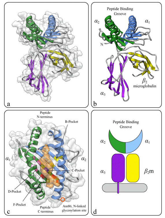
Human cells have on their surfaces particular proteins that are encoded by genes located on the short arm of chromosome 6 called the major histocompatibility complex or MHC. the MHC genes encode human leukocyte antigens or HLAs. HLA proteins are extremely variable from person to person, and this seems to be the case because the more variation we have in our HLA proteins, the better job the immune system does recognizing foreign invaders.

Each individual expresses MHC genes from each chromosome. Therefore, your cells contain a mosaic of surface proteins, some of which are encoded by the HLAs encoded by the chromosome you inherited from your father and others of which are encoded by the chromosome your inherited from your mother.

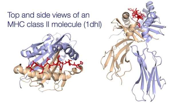
The MHC molecules are divided into 2 classes. Class I molecules are normally expressed on all nucleated cells, but class II molecules are expressed only on the so-called “professional antigen-presenting cells” or APCs. APCs include cells that have names like dendritic cells, activated macrophages, and B cells. T lymphocytes only recognize foreign substances when they are bound to an MHC protein. Class I molecules present antigens from within the cell, which includes bits from viruses, tumors and things like that. Class II molecules present extracellular antigens such as extracellular bacteria and so on to a subclass of T cells called T helper cells, which express a molecule called “CD4” on their cell surface.

MHC-Class-I-Topology_3mhc_class2

All this might seem very confusing, but it is vital to ensuring that the organ is properly received by the organ recipient. Some types of MHC are very different and will elicit robust immune responses against them, but others are not as different and can be rather well tolerated. How does the doctor which are which? Through three tests: 1) Blood type is the first one. If this does not match, you are out of luck; 2) lymphocytotoxicity assay in which blood from a patient is tested to determine if it reacts with lymphocytes from the blood of the donor. A positive crossmatch is a contraindication to transplantation because of the risk of hyperacute rejection. This is used mainly in kidney transplantation; 3) Panel-reactive antibody (PRA) screens in which the the serum of a patient is screened for antibodies against the lymphocytes from the donor. The presence of such antibodies is contraindicated for transplantation. Finally, there is a test that is not used a great called the mixed lymphocyte reaction test that uses lymphocytes from the blood of the organ donor and the organ recipient to see if they activate one another. This test takes a long time and can be difficult to interpret.

Once the patient receives the transplant, they are usually put on immunosuppressive drugs. These drugs include cyclosporine, tacrolimus, sirolimus, mycophenolate, and azathioprine. Each of these drugs has a boatload of side effects that range from hair loss, diabetes mellitus, nerve problems, increased risk of illness and tumors, and so on. None of these side effects are desirable, especially since the drug must be taken for the rest of your life after you receive the transplant.

Enter a new paper from University Hospital in Regensburg, Germany from the laboratory of Marc Dahkle that used particular stem cells from bone marrow to induce toleration of grafted heart tissue in laboratory animals without any drugs. This paper was published in Stem Cells Translational Medicine and is potentially landmark in what it shows.

In this paper, Dahkle and his colleagues used stem cells from the bone marrow known as multipotential adult progenitor cells or MAPCs. MAPCs have been thought to be a subtype of mesenchymal stem cell in the bone marrow because they have several cell surface markers in common. However, there are some subtle differences between these two types of cells. First of all, the MAPCs are larger than their mesenchymal stem cell counterparts. Secondly, MAPCs can be cultured more long-term, which increases the attractiveness of these cells for therapeutic purposes.

In this paper, the Dahkle group transplanted heart tissue from two unrelated strains of rats. Four days before the transplantation, the donor rats received an infusion of MAPCs into their tail veins. There were a whole slew of control rats that were used as well, but the upshot of all this is that the rats that received the MAPCs before the transplantation plus a very low dose of the immunosuppressive drug mycophenolate did not show any signs of rejection of the transplanted heart tissue. If that wasn’t enough, when the transplanted heart tissue was then extirpated and re-transplanted into another rat, those grafts that came from MAPC-treated rats survived without any drugs, but those that came from non-MAPC-treated rats did not.

Because control experiments showed that the rats treated with cyclosporine did not reject their grafts, Dahkle and others used this system to determine the mechanism by which MAPCs prevent immune rejection of the grafted tissue. They discovered that the MAPCs seem to work though a white blood cell called a macrophage. Somehow, the MAPCs signal to the macrophages to suppress rejection of the graft. If a drug (clodronate) that obliterates the macrophages was given to the rats with the MAPCs, the stem cells were unable to suppress the immunological rejection of the graft.

In this paper, the authors conclude that “When these data are taken together, our current approach advances the concept of cell-based immunomodulation in solid organ transplantation by demonstrating that third-party, adherent, adult stem cells from the bone marrow are capable of acting as a universal cell product that mediates long-term survival of fully allogeneic organ grafts.” Revolutionary is a good word for this findings of this paper.  Hopefully, further pre-clinical trials will eventually give way to clinical trials in human patients that will allow human patients to have their lives saved by an organ transplant without the curse of taking immunosuppressive drugs for the rest of their lives.

Stem Cell Gene Therapy for Sickle Cell Disease Moves Toward Clinical Trials


UCLA stem cell researchers and “gene jockeys” have successfully proven the efficacy of using genetically engineered hematopoietic (blood cell-making) stem cells from a patient’s own bone marrow to treat sickle-cell disease (SCD).

In a study led by Donald Kohn, professor of pediatrics and microbiology at the UCLA Eli & Edythe Broad Center of Regenerative Medicine & Stem Cell Research, an “anti-sickling” gene was introduced into the hematopoietic stem cells (HSCs) from patients with SCD. Because the HSCs divide continuously throughout the life of the individual, all the blood cells they make will possess the anti-sickling gene and will therefore no sickle. This breakthrough gene therapy technique is scheduled to begin clinical trials by early 2014.

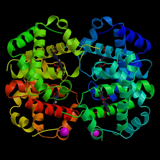
SCD results from a specific mutation in the beta-globin gene. Beta-globin is one of the two proteins that makes the multisubunit protein hemoglobin. Hemoglobin is a four-subunit protein that ferries oxygen from the lungs to the tissues and carbon dioxide from the tissues to the lungs. It is tightly packed into each red blood cell.

The structure of hemoglobin, each subunit is in a different color.

Hemoglobin has a very high affinity for oxygen when oxygen concentrations are high, but a low affinity for oxygen when oxygen concentrations are low. Therefore, hemoglobin does a very good job of binding oxygen when it is in the lungs, where oxygen is plentiful, and a very good job of releasing oxygen in the tissues, where oxygen is not nearly as plentiful. This adaptive ability displayed by hemoglobin is the result of cooperativity between the four polypeptide chains that compose hemoglobin. Two of these polypeptide chains are alpha-globin proteins and the other two a beta globin proteins. Hemoglobin acts as though it is an alpha-beta dimer, or as though it is composed of two copies of an alpha-globin.beta-globin pair. The interactions between these polypeptide chains and the movement of the hemoglobin subunits relative to each other creates the biochemical properties of hemoglobin that are so remarkable.

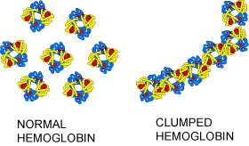
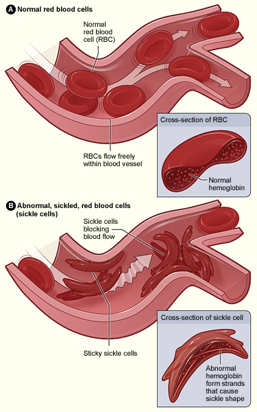
A mutation in the beta-globin gene that substitutes a valine residue where there should be a glutamic acid residue (position number 6), creates a surface on the outside of the beta-globin subunit that does not like water, and when oxygen concentrations drops, the mutant hemoglobin molecule changes shape and this new water-hating surface becomes a site for protein polymerization.

Sickle cell hemoglobin

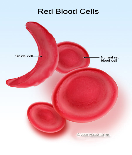
The mutant hemoglobin molecules for long, stiff chains that deform the red blood cells into a quarter moon-shaped structure that clogs capillaries.

Sickle cell RBCs


This new therapy seeks to correct the genetic mutation by inserting into the genome of the HSC that makes the abnormal red blood cells a gene for beta-globin that encodes a normal version of beta globin rather than a version of it that causes sickle-cell disease.  By introducing those engineered HSCs back into the bone marrow of the SCD patient, the engineered HSCs will make normal red blood cells that do not undergo sickling under conditions of low oxygen concentration.

Dr. Kohn noted that the results from his research group “demonstrate that our technique of lentiviral transduction is capable of efficient transfer and consistent expression of an effective anti-sickling beta-globnin gene in human SCD bone marrow progenitor cells, which improved the physiologic parameters of the resulting red blood cells.” Dr. Kohn’s statement may lead the reader to believe that this was done in a human patient, but that is not the case.  All this work was done in culture and in laboratory animals.

Kohn and his co-workers showed that in laboratory experiments, genetically engineered HSCs from SCD patients produced new non-sickled blood cells at a rate that would effectively allow SCD patients to show significant clinical improvement.  These new red blood cells also survived longer than those made by the nonengineered SCD HSCs.  The in vitro success of this technique has convinced the US Food and Drug Administration to grant Kohn the right to conduct clinical trials in SCD patients by early next year.

SCD affects more than 90,000 patients in the US, but it most affects people of sub-saharan African descent.  As stated before, the mutation that causes SCD produces red blood cells that are stiff, long, and get stuck in the tiny blood vessels known as capillaries that feed organs.  SCD causes multi-organ dysfunction and failure and can lead to death.

Sickle_cell_01

Treatment of SCD include bone marrow transplants, but immunological rejection of such transplants remains a perennial problem.   The success rate of bone marrow transplants is low and it is typically restricted to those patients with very severe disease who are on the verge of dying.

If Kohn’s clinical trials are successful, this stem cell-based treatment will hopefully become the gold standard for treatment of patients with SCD.  One potential problem with this technique is the use of lentiviral vectors to introduce a new gene into the HSCs.  Because lentiviruses are retroviruses, they insert their DNA into the genome of the host cells.  Such insertions can produce mutations, and it will be incumbent on Kohn and his colleagues to carefully screen each transformed HSC line to ensure that the insertion is not problematic and that the transformed cells are not sick or potentially tumorous.  However, such a vector is necessary in order to ensure permanent residence of the newly-introduced gene.

Even with these caveats, Kohn’s SCD treatment should go forward, and we wish all the best to Dr. Kohn, his team, and to the patients treated in this trial.

Increased Flexibility in Induced Pluripotent Stem Cell Derivation Might Solve Tumor Concerns


Regenerative medicine depends on stem cells for the promises that it can potentially deliver to ailing patients. Training stem cells to repair injured tissues with custom-grown tissue substitutes and to replace dead cells are some of the goals of regenerative medicine. A major player in regenerative medicine is induced pluripotent stem cells (iPSCs), which are made from a patient’s own tissues. Because iPSCs are derived from a patient’s own cells, their chance of being rejected by the patient’s own immune system is rather low. Unfortunately, Shinya Yamanaka’s formula for making iPSCs, for which he was awarded last year’s Nobel Prize, utilizes a strict recipe that uses a precise combination of genes, some of which increase the risk of cancer risk, and, therefore, restricts their full potential for clinical application.

From left: Emmanuel Nivet and Juan Carlos Belmonte. Seated: Ignacio Sancho Martinez. (Source: Salk Institute for Biological Studies)

However, the laboratory Juan Carlos Izpisua Belmonte and his colleagues at the Salk Institute have published a paper in the journal Cell Stem Cell that shows that the recipe for iPSCs is much more versatile than originally thought. For the first time, Izpisua Belmonte and his colleague have replaced a gene that was once thought to be impossible to substitute in the production of iPSCs. This creates the potential for more flexible recipes that should speed the adoption of iPSCs for stem cell-based therapies.

Pluripotent stem cells come from two main sources. Embryonic stem cells (ESCs) are derived from early human blastocyst-stage embryos. The cells of the inner cell mass are extracted and these immature cells that have never differentiated into specific cell types, and are cultured, grown, and propagated to form an embryonic stem cell line. Secondly, induced pluripotent stem cells or iPSCs, are derived from mature cells that have been reprogrammed back into an undifferentiated state. In 2006 by Yamanaka introduced four different genes into a mature cell to reprogram the cell to pluripotency. This pluripotent cell can be cultured and grown into an iPSCs line. Because of Yamanaka’s initial success in iPSC production, most stem cell researchers adopted his recipe, even though variations on his protocol have been examined and used.

Izpisua Belmonte and his colleagues used a fresh approach for the derivation of iPSCs. They played around with the Yamanka protocol and in doing do discovered that pluripotency (the stem cell’s ability to differentiate into nearly any kind of adult cell) can also be programmed into adult cells by “balancing” the genes required for differentiation. What genes? Those genes that code for “lineage transcription factors,” which are proteins that direct stem cells to differentiate first into a particular cell lineage, or type, such as a blood cell versus a skin cell, and then finally into a specific cell, such as a white blood cell.

“Prior to this series of experiments, most researchers in the field started from the premise that they were trying to impose an ’embryonic-like’ state on mature cells,” says Izpisua Belmonte, who holds the Institute’s Roger Guillemin Chair. “Accordingly, major efforts had focused on the identification of factors that are typical of naturally occurring embryonic stem cells, which would allow or further enhance reprogramming.”

Despite these efforts, there seemed to be no way to determine through genetic identity alone that cells were pluripotent. Instead, pluripotency was routinely evaluated by functional assays. In other words, if it acts like a stem cell, it must be a stem cell.

That condition led the team to their key insight. “Pluripotency does not seem to represent a discrete cellular entity but rather a functional state elicited by a balance between opposite differentiation forces,” says Izpisua Belmonte.

Once they understood this, they realized the four extra genes weren’t necessary for pluripotency. Instead, adult cells could be reprogrammed by altering the balance of “lineage specifiers,” genes that were already in the cell that specified what type of adult tissue a cell might become.

“One of the implications of our findings is that stem cell identity is actually not fixed but rather an equilibrium that can be achieved by multiple different combinations of factors that are not necessarily typical of ESCs,” says Ignacio Sancho-Martinez, one of the first authors of the paper and a postdoctoral researcher in Izpisua Belmonte’s laboratory.

Izpisua Belmonte’s laboratory showed that more than seven additional genes can facilitate reprogramming adult cells to iPSCs. Most importantly, for the first time in human cells, they were able to replace a gene from the original recipe called Oct4, which had been replaced in mouse cells, but was still thought indispensable for the reprogramming of human cells. Their ability to replace it, as well as SOX2, another gene once thought essential that had never been replaced in combination with Oct4, demonstrated that stem cell development must be viewed in an entirely new way. In point of fact, Belmonte’s group showed that genes that specify mesendodermal lineage can replace OCT4 in human iPSC generation, and ectodermal lineage specifiers are able to replace SOX2 in hiPSC generation. Simultaneous replacement of OCT4 and SOX2 allows human cell reprogramming to iPSCs

“It was generally assumed that development led to cell/tissue specification by ‘opening’ certain differentiation doors,” says Emmanuel Nivet, a post-doctoral researcher in Izpisua Belmonte’s laboratory and co-first author of the paper, along with Sancho-Martinez and Nuria Montserrat of the Center for Regenerative Medicine in Barcelona, Spain.

Instead, the successful substitution of both Oct4 and SOX2 shows the opposite. “Pluripotency is like a room with all doors open, in which differentiation is accomplished by ‘closing’ doors,” Nivet says. “Inversely, reprogramming to pluripotency is accomplished by opening doors.”

This work should help to overcome one of the major hurdles in the widespread adoption of iPSC-based therapies; namely, that the original four genes used to reprogram stem cells had been implicated in cancer. “Recent studies in cancer, many of them done by my Salk colleagues, have shown molecular similarities between the proliferation of stem cells and cancer cells, so it is not surprising that oncogenes [genes linked to cancer] would be part of the iPSC recipe,” says Izpisua Belmonte.

With this new method, which allows for a customized recipe, the team hopes to push therapeutic research forward. “Since we have shown that it is possible to replace genes thought essential for reprogramming with several different genes that have not been previously involved in tumorigenesis, it is our hope that this study will enable iPSC research to more quickly translate into the clinic,” says Izpisua Belmonte.

Other researchers on the study were Tomoaki Hishida, Sachin Kumar, Yuriko Hishida, Yun Xia and Concepcion Rodriguez Esteban of the Salk Institute; Laia Miquel and Carme Cortina of the Center of Regenerative Medicine in Barcelona, Spain.

Biowire Technology Matures Stem Cell-Derived Heart Cells


Heart research has taken yet another step forward with the invention of a new technique for maturing human heart cells in culture.

Researchers from the University of Toronto have created a fast and reliable method of creating mature human heart muscle patches in a variety of sizes. This technique applies pulsed electric current to the cells that mimics the heart rate of fetal humans.

Milica Radisic, an associate professor at the Institute of Biomaterials and Biomedical Engineering (IBBME), explained the significance of her new discovery: “You cannot obtain human cardiomyocytes (heart cells) from human patients.” However heart cells are vitally important for testing the safety and efficacy of heart drugs, and because human heart muscle cells do not normally divide robustly and form large swaths of heart tissue in culture, finding enough human heart tissue for pharmacological and toxicological test tests has been rather difficult. Tho circumvent this problem, researchers have been using heart muscle cells made from induced pluripotent stem cells (iPSCs). Unfortunately, once these cells are differentiated into heart muscle cells, they form highly immature heart muscle cells that beat too fast to work as a proper model system for adult human heart cells.

As Radisic put it: “The question is, if you want to test drugs or treat adult patients, do you want to use cells that look and function like fetal cardiomyocytes? Can we mature these cells to become more like adult cells?”

Radisic and her co-workers designed the “biowire” culture system for stem cell-derived cardiomyocytes. This system can mature heart muscle cells in culture in a reliable and reproducible manner.

The technique seeds human heart muscle cells along a silk suture, much like the kind used to sew up patients after surgery. The suture directs cells to grow along its length, after which they a treated to cycles of electric pulses. The biowire provides the pulses and acts like a stripped-down pacemaker. The biowire induces the heart muscle cells to increase in size and beat like more mature heart tissue. However the manner in shich the pulses are applied turns out to be very important. Radisic and her team discovered that if the cells were ramped up from zero pulses to 180 pulses per minute to 360 beats per minute, it mimicked the conditions that occur naturally in the developing heart. The fetal heart increases its heart rate prior to birth, and by ramping up the rate at which the pulses were delivered, Radisic and her team exposed the heart cells to the same kind of environment they would have experienced in the fetal heart.

“We found that pushing the cells to their limits over the course of a week derived the best effect,” said Radisic.

Growing the cells on sutures brings an added bonus: They can be sewn directly into a patient, which makes the biowires fully transplantable. Also, the cells can be grown on biodegradable sutures as well, which has practical implications for health care.

“With this discovery we can reduce the costs on the health care system by creating more accurate drug screening.” This discovery brings heart research one step closer to viable heart patches for replacing dead areas of the heart.

The paper’s first author, Sarah Nunes, said this: “One of the greatest challenges of tgransplanting these patches is getting the cells to survive, and for that they need blood vessels. Our next challenge is to put the vascularization together with cardiac cells.” Nunes is a cardiac and a vascular specialist.

Radisic enthusiastically labeled the new technique as a “game changer” in the field of cardiac medicine and it is a sign of how far the field has come in a very short time.

“In 2006 science saw the first derivation of induced pluripotent stem cells from mice. Now we can turn stem cells into cardiac cells and make relatively mature tissue from human samples, without ethical concerns.”

The vascularization part of this should be rather easy, since bone marrow-derived endothelial progenitor cells (EPCs) have been shown to make blood vessels in the heart. Putting these together with the heart patch should provide a winning combination

Inching toward human trials, but definitely making progress!!

Learning About Limb Regeneration from Fingernails


Fingertip amputation in mammals results in regeneration of the nail, the attendant nerves, and even the damaged bone. Humans can also regenerate a fingertip in as little as two months. This seemingly simple regenerative event remains poorly understood.

However work from the NYU Langone Medical Center has provided a greater understanding of this somewhat opaque event. By using genetically engineered mice, the NYU team was able to elucidate a chain of events that unfolds after finger amputation.

This may seem like a terribly small thing, but understanding the regeneration of a finger tip can lead to augmentation of this process so that eventually entire fingers can be regenerated and even entire limbs.

“Everyone knows that fingernails keep growing, but no one really knows why,” said lead author Mayumi Ito, assistant professor of dermatology in the Ronald O. Perelman Department of Dermatology at NYU School of Medicine. Also, the connection between the regenerative ability of the bone and surrounding to the growth and/or regeneration of the nail is equally poorly understood.

Ito and others have discovered an important clue, and that is a population of stem cells in the nail matrix. The nail matrix contains a bed that is rich in nerve termini and blood vessels that stimulate nail growth.

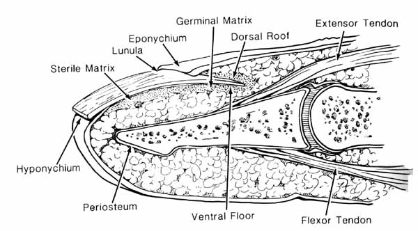
To review the structure of the nail, the nail plate consists of the hard visible part of the nail. The nail plate is composed of hard, keratinized, squamous cells that are loosely attached to the germinal matrix but strongly attached to the sterile matrix. The nail matrix is the tissue that a nail [nail plate] protects. It lies beneath the nail and contains nerves, lymph and blood vessels. The matrix is responsible for producing cells that become the nail plate. It has two parts: the sterile matrix and the germinal matrix.

anatomy_nail

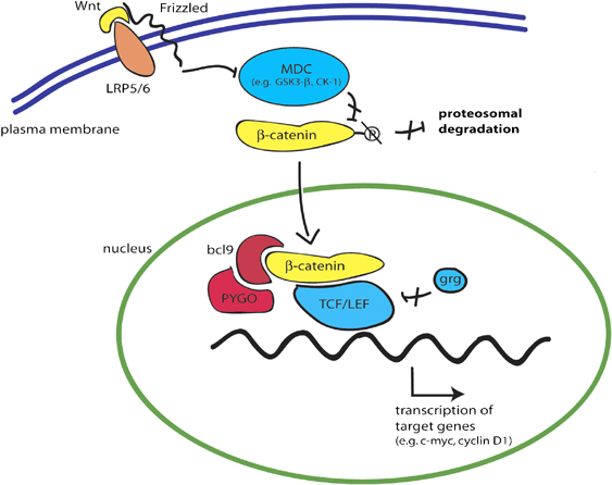
The stem cell population lies within the nail matrix, and these stem cells depend on a family of signaling proteins known as “Wnt” proteins. Wnt proteins are secreted glycoproteins that bind to Frizzled receptors. The Frizzled receptors bind Wnts and cause the polymerization of the Dsh or Disheveled protein at the cell membrane, and this inhibits GSK-3, a protein kinase. GSK-3 places phosphate groups on beta-catenin, and this marks beta-catenin for destruction. Once GSK-3 is inhibited, beta-catenin levels increase and it moves into the nucleus where it combines with Tcf proteins to activate the transcription of target genes.

Wnt signaling pathway

Wnt proteins play a crucial role in hair and tissue regeneration, and now they appear to play a truly vital role in bone regeneration as well.

Ito recounted her experiments: “When we blocked the Wnt-signaling pathway in mice with amputate fingertips, the nail and bone did not grow back as they normally would.”

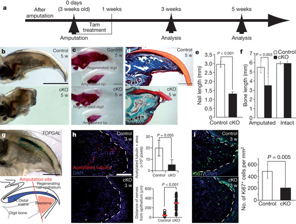
a, Experimental scheme. Three-week-old K14–Cre-ER;β-catenin conditional knockout (cKO) mice and littermates were treated with Tam for 7 days immediately after distal-tip amputation, and analysed at the indicated time points. b, Whole-mount transparent specimen of a regenerated digit 5 weeks after amputation. c, Whole-mount alizarin red analysis. d, Trichrome staining. e, f, Quantification analyses of the nail length and the bone length 5 weeks after amputation. g, Analysis of Wnt activation in regenerating nail epithelium using TOPGAL at 3 weeks after amputation. The lower panel is a schematic illustration of the upper panel. h, Quantitative analyses of the distance between nerve tip and wound epidermis and the innervations at 3 weeks after amputation. i, Proliferation analyses by Ki67 immunohystochemistry at 3 weeks after amputation. Red bars in h, right panel, indicate the averages. Dashed lines indicate the border between nail epithelium and connective tissue. Asterisks in part h, bottom panel, indicate autofluorescence from blood cells. Data are presented as the mean ± s.d. Scale bars, 500 μm (b–d); and 100 μm (h).

When Ito and her team manipulated the Wnt pathway they discovered that they could stimulate regeneration of bone and tissue just beyond the fingernail. “Amputations of this magnitude ordinarily do not grow back,” noted Ito.

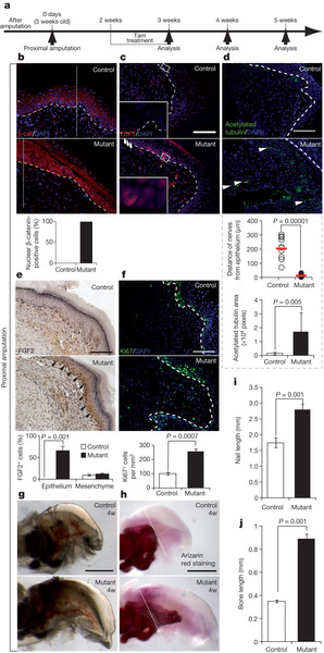
a, Experimental scheme. Three-week-old K14–Cre-ER;β-cateninfl/ex3 (mutant) mice and littermate controls were treated with Tam for 7 days starting from 2 weeks after amputation at the proximal level. b–f, Immunohistochemical analyses with indicated markers 3 weeks after amputation. g, Whole-mount transparent specimen of regenerated digits. h, Whole-mount alizarin red analysis. i, j, Quantification analyses of the nail (i) and bone length (j) 4 weeks after amputation. Red bars in d show the averages. Arrowheads in c and e, bottom panels, indicate TCF1− proximal matrix and FGF2+ epidermis, respectively. Arrowheads in d point to nerves. Fine dotted lines in b and h indicate the amputation plane. Dashed lines indicate the border between epidermis and connective tissue. Quantified data are presented as the mean ± s.d. Scale bars, 100 μm (b–f); and 500 μm (g and h).

These findings suggest that Wnt signaling is essential for fingertip regeneration, and indicate that the way to develop therapies for regenerating lost limbs is to more deeply understand Wnt signaling and its role in limb regeneration. Some 1.7 million people in the US alone live with amputations. Therefore, research of this type could prove remarkably useful.

Recovery of the Brain After a Stroke


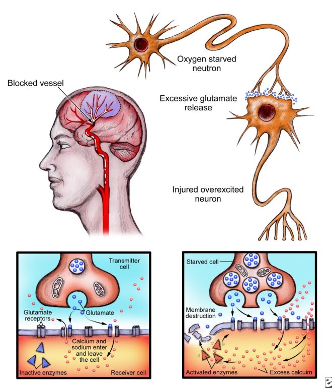
A stroke results when the brain suffers from “ischemia” or a lack of blood flow for an extended period of time. Blockage in the small vessels that feed blood to the brain can cause a trans-ischemic attack (TIA) or stroke. The lack of oxygen causes localized death of brain cells. The dying cells dump a whole gaggle of molecules into the spaces surrounding nearby brain cells, and these cell-derived molecules can actually poison surrounding cells, thus increasing the area that dies as a result of a stroke.

Stroke pathology

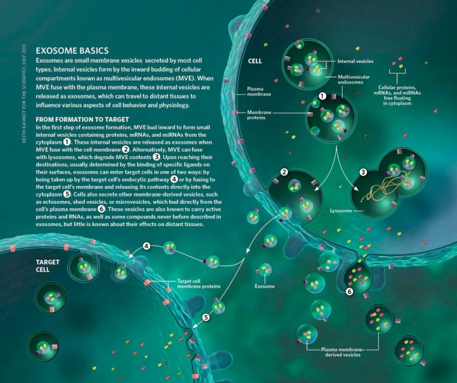
New work from by Henry Ford Hospital researchers in Detroit, Michigan suggests that some of the molecules released by brain cells during a stroke might actually help the brain heal after a stroke. Small RNA molecules or microRNAs that are packaged into lipid-bound vesicles in cells known as exosomes are released by stem cells after a stroke and seem to contribute to neurological recovery.

Exosomes are secreted vesicles that were first discovered nearly 30 years ago. They were, at first, considered little more than garbage cans whose job was to discard unwanted cellular components. However, once cell biologists began to study these little structures, evidence began to accumulate that these dumpsters also act as messengers that convey information to distant tissues. Exosomes contain cell-specific payloads of proteins, lipids, and genetic material that are transported to other cells, where they alter function and physiology.

Exosome_Basics

Therefore, it is little wonder that exosomes can also transport microRNAs. In this present study from the laboratory of Michael Chopp, rats were given experimentally induced strokes, and then the neurological recovery of the rats was examined at the molecular level.

Chopp and his colleagues first isolated mesenchymal stem cells (MSCs) from the bone marrow of their laboratory rats. Then they genetically engineered these MSCs to release exosomes laden with specific microRNAs; in particular miR-133b.

MicroRNAs are a class of post-transcriptional regulators. Since they are usually only about 22 base pairs in length, they are far too short to encode anything. microRNAs usually bind to complementary sequences in the 3’ untranslated region of messenger RNAs, and this binding silences the RNA, which simply means that the RNA cannot be recognized by ribosomes and will not be translated into protein, or that the RNA is degraded by special enzymes that target RNAs bound by microRNAs. Single microRNAs target hundreds genes at a time, and some 60% of all genes are regulated by microRNAs. MicroRNAs are abundantly present in all human cells. They are also highly conserved in organisms ranging from the unicellular algae Chlamydomonas reinhardtii to mitochondria in vertebrates, which suggest that they are a vital part of genetic regulation throughout the plant and animal kingdoms.

The Actions of Small Silencing RNAs (A) Messenger RNA cleavage specified by a miRNA or siRNA. Black arrowhead indicates site of cleavage. (B) Translational repression specified by miRNAs or siRNAs. (C) Transcriptional silencing, thought to be specified by heterochromatic siRNAs.
The Actions of Small Silencing RNAs
(A) Messenger RNA cleavage specified by a miRNA or siRNA. Black arrowhead indicates site of cleavage.
(B) Translational repression specified by miRNAs or siRNAs.
(C) Transcriptional silencing, thought to be specified by heterochromatic siRNAs.

The microRNA known as miR-133b has been shown to enhance the death of prostate cancer cells when they are delivered to them (see Patron JP, Fendler A, Bild M, Jung U, Müller H, et al. (2012) MiR-133b Targets Antiapoptotic Genes and Enhances Death Receptor-Induced Apoptosis. PLoS ONE 7(4): e35345. doi:10.1371/journal.pone.0035345). However, because different cell types show different responses to the same reagents, exposing brain cells to this microRNA after a stroke might elicit a very different response.

By raising or lowering the amount of miR-133b in MSCs, Chopp and his colleagues were able to determine the effects of miR-133b on brains cells after a stroke. Chopp and others injected their genetically engineered MSCs into the bloodstream of rats 24 hours after inducing a stroke in these animals. When the exosomes of the MSCs were enriched in miR-133b, the neurological recovery in the rats was amplified, but when injected MSCs were deprived of miR-133b, the neurological recovery was substantially less.

To measure neurological recovery, researchers separated the disabled rats into several groups and injected each groups with saline, nongenetically-engineered MSCs, MSCs with low levels of miR-133b, and MSCs with high levels of miR-133b. The rats were given behavioral tests 3, 7, and 14 days after treatment. These tests measured the gait of the animals on a grid to determine if the rats could walk on an unevenly spaced grid (foot-fault test). The second test determined how long it took the rats to remove a piece of adhesive tape that was stuck to their front paws.

in every test, the rats injected with miR-133b-enriched MSCs showed superior levels of neurological recovery. Autopsies of these same animals revealed that the rats treated with miR-133b-enriched MSCs had enhanced rewiring of the brain and axonal outgrowth. In the areas of the brain adversely affected by the stroke, the rats showed increased axonal plasticity and neurite remodeling.

Most stroke victims recover some ability to use their hands and other body parts on a voluntary basis, but almost half of all stroke victims are left with some weakness on one side of their body and many are permanently disabled by the stroke.

No treatment presently exists for improving or restoring this lost motor function in stroke patients, mainly because of mysteries about how the brain and nerves repair themselves.

Chopp said, “This study may have solved one of these mysteries by showing how certain stem cells play a role in the brain’s ability to heal itself to differing degrees after stroke or other trauma. Chopp also serves as the scientific director of the Henry Ford Neuroscience Institute.

Stem Cell-Promoting Gene Also Promotes the Growth of Head and Neck Cancer


Nanog is a very funny name for a gene, but the Nanog gene is an essential part of the cellular machinery that keeps embryonic stem cells from differentiating and maintains them in a pluripotent state. Unfortunately, Nanog also has other roles if it is mis-expressed and that includes in the genesis of cancers of the head and neck.

Nanog function during development

This study emerged from work done by researchers at the Ohio State University Comprehensive Cancer Center – Arthur G. James Cancer Hospital, and Richard J. Solove Research Institute or OSUCCC-James. Since Nanog has been studied in some depth, understanding Nanog activity might provide vital clues in the design of targeted drugs and reagents for treating particular cancers.

“This study defines a signaling axis that is essential for head and neck cancer progression, and our findings show that this axis may be disrupted at three key steps,” said Quintin Pan, associate professor of otolaryngology at OSUCCC-James and principal investigator in this research effort. “Targeted drugs that are designed to inhibit any or all of these three steps might greatly improve the treatment of head and neck cancer.”

What kind of signaling axis is Dr. Pan referring to? An enzyme called protein kinase C-epsilon or PKC-epsilon can place phosphate groups on the Nanog protein. Phosphate groups are negatively charged and are also quite bulky. Attaching such chemical groups to a protein can effectively change its structure and function. In the case of Nanog, phosphorylation of stabilizes it and activates it.

Phosphorylated Nanog proteins can bind together to form a dimer, which attracts a third protein to it; p300. This third protein, p300, in combination with the paired Nanog proteins acts as a potent activator of gene expression of particular genes, in particular a gene called Bmi1. When expressed at high levels, Bmi1 stimulates the proliferation of cells in an uncontrolled fashion.

Bmi1 - Nanog interaction

“Our work shows that the PKCepsilon/Nanog/Bmi1 signaling axis is essential to promote head and neck cancer,” Pan said. “And it provides initial evidence that the development of inhibitors that block critical points in this axis might yield a potent collection of targeted anti-cancer therapeutics that could be valuable for the treatment of head and neck cancer.”

Stem Cell Gene Provides Target for Cancer Treatment


A gene called SALL4 encodes a zinc finger transcription factor protein that helps stem cells maintain their undifferentiated state and continue dividing. Cells tend to only express SALL4 during embryonic development, but in almost all cases of acute myeloid leukemia, and in 10-30% of liver, gastric, ovarian, endometrial, and breast cancers, SALL4 is re-expressed. This is solid evidence that SALL4 plays a central role in tumor formation.

Harvard Stem Cell Institute (HSCI)-affiliated labs in Singapore and Boston have shown that knocking out the SALL4 gene in mouse tumors leads to a cessation of tumor growth. Additionally, designing small molecules that inhibit SALL4 activity also treat the cancer and cause cessation of tumor growth and shrinkage of the tumor.

“Our paper is about liver cancer, but it is likely true about lung cancer, breast cancer, ovarian cancer, many, many cancers,” said HSCI Blood Diseases Program leader Daniel Tenen, who also directs a laboratory at the Cancer Science Institute of Singapore (CSI Singapore). “SALL4 is a marker, so if we had a small molecule drug blocking SALL4 function, we could also predict which patients would be responsive.”

Studying the therapeutic potential of a transcription factor is unusual in the field of cancer research. Transcription factors are typically avoided because of the difficulty of developing drugs that safely interfere with genetic targets. Most cancer researchers focus their attention on kinases (enzymes that attach phosphate groups to other molecules).

However, inquiry into the basic biology of the SALL4 gene by HSCI researchers has shown that there is another way to interfere with its activity in cancer cells. The SALL4 protein turns off a tumor suppressor gene, and this causes the cell to divide uncontrollably. By targeting the SALL4 protein with synthetic molecules that inhibit its activity, they could halt the growth of the tumors.

“The pharmaceutical companies decided that if it is not a kinase, and it is not a cell surface molecule, then it is ‘undruggable,'” said Tenen. “To me, if you say anything in ‘undoable,’ you are limiting yourself as a biomedical scientist.”

Earlier this year, Tenen’s co-author, HSCI-affiliated faculty member Li Chau, assistant professor of pathology at Harvard Medical School and Brigham and Woman’s Hospital, published a report that synthetic SALL4 inhibitors have treatment potential in leukemia cells.

Chai took blood samples from patients with acute myeloid leukemia, and treated the leukemia cells with this synthetic inhibitor and then transplanted that blood back into the leukemic mice. The cancer showed gradual regression.

“I am excited about being on the front line of this new drug development,” said Chai. “As a physician-scientist, if I can find a new class of drug that has very low toxicity to normal tissues, my patients can have a better quality of life.”

Chai and Tenen are working with HSCI Executive Committee member Lee Rubin from the Harvard Institute of Chemistry and Biology, and James Bradner from the Dana Farber Cancer Institute (another HSCI-affiliated faculty member), to help them with the drug development part of their project. Demonstrating the potential of SALL4-interfering compounds is labor intensive, but might also be efficacious for the treatment of other cancers.

“I think as academics, we seek to engage drug companies because they can do these types of things better than we can,” said Tenen. “But, also as an academic, I want to go after the important biologic targets that are not being sought after by the typical drug company – because if we do not, who will?”

How Neural Stem Cells Create New and Varied Neurons


A new study in fruit flies has elucidated a mechanism in neural stem cells by which these types of stem cells generate the wide range of neurons that they form.

Chris Doe, a professor of biology from the Institute of Neuroscience at the University of Oregon, and his co-authors have used the common fruit fly Drosophila melanogaster to investigate the cellular mechanism by which neural stem cells make their distinctive progeny.

As Doe put it, “The question we confronted was ‘How does a single kind of stem cell, like a neural stem cell, make all kinds of neurons?”

Researchers have known for some period of time that stem cells have the capacity to produce new cells, but the study by Doe’s group shows how a select group of stem cells can create progenitor cells that can generate numerous subtypes of cells.

Doe’s study builds on previous studies in which Doe and his colleagues identified the specific set of stem cells that generated neural precursors. These so-called “intermediate neural progenitors” or INPs can expand to form several different new cell types. However, this study did not account for the diversity of the cells generated even if it did account for the number of cells generated (see Boone JQ, Doe CQ, Dev Neurobiol. 2008 Aug;68(9):1185-95).

“While it’s been known that individual neural stem cells or progenitors could change over time to make different types of neurons and other types of cells in the nervous system, the full extent of this temporal patterning had not been described for large neural stem cell lineages, which contain several different kinds of neural progenitors,” according to this study’s first author Omar Bayraktar.

The cell types discovered in this study have analogs in the developing human brain and the research has potential applications for human biologists who want to know how neurons form in the human brain.

The paper from Doe’s lab was published along another study on the generation of diverse neurons by a group from New York University. These two papers provide new insight into the means by which neural stem cells generate the wide range of neurons found in the brains of fruit flies and humans.

In their study, Bayraktar and Doe specifically examined stem cells in fruit fly brains known as type II neuroblasts, which generate INPs. However, in this study, the type II neuroblasts were shown to generate INPs, which then go on to form distinct neural subtypes. Even though previous work showed that INPs went on to form about 100 new neurons, in this paper, the INPs were shown to make about 400-500 new neurons.

Another interesting finding was that the gene expression patterns of INPs, which began with three different transcription factors (Dichaete, Grainy Head, and Eyeless). These transcription factors lay the groundwork for INP differentiation, but once INP formation occurs, a new transcriptional program is extended that extends the types of neurons that INPs can form. Such nested transcriptional programs are also common during the specification of neural stem cell progeny in humans brains, with many of the same transcription factors playing a central role in neuron specification.

“If human biologists understand how the different types of neurons are made, if we can tell them ‘This is the pathway by which x, y, and Z neurons are made,’ then they may be able to reprogram and redirect stem cells to make these precise neurons,” Doe said.

However, the mechanism described in this paper has its limits. Eventually the process of generation new cells stops. One of the next questions to answer will be what makes the mechanism turn off, according to Doe.

“This vital research will no doubt capture the attention of human biologists,” said Kimberly Andrews Espy, who is vice-president for research and innovation and the dean of the UP graduate school. “Researchers at the University of Oregon continue to further our understanding of the processes that undergird development to improve the health and well-being of people throughout the world.”

See Bayraktar OA, Doe CQ. Combinatorial temporal patterning in progenitors expands neural diversity. Nature. 2013 Jun 27;498(7455):449-55. doi: 10.1038/nature12266.

Exporting Tissue Engineering


Professor György K.B. Sándor from the Finland Distinguished Professor Program or FiDiPro believes that tissue engineering has the ability to become a new global export item.

FiDiPro is a joint funding program of the Academy of Finland and Tekes (the Finnish Funding Agency for Technology and Innovation) that enables top researchers to do work in Finland for a fixed period of time.

Sándor is a Canadian professor who specializes in oral and maxillofacial surgery who has participated in FiDIPro. Sandor’s research program examines bone regeneration, hyperbaric oxygen, tissue regeneration, and stem cells. He works at the BioMediTech research institute which is run by the University of Tampere and the Tampere University of Technology. BioMediTech is an innovation center that combines biomedical research with new technologies.

The goal of Sándor’s research program is to produce bone and cartilage by means of tissue engineering techniques that grow tissue-derived stem cells. Some people are missing bone at birth as a result of a developmental disorder, or, in some cases, bone defects from accidents, and various inflammatory diseases can cause bone loss. Particular surgeries that require bone removal can also cause bone loss,

Tissue engineering can produce tailored, living spare parts for human beings. If the protocols and methods of tissue engineering can be up-scaled appropriately, it could become the third alternative form of treatment alongside more traditional forms of treatment for such conditions that include surgery and drug treatments.

“Tissue-derived stem cells can be isolated from the patient’s own tissue. In that way, they will not cause a rejection reaction. Compared to tissue stem cells, human embryonic stem cells have a greater ability to differentiate into different cell types, In practice, that means that all cell types can be used,” Sándor said.

Sandor noted that Finland is a forerunner in developing bone engineering techniques. “At the moment expertise in the field is concentrated in Finland, but it also generated global interest in other medically advanced countries,” said Sándor .

In the near future, large numbers of patients might travel to Finland to receive tissue engineering-based treatments. As such forms of treatment increase and are perfected, expertise in tissue engineering can be exported for use on a larger scale.

“We have proven with more than 20 clinically successful operations that tissue engineering works,” Sándor said.

Sándor considers the research community in Tampere to be unique when it comes to the way it is run and functions. One of the key reasons why Sandor decided to stay and continue his research in Finland even after his experience with FiDiPro came to an end.

“In the field, BioMediTech is a unique concentration of researchers and expertise. In the Pirkanmaa region, also the cooperation between research, industry, and administration works well. That enables efficient decision-making which, in turn, contributes to the creation of new innovations,” he said.

“Cooperation with colleagues is smooth too. That was the determining factor in my decision to stay in Finland. Each day is like a new adventure.”