Misrepresentation of the Embryological Facts of Cloning by Reporters


Wesley Smith at National Review Online has been keeping tabs on the reporting of the Cell paper by Shoukhrat Miltalipov from the Oregon Health and Science University. The misrepresentation has been extensive but it’s not really all that surprising given the ignorance and lack of clear thinking on this issue. Nevertheless, Smith has kept up his yeoman’s work, cataloging the factual errors for reporters in multiple publications.

For his first example, see here, where Loren Grush on Fox News.com wrote:

Through a common laboratory method known as somatic cell nuclear transfer (SCNT), ONPRC scientists, along with researchers at Oregon Health & Science University, essentially swapped the genetic codes of an unfertilized egg and a human skin cell to create their new embryonic stem cells…The combination of the egg’s cytoplasm and the skin cell’s nucleus eventually grows and develops into the embryonic stem cell.

Grush, as Smith points out, is quite wrong. Introducing a nucleus from a body cell into the unfertilized egg and inducing it does not turn the egg into embryonic stem cells, but turns it into a zygote. The zygote them undergoes cleavage (cell division) until it reaches the early/mid blastocyst stage 5-6 days later, then immunosurgery is used to isolated the inner cell mass cells, after which they are cultured. Somatic cell nuclear transfer is a stand-in for fertilization. It produces an embryo and all the redefinition in the world will not change that.

Next comes my favorite newspaper, the Wall Street Journal, which normally has decent to pretty good scientific reporting, but this one story from Gautam Naik contains a real howler:

Scientists have used cloning technology to transform human skin cells into embryonic stem cells, an experiment that may revive the controversy over human cloning. The researchers stopped well short of creating a human clone. But they showed, for the first time, that it is possible to create cloned embryonic stem cells that are genetically identical to the person from whom they are derived.

As Smith points out, Miltalipov and others did not stop short of creating a human clone, then explicitly made a cloned human embryo and therefore made a cloned young human being.

Then there is this humdinger from an online Australian news report:

US researchers have reported a breakthrough in stem cell research, describing how they have turned human skin cells into embryonic stem cells for the first time. The method described on Wednesday by Oregon State University scientists in the journal Cell, would not likely be able to create human clones, said Shoukhrat Mitalipov, senior scientist at the Oregon National Primate Research Center. But it is an important step in research because it doesn’t require the use of embryos in creating the type of stem cell capable of transforming into any other type of cell in the body.

Oh my gosh, folks the paper describes the production of cloned embryos expressly for the purpose of dismembering them and destroying them. This “doesn’t require the use of embryos” crap reveals a very basic ignorance of how the experiment was done. See Smith’s excellent post for more details.

Then there is this story from one of my least favorite papers, the LA Times:

Some critics continue to argue that it’s unethical to manipulate the genetic makeup of human eggs even if they’re unfertilized, and others warn about potential harm to egg donors. The biggest ethical issue for the OHSU team, though, is that it artificially created a human embryo, albeit one that was missing the components needed for implantation and development as a fetus.

Come on people! The cloned embryo does not have the components needed to implant because there is no womb into which it can be implanted. Dolly was made the same way. Surely Dolly had the components required to implant.  The problem here is one of will, since these embryos were made to be destroyed. Not capacity. What was done to those embryos was dismemberment. Would we object if they were toddlers?

Just to show that obfuscation is not wholly an American news feature, there is this story from the German newspaper Deutche Welle:

Scientists, for the first time, have cloned embryonic stem cells using reprogrammed adult skin cells, without using human embryos…The process used by Mitalipov is an important step in research because it does not require killing a human embryo–that is, a potential human being–to create transformative stem cells.

As Smith points out, this research made a human embryo that was then killed to make embryonic stem cells. Calling this research humane is to redefine humane to the point of absurdity.

Finally this jewel of blithering ignorance from bioethicist Jonathan Moreno in his column in the Huffington Post:

Despite some confused media reports, the Oregon scientists did not clone a human embryo but a blastocyst that lacks some of the cells needed to implant in a uterus.

And you wonder why people like me have lost all faith in American bioethics. As a developmental biologist, this one just grates on me.  A blastocyst has two cell populations; an outer trophectoderm composed of trophoblast cells that will form the placenta and the inner cell mass cells on the inside of the embryo, which will form the embryo proper and a few placental structures. To be a blastocyst is to have the equipment to implant.

To drive the nail into the coffin, Smith quotes the father of embryonic stem cells James Thomson from an MSNBC interview:

See, you are trying to redefine it away…If you create an embryo by nuclear transfer, if you gave it to somebody who didn’t know where it came from, there would be no test you could do on that embryo to say where it came from. It is what it is. By any reasonable definition, at least as some frequency, you are creating an embryo. If you try to redefine it away, you are being disingenuous.

Check out Smith’s posts. They are all worth reading. Maybe the press will learn some embryology, but I doubt it.

Postscript:  Brendan P. Foht writes at the Corner on National Review Online that in 2010 Shoukhrat Mitalipov, the leader of the Oregon cloning team, reported that he had achieved a single pregnancy using cloned monkey embryos that were made with exactly the same technology as was employed with human eggs in his 2013 Cell paper.  The fetus developed long enough to have a heartbeat detectable through ultrasound. Although the pregnancy failed after 81 days (about half the normal gestation period for that species), the fact that a pregnancy would develop so far indicates that reproductive cloning of primates is in principle possible.  This definitively shows that all this talk about the embryos made in Mitalipov’s lab not being able to implant is pure drek.

Mesenchymal Stem Cells Engineered to Express Tissue Kallikrein Increase Recovery After a Heart Attack


Julie Chao is from the Department of Biochemistry and Molecular Biology, at the Medical University of South Carolina. Dr. Chao and her colleagues have published a paper in Circulation Journal about genetically modified mesenchymal stem cells and their ability to help heal a heart that has just experienced a heart attack.

Several laboratories have used mesenchymal stem cells (MSCs), particularly from bone marrow, to treat the hearts of laboratory animals that have recently experienced a heart attack. However, heart muscle after a heart attack is a very hostile place, and implanted MSCs tend to pack up and die soon after injection. Therefore, such injected cells do little good.

To fix this problem, researchers have tried preconditioning cells by growing them in a harsh environment or by genetically engineering them with genes that can increase their tolerance of harsh environments. Both procedures have worked rather well. In this paper, Chao and her group engineered bone marrow-derived MSCs to express the genes that encode “tissue kallikrein” (TK). TK circulates throughout our bloodstream but several different types of cells also secrete it. It is an enzyme that degrades the protein “kininogen” into small bits that have several benefits. Earlier studies from Chao’s own laboratory showed that genetically engineering TK into the heart improved heart function after a heart attack and increased the ability of MSCs to withstand harsh conditions (see Agata J, Chao L, Chao J. Hypertension 2002; 40: 653 – 659; Yin H, Chao L, Chao J. Journal of  Biol Chem 2005; 280: 8022 – 8030). Therefore, Chao reasoned that using MSCs engineered to express TK might also increase the ability of MSCs to survive in the post-heart attack heart and heal the damaged heart.

In this paper, Chao and others made adenoviruses that expressed the TK gene. Adenoviruses place genes inside cells, but they do not integrate those genes into the genome of the host cell. Therefore, they are safer to use than retroviruses. Chao and others used these TK-expressing adenoviruses to infect tissue and MSCs.

When TK-expressing MSCs were exposed to low-oxygen conditions, like what cells might experience in a post-heart attack heart, the TK-expressing cells were much heartier than their non-TK-expressing counterparts. When injected into rat hearts 20 minutes after a heart attack had been induced, the TK-expressing MSCs showed good survival and robust TK expression. Control hearts that had been injected with non-TK-expression MSCs or had not been given a heart attack showed no such elevation of TK expression.

There were also added bonuses to TK-expressing MSC injections. The amount of inflammation in the hearts was significantly less in the hearts injected with TK-expressing MSC injections compared to the controls. There were fewer immune cells in the heart 1 day after the heart attack and the genes normally expressed in a heart that is experiencing massive inflammation were expressed at lower levels relative to controls, if they were expressed at all.

Reduced inflammation by TK-MSC administration was determined by (C) ED-1 immunohistochemical staining, (D) monocyte/macrophage quantification, (E) neutrophil quantification, and gene expression of (F) TNF-α, (G) ICAM-1, and (H) MCP-1. ED-1-positive cells are indicated by arrows. Original magnification, ×200. Data are mean ± SEM (n=5–8). *P<0.05 vs. other MI groups; **P<0.05 vs. MI/Control group. MSC, mesenchymal stem cell.
Reduced inflammation by TK-MSC administration was determined by (C) ED-1 immunohistochemical staining, (D) monocyte/macrophage quantification, (E)
neutrophil quantification, and gene expression of (F) TNF-α, (G) ICAM-1, and (H) MCP-1. ED-1-positive cells are indicated by arrows.
Original magnification, ×200. Data are mean ± SEM (n=5–8). *P

Another major bonus to the injection of TK-expressing MSCs into the hearts of rats was that these cells protected the heart muscle cells from programmed cell death. To make sure that this was not some kind of weird artifact, Chao and her team placed the TK-expressing MSCs in culture with heart muscle cells and then exposed them to low-oxygen tension conditions. Sure enough, the heart muscle cells co-cultured with the TK-expressing MSCs survived better than those co-cultured with non-TK-expressing MSCs.

TK-MSCs protect against cardiac cell apoptosis at 1 day after myocardial infarction (MI) and in vitro. TK-MSC administration reduced apoptosis in the infarct area at 1 day after MI, as determined by (A) TUNEL staining, (B) quantification of apoptotic cells, and (C) caspase-3 activity. Original magnification, ×200. Data are mean ± SEM (n=5–8). *P<0.05 vs. other MI groups. Cultured cardiomyocytes treated with 0.5 ml of TK-MSC-conditioned medium exhibit higher tolerance to hypoxia-induced apoptosis, as evidenced by (D) Hoechst staining,
TK-MSCs protect against cardiac cell apoptosis at 1 day after myocardial infarction (MI) and in vitro. TK-MSC administration
reduced apoptosis in the infarct area at 1 day after MI, as determined by (A) TUNEL staining, (B) quantification of apoptotic
cells, and (C) caspase-3 activity. Original magnification, ×200. Data are mean ± SEM (n=5–8). *Pcardiomyocytes treated with 0.5 ml of TK-MSC-conditioned medium exhibit higher tolerance to hypoxia-induced apoptosis, as
evidenced by (D) Hoechst staining,

Finally, when the hearts of the rats were examined 2 weeks after the heart attack, it was clear that the enlargement of the heart muscle (so-called “remodeling”) occurred in animals that had received non-TK-expressing MSCs or had received no MSCs at all, but did not occur in the hearts of rats that had received injections of TK-expressing MSCs. The heart scar was also significantly smaller in the hearts of rats that had received injections of TK-expressing MSCs, and had a greater concentration of new blood vessels. Apparently, the TK-expressing MSCs induced the growth of new blood vessels by recruiting EPCs to the heart to form new blood vessels.

In conclusion, the authors write that “MSCs genetically-modified with human TK are a potential therapeutic for ischemic heart diseases.”

Getting FDA approval for genetically engineered stem cells will not be easy, but TK engineering seems much safer than some of the other modifications that have been used. Also the vascular and cardiac benefits of this gene seem clear in this rodent model. Pre-clinical trials with larger animals whose cardiac physiology is more similar to humans is definitely warranted and should be done before any talk of human clinical trials ensues.

Adult Mammals Lack the Stem Cell Activity to Make New Eggs


Recent research in mice and humans have discovered a stem cell population in ovaries that can form eggs. However, this discovery begs a question: namely, why do adult female mammals run out of eggs in their lifetime if they have a stem cell population that can produce eggs?

New research from the Carnegie Institute for Science demonstrates that adult mice do not use stem to produce new eggs, thus answering this apparent conundrum.

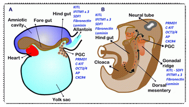
Before birth, mouse and human ovaries contain an abundant supply of germ cells that originate from primordial germ cells that form from the inner layer of the primary umbilical vesicle (otherwise known as the yolk sac).  Between the time when the embryo is four to six weeks old, the primordial germ cells (PGCs) migrate from the wall of the primary umbilical vesicle to the gut tube.  From the gut tube, the PGCs migrate to the dorsal body wall by means of the mesentery that suspends the gut from the body wall.  Once in the body wall, the PGCs come to rest on either side of the midline in the loose mesenchymal tissue just inside the membranous lining of the body cavity (known more technically as the coelomic cavity).

PGC Migration Pathway2

Most of the PGCs populate the region of the body wall at the level that will form the gonads.  During their migration, PGCs continue to multiply by means of mitosis, which increase their numbers substantially.  Some PGCs may become stranded during their migration, coming to rest at extragonadal sites.  Occasionally, stray germ cells of this type may give rise to a type of tumor called a teratoma.

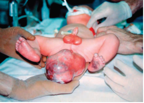
Teratoma
Teratoma

Once in their final location, the PGCs will stimulate the formation of the genital or gonadal ridge.

In females, PGCs (which are now called gonocytes) undergo a few more mitotic divisions after they are surrounded by the somatic support cells and become intimately associated with them.  The gonocytes differentiate into oogonia, and by the 5th month of fetal development all oogonia initiate meiosis.  After they initiate meiosis, the oogonia are called primary oocytes.  However, during an early phase of meiosis all sex cells enter a state of dormancy, and they remain in meiotic arrest as primary oocytes until sexual maturity.  Beginning at puberty, each month a few ovarian follicles resume development in response to the monthly surge of pituitary gonadotropic  hormones, but usually only one primary oocyte matures into a secondary oocyte and is ovulated. This oocyte enters a second phase of meiotic arrest and does not actually complete meiosis unless it is fertilized. These monthly cycles continue until the onset of menopause at approximately 50 years of age.

Near the time of birth, the ovaries of mice and humans contain an abundant supply of eggs that will be released from follicles during ovulation each menstrual cycle.  At the birth of the baby, she will possess a large reserve of primordial follicles that contain a single egg surrounded by supporting follicle cells.  Evidence of new follicle production is absent after birth.  Therefore, it has long been thought that the supply of follicles is fixed at birth and eventually is exhausted at menopause.

During the last decade, researchers have found primordial follicles in adult mouse ovaries that turn over and claimed that adult germ-line stem cells constantly resupply the follicle pool and sustain ovulation.  These claims were based on observations of ovarian tissue and one the behavior of extremely rare ovarian cells after these cells were cultured for some time in the laboratory.  Such criteria are subjective, especially in light of the fact that culturing cells for long periods of time in the laboratory can effectively reprogram them.

At Carnegie, Lei Lei and Allan Spradling used a technique that tracks individual cells and their progeny within living tissue over a specific time course.  The cells are marked with a gene, and this gene is inherited by the progeny of that cell, thus allowing the careful tracking of all the progeny of that cell or those cells.  This technique is called “lineage tracking” and it is a very popular technique in developmental and cell biology.

By subjecting primordial follicles to lineage tracking, Lei and Spradling showed that germ-line stem cell activity cannot be detected in mice.  Furthermore, primordial follicles are stable, and even if half the existing follicles die off, no germ-line stem cell activity is detectable.  This research does not prove that there are no germ-line stem cell divisions within the ovary of the mouse, but it does place an upper limit on the divisions of the germ-line stem cell population of one division every two weeks at the most, which is biologically insignificant.

What then can be said about the germ-line stem cell cultures isolated in the laboratory?  According to Alan Spradling, the cells “likely arise by dedifferentiation in culture,” and “the same safety and reliability concerns would apply as to any laboratory-generated cell type that lacks a normal counterpart” in the body.

This should be a warning to those conclusions that are solely derived from experiments conducted in culture alone and not in a living creature as well.

Neurons Made from the Skin Cells of Down Syndrome Patients Show Reduced Connectivity


The most common form of intellectual disability in the United States is caused by Down syndrome (DS). DS results when babies are born with an extra copy of an extra piece of chromosome 21. Individuals with DS show various types of intellectual deficits and other health problems as well, such as heart problems, poor muscle tone, an under-active thyroid, respiratory infections, hearing problems, celiac disease, eye conditions, depression or behavior problems associated with attention-deficit hyperactivity disorder or autism.

Even though Down syndrome patients have symptoms and health problems that are well described, how the extra chromosome causes such widespread effects is still largely mysterious.

In recently published research, Anita Bhattacharyya, who is a neuroscientist at the Waisman Center at the University of Wisconsin-Madison, reported that brain cells that were grown from skin cells taken from individuals with Down syndrome.

“Even though Down syndrome is very common, it’s surprising how little we know about what goes wrong in the brain,” says Bhattacharyya. “These new cells provide a way to look at early brain development.”

The skin cells taken from DS patients were grown in culture and genetically engineered to so that a fraction of them were transformed into induced pluripotent stem cells (iPSCs). Since iPSCs can be differentiated into any adult cell type, Bhattacharyya’s lab, working with collaboration with Su-Chun Zhang and Jason Weick, grew those iPSCs in culture and differentiated them into dorsal forebrain neurons, which they could test in the laboratory.

Neurophysiological tests of the DS neurons revealed that these neurons formed a reduced number of connections between them each other. Bhattacharyya says. “They communicate less, are quieter. This is new, but it fits with what little we know about the Down syndrome brain.” Brain cells communicate through connections called synapses, and the Down neurons had only about 60 percent of the usual number of synapses and synaptic activity. “This is enough to make a difference,” says Bhattacharyya. “Even if they recovered these synapses later on, you have missed this critical window of time during early development.”

Bhattacharyya and colleagues also examined the genes that were affected in the Down syndrome stem cells and neurons. They discovered that those genes on the extra chromosome were increased 150 percent, which is consistent with the contribution of the extra chromosome.

However, the output of about 1,500 genes elsewhere in the genome was strongly affected. “It’s not surprising to see changes, but the genes that changed were surprising,” says Bhattacharyya. The predominant increase was seen in genes that respond to oxidative stress, which occurs when molecules with unpaired electrons called free radicals damage a wide variety of tissues.

“We definitely found a high level of oxidative stress in the Down syndrome neurons,” says Bhattacharyya. “This has been suggested before from other studies, but we were pleased to find more evidence for that. We now have a system we can manipulate to study the effects of oxidative stress and possibly prevent them.”

DS includes a range of symptoms that might result from oxidative stress, Bhattacharyya says, including accelerated aging. “In their 40s, Down syndrome individuals age very quickly. They suddenly get gray hair; their skin wrinkles, there is rapid aging in many organs, and a quick appearance of Alzheimer’s disease. Many of these processes may be due to increased oxidative stress, but it remains to be directly tested.”

Oxidative stress could be especially significant, because it appears right from the start in the stem cells. “This suggests that these cells go through their whole life with oxidative stress,” Bhattacharyya adds, “and that might contribute to the death of neurons later on, or increase susceptibility to Alzheimer’s.”

Other researchers have created neurons with DS from induced pluripotent stem cells, Bhattacharyya notes. “However, we are the first to report this synaptic deficit, and to report the effects on genes on other chromosomes in neurons. We are also the first to use stem cells from the same person that either had or lacked the extra chromosome. This allowed us to look at the difference just caused by extra chromosome, not due to the genetic difference among people.”

The research, published the week of May 27 in the Proceedings of the National Academy of Sciences, was a basic exploration of the roots of Down syndrome. Bhattacharyya says that while she did not intend to explore treatments in this work, she did note that “we could potentially use these cells to test or intelligently design drugs to target symptoms of Down syndrome.”

Circulating Factor Rejuvenates the Hearts of Older Mice


Two researchers associated with the Harvard Stem Cell Institute, one of whom is a practicing cardiologist at Brigham and Women’s Hospital, and a cell biologist have identified a protein in the blood of mice and humans decreases during aging and may prove to be the first effective treatment for the form of age-related heart failure that affects millions of Americans.

Growth Differentiation Factor 11 or GDF11 circulates throughout the bloodstream of humans and mice. Injections of GDF11 into old mice that have developed thickened heart walls in a manner similar to aging humans, the hearts were reduced in size and thickness, and resembled the healthy hearts of younger mice.

Even more important than the implications for the treatment of diastolic heart failure, the finding by Richard T. Lee, from Harvard Medical School and Amy Wagers, who is a professor in Harvard’s Department of Stem Cell and Regenerative Medicine, might rewrite our understanding of aging. These findings were published in the prestigious, international journal Cell.

“The most common form of heart failure [in the elderly] is actually a form that’s not caused by heart attacks but is very much related to the heart aging,” said Lee, who, like Wagers, is a principal faculty member at HSCI.

“In this study, we were able to show that a protein that circulates in the blood is related to this aging process, and if we gave older mice this protein, we could reverse the heart aging in a very short period of time,” Lee said. “We are very excited about it because it opens a new window on the most common form of heart failure.”
He added, “This is the coolest thing I’ve ever been a part of.”

Doug Melton, HSCI co-director, called the discovery “huge. It’s going to change the way we think about aging.”

Lee, who practices medicine as a cardiologist, noted that he has approximately 300 patients, and of these “I think I have about 20 who are suffering from this type of heart failure, which we sometimes call diastolic heart failure,” said Lee. “They come into the hospital, have a lot of fluid taken off, then they’ll go home. Then they come back again. It’s really frustrating because we don’t have any drugs to treat this. We need to work as hard as we can to figure out if this discovery can be turned into a treatment for heart failure in our aging patients.”

The Lee and Wagers labs would like to move GDF-11 toward clinical trials. Lee predicts that they might be able to begin these studies in four to five years. However, they need to determine the other tissues that are affected by GDF11.

Wagers was a postdoctoral research fellow at Stanford University, where she learned how to work with the “parabiotic” mouse system. Parabiosis refers to two animals that share a common circulatory system. Wagers used this system to link the circulatory system of young mice with that of older mice. When she did that, she and her colleagues discovered that soluble factors in the blood of young animals have a rejuvenating effect on various tissues in older animals. In particular, the spinal cord and the musculature of the older animals showed marked improvements.

“As we age, there are many changes that occur in different parts of the body,” Wagers said, “and those changes are often associated with a decline in the function of our bodies. One of the interests of my laboratory is in understanding why this happens and whether it is an inevitable consequence of aging, or if it might be reversible.

“In this study, we compared young and old animals and identified a substance in the blood that is present at high levels when you’re young and lower levels when you’re old. We further found that when we supplemented the low levels of this substance that were present in old animals to the levels normally seen in youth, this could have a dramatic effect on the heart.

“It’s been observed for many, many years that when aging occurs it affects multiple body systems sort of in a semi-synchronous way,” Wagers said, “and this suggests that there may be some common signal that drives the body’s response to getting older. We hypothesized that this common signal might be a substance that was traveling in the bloodstream, because the bloodstream accesses organs throughout the body.”

“I think Amy and I started thinking about something like this almost five years ago,” said Lee, who added that he and Wagers were brought together by HSCI. “Without the Harvard Stem Cell Institute, this never would have happened,” he said.

Lee and Wagers conducted their first experiment about four years ago, and the results were startling, Lee said. “A fellow named Francesco Loffredo was examining the hearts of the aging mice. He came to me and said, ‘You don’t have to analyze it; you can see it with the naked eye.’ I couldn’t believe that, and I said ‘Go back, analyze it, and do it blinded.’ Then I looked at the hearts, and I could see he was correct,” Lee recalled.

“When we started these experiments, I actually was thinking that there would not be a response,” Wagers said. “We had been using similar kinds of approaches in other tissues, regenerative tissues, tissues that we know have the capacity to heal themselves after they’ve been injured. But the heart is not well known for doing that, and so I was quite convinced that there would be no response. When I saw the dramatic difference in heart size that was very apparent after this exposure of an old animal to young blood, it was very clear that we had to figure out what was going on,” she said.

“The blood is full of all kinds of things,” the biologist said, “and trying to narrow down what might be the responsible factor was going to be a big challenge. I think that’s where the collaboration was so wonderful; in that we could take advantage of the expertise in both of our laboratories to really home in on what might be the responsible substance.”

Lee explained, “We thought it was interesting right away, and we repeated it right away. But we had to show that this was not a blood pressure effect, that the young mice didn’t just cause the old mice to have lower blood pressure. We had to build a custom device to measure blood pressures off their tails. It took a year to do the analysis to show that it was not a blood pressure effect.

“After about 2½ years we were convinced, and said, ‘we really have to identify this factor.’ It took about six months to find something, and another year to be convinced that it was real,” Lee said. “We looked at lipids; we looked at metabolites. Then we set up a collaboration with a startup company in Colorado, called SomaLogic that had an interesting technology for analyzing factors in blood. And by working closely with SomaLogic, we found the likely factor.”

They discovered that at least one of the factors responsible for heart rejuvenation was GDF-11, “a member of a very important family of proteins called TGF-beta proteins, for transforming growth factor. There are around 35 members of the family,” Lee said. “Some have been very well studied, and this is one that is relatively obscure.”

The work was supported in part by HSCI, the National Institutes of Health, and the American Heart Association.

Human Brain Cells Made in the Lab that Grow in the Mouse Brain


The laboratory of Arnold Kriegstein, who serves as the director of the Broad Center of Regenerative Medicine and Stem Cell Research at UC San Francisco has made a vitally important type of brain cell from human pluripotent stem cells that smoothly integrates into the brains of laboratory animals. Such a discovery could potentially provide cells that could treat epileptics, Parkinson’s disease or even Alzheimer’s disease.

Medial ganglionic eminence (MGE) cells are a unique type of progenitor cell in the developing brain that guides cell and axon migration. The MGE is located between the thalamus and the caudate nucleus in the developing brain and it facilitates tangential cell migration during embryonic development in the brain.

In the developing brain, cells move radially, along special glial cells that act as tracts for the migrating cells, or tangentially between the radial glial cells. Those cells that move tangentially (perpendicular to the radial glial cells) are specially designated to form GABAminergic neurons; that is neurons that use gamma-amino-butyric acid as their neurotransmitter. However, the MGE also contributes cells to the basal ganglia, which helps control voluntary movement, and guides those axons that grow from the thalamus into the cerebral cortex, or, conversely, those axons that grow from the cerebral cortex to the thalamus. The MGE is a transient structure, and after one year of age, the MGE disappears.

medial ganglionic eminence

Making MGE cells from pluripotent stem cells has been one of the holy grails of developmental neurobiology. Now Kirgstein’s laboratory has succeeded in doing just that.   By subjecting human embryonic stem cells and induced pluripotent stem cells to a complex and extensive differentiation procedure, Kirgstein and his coworkers succeeded in producing large quantities of MGE progenitors that readily matured into forebrain interneurons.  They treated pluripotent stem cells with several growth factors, but more importantly, they timed the delivery of these factors to shape their developmental path.  By conducting neurophysiological experiments on the cells as they differentiated them, Kirgstein and coworkers discovered that they could effectively determine if they had properly derived GABAminergic interneurons.  Jiadong Chen in Kirgsteins’s laboratory showed that the MGE-like progenitors formed proper synapses or connections with other neurons and responded appropriately when stimulated.  Also, as the interneurons matured into more adult-like interneurons, their neurophysiology became more adult-like.  

When grown in the laboratory in culture or when injected into the brains of mice, these MGE-like cells developed into GABAergic interneuron subtypes that displayed the properties of mature GABAminergic neurons.  Also, the cells kept these properties for up to 7 months, and therefore, faithfully mimicked endogenous human neural development.

When injected into mouse brains, the MGE-like progenitors integrated into the brain and formed connections with existing cells.  According to Kirgstein, this property of these cells and their behavior in living tissue makes them prime candidates to test interneuron malfunction that is characteristic of human diseases.  They might also provide material to treat patients who suffer from neurological diseases that affect interneuron function.

According to Kirgstein, “We think that this one type of cell may be useful in treating several types of neurodevelopmental and neurodegenerative disorders in a targeted way.”

In earlier work, Kirgstein implanted mouse MGE cells into the spinal cord of mice that suffered from neuropathic pain.  The implanted cells reduced the pain of those mice, suggesting that they can be used to treat other neurological conditions such a spasticity, Parkinson’s disease and epilepsy.

The first author of this paper, Cory Nicholas said, “The hope is that we can deliver these cells to various places within the nervous system that have been overactive and that they will functionally integrate and provide regulated inhibition.”

Baby from Ohio Saved With An Airway Splint Made by A 3-D Printer


A baby boy from Ohio, Kaiba (KEYE’-buh) Gionfriddo, was born with a trachea (windpipe) that was fragile and kept collapsing. Without precious oxygen, he choked and passed out. Even though the physicians attending him thought about using an airway splint to open his airway, they had yet to implant it. Kaiba was not getting any better, and without a way to get him the oxygen that his little body desperately needed, he did not have much time. All he could do was lie in a hospital bed on a breathing machine.

Kaiba Gionfriddo

To solve this problem, the doctors used plastic particles and a 3-D laser printer to generate an airway splint to deliver oxygen to his lungs. This is a technological first and is the latest advance in the quickly advancing field of regenerative medicine that tries to make human body parts in the lab.

The even more stupendous aspect of this feat is that the production of the tracheal tube only too k one day. Yes, in a single day they “printed out” 100 tiny tubes by employing computer-guided lasers to stack and fuse thin layers of plastic to form various shapes and sizes. The next day, with special permission from the US Food and Drug Administration, they implanted one of these tubes in Kaiba. Needless to say, this is the first time such a treatment has even been done.

Suddenly, Kaiba, whom doctors said would probably never leave the hospital alive, could breathe normally for the first time. Kaiba was 3 months old when the operation was done last year and is nearly 19 months old now. He is about to have his tracheotomy tube removed since it was placed in his throat when he was a couple of months old. He no longer needs a breathing machine and has had not had a single breathing crisis since coming home a year ago.

“He’s a pretty healthy kid right now,” says Dr. Glenn Green, a pediatric ear, nose and throat specialist at C.S. Mott Children’s Hospital of the University of Michigan in Ann Arbor, where the operation was done. This remarkable feat of tissue engineering is described in the New England Journal of Medicine.

Independent experts have highly praised this and the potential 3-D printing provides for creating and quickly manufacturing body parts to solve unmet medical needs.

“It’s the wave of the future,” says Dr. Robert Weatherly, a pediatric specialist at the University of Missouri in Kansas City. “I’m impressed by what they were able to accomplish.”

So far, only a few adults have had trachea, or windpipe transplants, and these are usually used to replace windpipes destroyed by cancer. The windpipes came from dead donors or were lab-made, sometimes using stem cells. Last month, a 2-year-old girl born without a windpipe received one grown from her own stem cells on a plastic scaffold at a hospital in Peoria, Ill.

Kaiba, however, had a different problem; namely an incompletely formed bronchus. The bronchi are the tubes that branch from the windpipe to the lungs. Approximately 2,000 babies are born with such defects each year in the United States and most outgrow them by age 2 or 3, as they grow and mature and their respiratory tract replaces the lost tissues.

In severe cases, parents learn of the defect when the child suddenly stops breathing and dies. That almost happened when Kaiba was 6 weeks old at a restaurant with his parents, April and Bryan Gionfriddo, who live in Youngstown, in northeast Ohio. “He turned blue and stopped breathing on us,” and his father did CPR to revive him, April Gionfriddo says.

More episodes followed, and Kaiba had to go on a breathing machine when he was 2 months old. Doctors told the couple his condition was grave. “Quite a few of them says he had a good chance of not leaving the hospital alive. It was pretty scary,” his mother says. “We pretty much prayed every night, hoping that he would pull through.”

Fortunately a physician at Akron Children’s Hospital named Dr. Marc Nelson suggested the experimental work in Michigan in which researchers were testing airway splints made from biodegradable polyester that are sometimes used to repair bone and cartilage.

Kaiba had the operation on Feb. 9, 2012. The splint was placed around his defective bronchus, which was stitched to the splint to keep it from collapsing. The splint has a slit along its length so it can expand and grow as the child does — something a permanent, artificial implant could not do.

The plastic from which the splint is made is designed to degrade and gradually be absorbed by the body over three years, as healthy tissue forms to replace it, according to the biomedical engineer who led the work, Scott Hollister.

Green and Scott Hollister have a patent pending on the device and Hollister has a financial interest in a company that makes scaffolds for implants.

Dr. John Bent, a pediatric specialist at New York’s Albert Einstein College of Medicine, says only time will tell if this proves to be a permanent solution, but he praised the researchers for persevering to develop it.

“I can think of a handful of children I have seen in the last two decades who suffered greatly … that likely would have benefited from this technology,” Bent says.

Bioengineered Trachea Implanted into a Child


Hannah Genevieve Warren was born in 2010 in Seoul, South Korea with tracheal agenesis, which is to say that she was born without a trachea. Hannah had a tube inserted through her esophagus to her lungs that allowed her to breathe. Children with tracheal agenesis usually die in early childhood, 100% of the time. No child with this condition has ever lived past six years of life. Hannah spent the first two years of her life at the Seoul National Hospital before she was transported to Illinois for an unusual surgery.

While at the Children’s Hospital of Illinois, on April 9, 2013, Hannah had a bioengineered trachea transplanted into her body. This trachea was the result of a remarkable feat of technology called the InBreath tracheal scaffold and bioreactor system that was designed and manufactured by Harvard Bioscience, Inc. Harvard Bioscience, or HBIO, is a global developer, manufacturer and marketer of a broad range of specialized products, primarily apparatus and scientific instruments, used to advance life science research and regenerative medicine.

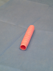
InBreath tracheal scaffold

Hannah’s tracheal transplant was the first regenerated trachea transplant surgery that used a biomaterial scaffold that manufactured by the Harvard Apparatus Regenerative Technology (HART) Inc., a wholly owned subsidiary of Harvard Bioscience. HART ensured that the scaffold and bioreactor were custom-made to Hannah’s dimensions. Then the scaffold was seeded with bone marrow cells taken from Hannah’s bone marrow, and the cells were incubated in the bioreactor for two days prior to implantation. Because Hannah’s own cells were used, her body accepted the transplant without the need for immunosuppressive (anti-rejection) drugs.

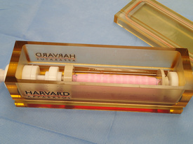
InBreath Bioreactor
InBreath Bioreactor

The surgeons who participated in this landmark transplant were led by Dr. Paolo Macchiarini of Karolinska University Hospital and Karolinska Institutet in Huddinge, Stockholm and Drs. Mark J. Holterman and Richard Pearl both of Children’s Hospital of Illinois. This surgery was approved by the FDA under an Investigational New Drug (IND) application submitted by Dr. Holterman.

Dr. Mark Holterman, Professor of Surgery and Pediatrics at University of Illinois College of Medicine at Peoria, commented: “The success of this pediatric tracheal implantation would have been impossible without the Harvard Bioscience contribution. Their team of engineers applied their talent and experience to solve the difficult technical challenge of applying regenerative medicine principles in a small child.”

David Green, President of Harvard Bioscience, said: “We would like to congratulate Dr. Macchiarini, Dr. Holterman, Dr. Pearl and their colleagues for accomplishing the world’s first transplant of a regenerated trachea in a child using a synthetic scaffold and giving Hannah a chance at a normal life. We also wish Hannah a full recovery and extend our best wishes to her family.”

Hannah’s surgery is the seventh successful implant of a regenerated trachea in a human using HART technology. Prior successes included the first ever successful regenerated trachea transplant in 2008, the first successful regenerated trachea transplant using a synthetic scaffold in 2011, and the commencement of the first clinical trial of regenerated tracheas in 2012. HART has plans to commence discussions with the FDA and EU regulatory authorities in the near future regarding the clinical pathway necessary to bring this new therapeutic approach to a wider range of patients who are in need of a trachea transplant.

Stem Cell Treatments for Large Ankle Cartilage Lesions


The Regenexx blog has called attention to to a study that shows that older patients with ankle problems can benefit from mesenchymal stem cell treatments of the ankle.

The paper referenced by Centeno on his blog was published in the American Journal of Sports Medicine 2013 May;41(5):1090-9, and is entitled, “Clinical outcomes of mesenchymal stem cell injection with arthroscopic treatment in older patients with osteochondral lesions of the talus.” The authors of this paper are Kim YS, Park EH, Kim YC, Koh YG. who all hale from the Department of Orthopedic Surgery, Yonsei Sarang Hospital, Seoul, South Korea.

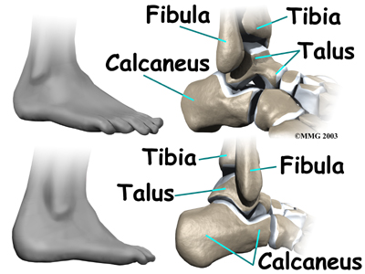
The paper notes that there are no generally accepted, ideal treatments for “osteochondral lesions of the talus”, which is in plain English means cartilage erosions of the large bone in the foot that articulates with the base of the lower leg bones. That bone, the talus, sits above the heel bone (calcaneus) and is covered with articular cartilage to absorb shocks that occur between blows from the fibula and tibia and the talus. Over time, wear and tear can erode this cartilage and the best way to go about fixing these osteochondral lesions of the talus or OLTs as they are called, is not at all clear. Centeno mentions in his blog that he and his colleagues have been treating OLTs with mesenchymal stem cells for some time (since 2006). In this paper, Kim and others tested the ability of bone marrow mesenchymal stem cells (MSCs) to provide relief from OLT.

ankle_anatomy_bones01

Kim and colleagues compared the outcomes of patients who had received MSC injections and arthroscopic marrow stimulation treatments against the outcomes of those patients who had received only arthroscopic marrow stimulation treatment alone.

In this study, from a starting group of 107 patients with OLTs that were treated arthroscopically, only those patients older than 50 years (65 patients) were included in this study. Kim and others divided the patients into 2 groups: 35 patients (37 ankles) were treated with arthroscopic marrow stimulation treatment alone (group A) and 30 patients (31 ankles) who underwent MSC injection along with arthroscopic marrow stimulation treatment (group B).

The clinical outcomes for these patients were assessed according to the visual analog scale (VAS) for pain, but there were other measurements as well. For example, how active were the patients? A Tegner score determined how active patients were, which is important because the more active a patient is, the less likely they are to be in pain. Other assessments included the American Orthopaedic Foot and Ankle Society (AOFAS) Ankle-Hindfoot Scale, which measures how well you walk and how much pain you experience when you do it, and other scores for the foot and how well it works

The outcomes for the study were as follows. Both groups showed a decrease in pain. Group A had a VAS score that started at 7.2 ± 1.1 and fell to 4.0 ± 0.7, whereas group B started at 7.1 ± 1.0 and decreased to 3.2 ± 0.9. Therefore the patients in group B showed a slightly greater decrease in pain over the non-MSC group.

As for the AOFAS score in each group, which measures ankle function and pain while using the ankle, again, both groups showed improvement. Patients in group A went from 68.0 ± 5.5 to 77.2 ± 4.8, and patients in group B went from 68.1 ± 5.6 to 82.6 ± 6.4. Thus the patients who received the MSC treatments used their ankles better than those patient who did not receive MSC treatments, and they also used their ankles with less pain.

A different measure of ankle function, the Roles and Maudsley score, also increased significantly in group A patients as opposed to group A patients at postsurgical follow up (1-4 years after the surgery). However, the real “money” finding of this research was that the patients who had received the MSC injections were significantly more active after surgery than the non-MSC patients. The activity score in patients from group A. Group A patients had a Tegner activity score that went from 3.5 ± 0.8 to 3.6 ± 0.6. That is not a significant increase. However the patients in group B, who had received the injections of their own MSCs into the ankle, had Tegner activity scores that improved from 3.5 ± 0.7 to 3.8 ± 0.7 (P = .041). Thus the patients who had received the MSC injections had less pain, better ankle function with less pain, better ankle and foot function 1-4 years after surgery, and were significantly more active after surgery.

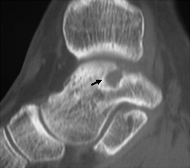
From these data, the authors concluded that injection of MSCs with marrow stimulation treatment was a treatment option in patients older than 50 years than marrow stimulation treatment alone. THis was especially the case if the OTL was larger than 109 square millimeters. Also, those patients in group A who had subchondral cysts did not fare well with their treatment, but there was no such correlation between a poor clinical outcome and the presence of a subchondral cyst in patients from group B.

subchondral cyst in the ankle

Therefore, even though MSC treatments for OTLs is still in its early stages of development, they seem to have the potential to treat of OLTs in patients older than 50 years. However, this study, while not tiny, is still not all that large, and larger studies are warranted.

A Model System for A Devastating Childhood Disease


A Japanese research team from Fukuoka, Japan, specifically from the Department of Pediatrics at the University of Fukuoka, Japan, have used induced pluripotent stem cell technology to make neurons from human patients who suffer from a rare, devastating condition known as Dravet syndrome as a model system.

Dravet syndrome (DS) causes difficult to control seizures within the first year or two of life and later causes cognitive deficits and autistic traits. Dravet’s syndrome is caused by genetic alterations in the SCN1A gene, which encodes the α-subunit of the voltage-gated sodium channel.

DS is very rare – 1/30,000 children, but the mutation is typically not inherited from either parent, but occurs spontaneously in the baby’s cells during development. The best model systems to date are genetically engineered mice, but the differences between human and mouse brains limits the usefulness of this model system.

To make a better model system, workers from the laboratory of Shinichi Hirose took skin biopsy samples from a DS patient, and converted those skin cells into induced pluripotent stem cells (iPSCs), which were then differentiated into neurons. In particular, the neurons that malfunction in DS patients are GABAminergic neurons, and by differentiating DS iPSCs into GABAminergic neurons, Hirose’s laboratory made a model system for DS patients that could be grown in a laboratory culture dish.

Hirose explained their results this way: “From research in mice we believed that SCN1A mutations affect GABAminergic neurons in the forebrain from signaling properly. From the human neurons we also found that GABAminergic neurons were affected by DS, especially during intense stimulation. These patient-specific cell provide an unparalleled insight into the mechanism behind DS and a unique platform for drug development.”

Perhaps such experiments could eventually lead to regenerative treatments for DS patients as well.

Turning Adult Cells into Early Stage Neurons and Bypassing the Pluripotent Cell Stage


Researchers at the University of Wisconsin, Madison have converted skin cells from monkeys and humans into early neural stem cells that can form a wide variety of nervous system-specific cells. This reprogramming did not require converting adult cells into induced pluripotent stem cells or iPSCs. Su-Chun Zhang, professor of neuroscience and neurology at the University of Wisconsin, Madison, served as the senior author of this research. Bypassing the ultraflexible iPSC stage was the key advantage in this research, accord to Zhang.

Zhang added, “IPSC cells [sic] can generate any cell type , which could be a problem for cell-based therapy to repair damage due to disease in the nervous system.” In particular, the absence of iPSCs greatly reduces the risk of tumor formation in the recipient of the stem cell therapy.

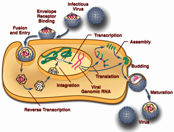
There is a second advantage to this procedure. Namely that iPSC generation usually requires the recombinant viruses that deliver genes to the adult cells. These viruses, retroviruses, insert their genes directly into the genomes of the host cell. While there are ways are using such viruses, the use of retroviruses is definitely the most popular strategy for converting adult cells into iPSCs.

Retroviral life cycle

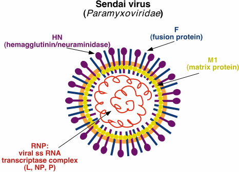
However, the procedure used in Zhang’s laboratory, utilized recombinant Sendai viruses that do not integrate their genes into the genome of the host cell, but expressed them transiently, after which, the exogenous genes are degraded.

Sendai virus
Sendai virus

Jaingfeng Lu, a postdoctoral researcher in Zhang’s lab, removed skin cells from monkeys and people, and exposed them to recombinant Sendai viruses that contained the four genes normally used to make iPSCs for 24 hours. Then Lu heated the cells to thirty-nine degrees to kill the viruses and prevent the cells from becoming iPSCs. However, 13 days later, Lu found that the cells had become induced neural progenitors or iNPs. When implanted into newborn mice, the iNPs grew normally and differentiated into neural cell types without forming any tumors.

While other researchers have managed to convert adult cells directly into neurons, Zhang admitted that he had a different goal. “our idea was to turn skin cells into neural progenitors, cells that can produce cells relating to the neural tissue. These progenitors can be propagated in large numbers.”

the research overcomes limitations of previous efforts, according to Zhang. The Sendai, which produces little more than a cold, is not a severe pathogen, does not integrate its genes into the genome of the host cell, does not cause tumors, and is considered safe, since it can be killed by heat within 24 hours. This illustrates how fevers in our bodies can kill off cold viruses. Secondly, the iNPs have a greater ability to grow in culture. Third, iNPs are far enough along in their differentiation so that they can only form nervous system-specific cell types. They cannot form muscle or live. However, the iNPs can form many more specialized cells.

Interestingly, the neurons produced from the iNPs had the characteristics of neurons normally formed in the back part of the brain, something that is potentially helpful. As Zhang noted, “For therapeutic use, it is essential to use specific types of neural progenitors. We need region-specific and function-specific neuronal types for specific neurological diseases.”

Progenitor cells grown from the skin of ALS or spinal muscular atrophy patients can be used to make a whole host of neural cells in order to model each disease and allow rapid drug screening. Such cells could also be used to treat patients with neurological disease too.

“These transplantation experiments confirmed that the reprogrammed cells indeed belong to ells of the intended brain regions and the progenitors produced the three major classes of neural cells: neurons, astrocytes, and oligodendrocytes. This proof-of-principle study highlights the possibility to generate [sic] many specialized neural progenitors for specific neurological disorders.”

Neural progenitors
Neural progenitors

Lu, Jianfeng, Liu, Huisheng, Huang, Cindy Tzu-Ling, Chen, Hong, Du, Zhongwei, Liu, Yan, Sherafat, Mohammad Amin, Zhang, Su-Chun.  Generation of Integration-free and Region-Specific Neural Progenitors from Primate Fibroblasts.  2013/05/02. Cell Reports 2211-1247. http://linkinghub.elsevier.com/retrieve/pii/S221112471300171X

Wesley Smith and Cloning


My favorite bioethicist, Wesley Smith said this about human cloning in his prescient book: A Consumer’s Guide to A Brave New World:

We can pursue biotechnology to treat disease and improve the human condition, while retaining sufficient humility and self-restraint to keep ourselves from endangering the intrinsic value of human life. Or, we can hubristically rush onto the very anti-human path warned against by Aldous Huxley, driven by our thirst for knowledge, vast profits, and obsession with control and vastly expanded life spans.

These issues are too important to be “left to the scientists.” Nor can we afford to allow the marketplace to determine what is right and what is wrong. The stakes are too high, the potential impact on each and every one of us too profound, to remain passive and indifferent to the decisions that are to be made. It is our duty to participate in the crucial cultural and democratic debates over biotechnology. The human future, quite literally, depends on it.

Prophetic and poignant – and DEAD RIGHT!!

The Archbishop of Denver Speaks Out: Cloning Kills the Smallest Among Us and the Next Victims WIll be Us


Samuel Aquila is archbishop of the Archdiocese of Denver, Colorado and has weighed in with regards to the cloning of human embryos. I am not a Roman Catholic, and Fr. Aquila is not a person whose religious authority I am obligated to accept de fide. Nevertheless, his stance on this subject is reasoned and was published on the National Review Online website here. It is well worth reading.

Human Stem Cells From Cloned Embryos: What Horrors WIll Follow?


First the news, then the commentary. Here’s the news:

In the May 14th edition of the international journal Cell, Shoukhrat Miltalipov from the Oregon Health and Science University, reported the derivation of human embryonic stem cells from cloned human embryos. This is the first time this has been successfully reported. In 2004, a South Korean researcher, Woo Suk Hwang, reported that his laboratory had succeeded in making patient-specific human embryonic stem cells from cloned embryos, but his papers were later shown to be completely fraudulent, and Hwang, in a word, walked. For more on this sad, sordid event, see my “Catastrophic Cloning Caper” here.

Many laboratories have tried and failed to get cloned human embryos to live long enough to get embryonic stem cells from them. The cloning procedure produces a very abnormal embryo that dies very early during development.

How did Mitalipov succeed when so many others before him had failed? Mitalipov honed his cloning protocol in work with early embryos from Rhesus macaques, and during this work, Mitalipov and his coworkers discovered that including caffeine with the mix of chemicals used during donor removal and transplantation into the host egg prevents the oocytes that have just had their nuclei removed from dividing prematurely, and if these oocytes are used in a cloning experiment, they survive longer than oocytes treated with standard cloning techniques.

“It was a huge battery of changes to the protocols over a number of different steps,” said Mitalipov. “I was worried that we might need a couple of thousand eggs to make all these optimizations, to find that winning combination.”

The procedure used in this paper, cloning, is more technically known as “somatic cell nuclear transfer” or SCNT. SCNT requires human eggs that are extracted from female volunteers of reproductive age who are given several drugs to hyperstimulate their ovaries, which then ovulate several eggs at a time. The eggs are harvested by means as aspiration, and used in SCNT.

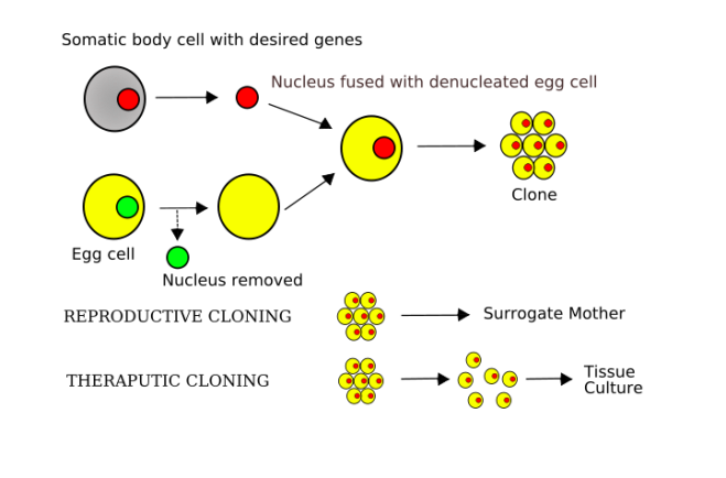
For SCNT, the egg nucleus is removed by means of a micropipette. The egg is ever so gently squeezed until the nucleus, which is usually off to one side in the egg, protrude through the cell membrane, and the nucleus is sucked off with the micropipette. Then a body cell; in this paper, fibroblasts from the skin were used, is laid next to the nucleus-less egg, and an electric current is pulsed through the two cells, which causes them to fuse. This fusion converts the egg, which used to have one set of every chromosome, into a cell that now has two sets of every chromosome, and the egg cell, begins to divide and recapitulate the events of early development. This is also referred to as cloning.

Somatic_cell_nuclear_transfer-image

Sperm and eggs have chromosomes that have been modified in specific ways. When the sperm and egg fuse, the process of fertilization begins, and the modifications to the chromosomes serve their purpose during the early stages of development, but those modifications and gradually undone as development proceeds. This phenomenon is known as genetic imprinting and it is very common in mammals. For a good paper on genetic imprinting see Wood AJ, Oakey RJ (2006) Genomic Imprinting in Mammals: Emerging Themes and Established Theories. PLoS Genet 2(11): e147. doi:10.1371/journal.pgen.0020147.

Since cloned embryos have a genome that is not properly imprinted, its development is hamstrung to one degree or another. Most researchers were unable to get cloned human embryos to survive past the 8-cell stage. However, by including caffeine in the SCNT medium during egg nucleus removal and transplantation of the donor nucleus into the host egg, enough of the cloned embryos survived to the 150-cell blastocyst stage to allow for the derivation of embryonic stem cells. Even though SCNT is an exceedingly inefficient process, Mitalipov was able to derive six embryonic stem cells lines from 128 eggs, which is about a 4% success rate.

George Daley of Boston Children’s Hospital and the Harvard Stem Cell Institute, who was not involved in the research, said of it: ““I think it is a beautiful piece of work.” He continued: “This group has become remarkably proficient at a very technically demanding procedure and [has] shown that SCNT-ESCs may in fact be a practical source of cells for regenerative medicine.”

Mitalipov and his group analyzed four of the cloned embryonic stem cell lines and found that their NT-hESCs could successfully differentiate into beating heart cells in culture dishes. Also, they could differentiate into a variety of cell types in teratoma tumors when transplanted into live, immunocompromised mice. These stem cells also had no chromosomal abnormalities, and displayed fewer problematic epigenetic leftovers from parental somatic cells than are typically seen in induced pluripotent stem cells (although, for the life of me, no one has shown that these epigenetic holdovers are a big problem for regenerative medicine). Mitalipov said more comparisons are required, however.

“We are now left to analyze the detailed molecular nature of SCNT-ES cells to determine how closely they resemble embryo-derived ES cells and whether they have any advantages over iPS cells,” added Daley. “iPS cells are easier to produce and have wide applications in research and regenerative medicine, and it remains to be shown whether SCNT-ES cells have any advantages.”

Mitalipov, however, did point out one fundamental difference between NT-ESCs and iPSCs: their nuclear genomes come from the donor cell, but NT-hESCs contain mitochondrial DNA (mtDNA) from the host egg cell. Therefore, SCNT reprograms the cell but also corrects any mtDNA mutations that the donor may carry. Thus, patient-specific NT-hESCs could be used to treat people with diseases caused by mitochondrial mutations. “That’s one of the clear advantages with SCNT,” Milatipov said.

The cells used for this cloning experiment came from infants.  It still remains for cloning to succeed with adult cells as the donor cells.

Now for the commentary:

Regular readers of this blog will already know that I am deeply opposed to human cloning in any form.  It is the equivalent of making people for spare parts.  This is immoral and barbaric.  I predicted some time ago (OK not so long ago, 4 years to be exact), that the technical problems with human cloning would be solved and scientists would one day clone a human embryo.  Now that it is here, I hope that people are as horrified by it as I am.

“Get over it.  It’s an embryo and a cloned one at that.” you might say.  But what if the malady that doctors want to cure is poorly served by embryonic stem cells made from cloned embryos and a cloned fetus is a better source of cells?  Do we allow gestation of the cloned embryo to the fetal stage so that we can dismember it and take its tissue?  Let’s bring this home.  What if the cells needed to come from a five-year old?  Can we justify that because the kid was cloned?

“But wait, that’s a five-year old and this is an embryo,” you say.  But you were once a blastocyst.  You did not pass through the blastocyst stage, you WERE a blastocyst.  The only difference between the blastocyst and you now is time, environment, degree of dependence, and size.  Are any of these differences morally significant when it comes to whether or not we can kill you?  Can we kill all the short people?  Can we kill all the younger people because they are not as well-developed?  Can we kill people who are dependent on others (that includes everyone mate, so put your hand down)?  Can we kill those in a different location (genocide anyone)?  None of these categories constitutes a good reason for terminating someone’s life.  Likewise, none of these changes renders you essentially different from who and what you are.  To kill someone at the earliest stages for their tissue is simple murder, and we use size, location, extent of development, location and degree of dependence to salve of consciences for doing it, but that won’t define what we are doing.

People will go on and on about the great advances that could lead to.  Sorry, I’m not buying that one.  Embryonic stem cells have been promising that one for the last 15 years with pert near little to nothing to show for it.  This discovery is a great technical advance, but it opens to door to reproductive cloning – an even bigger mistake, and fetus farming, in which we destroy our own children in the womb, not because they are in inconvenience to us, but because we want their tissues to save our lives.  Now children, rather than being a blessing, are merely tissues to be harvested.  We have become like the Greek gods from the stories of old who ate their own children.  May God forgive us.

A Living Patch for Damaged Hearts


Duke University scientists have constructed a three-dimensional human heart muscle patch that behaves much like natural heart muscle tissue. This advance could be used to either treat heart attack patients or to test new heart medicines.

This “heart patch” was grown in the laboratory from human cells, and the procedures used in this research overcame two large roadblocks. First the patch conducts electrochemical impulses at the same speed as normal adult human heart tissue and it contracts to the same degree as normal human heart tissue. In the past, heart tissue patches have conducted electrochemical impulses too slowly and contracted weakly.

The cell source used by the Duke University team were human embryonic stem cells. Thus, the heart patch would not be appropriate for human patients, since it would be rejected by the patient’s immune system. However, the procedures used in this research could also be applied to heart muscle cells made from induced pluripotent stem cells.

Nenad Bursac, associate professor of biomedical engineering at Pratt Engineering, said, “The structural and functional properties of these 3-D tissue patches surpass all previous reports for engineered human heart muscle. This is the closest man-made approximately of native human heart tissue to date.” Bursac also said that the approach does not involve genetic manipulation of the cells.

Bursac continued: “In past studies, human stem cell-derived cardiomyocytes (that is, heart muscle cells) were not able to both rapidly conduct electrical activity and strongly contract as well as normal cardiomyocytes. Through optimization of a three-dimensional environment for cell growth, we were able to ‘push’ cardiomyocytes to reach unprecedented levels of electrical and mechanical maturation.”

The rate of functional maturation is a procedural issue that has very practical implications. If clinicians want to make a heart patch for a patient, the time required to make the heart patch is important, since a heart patch that takes too long to make is of no clinical use to heart patients. In the developing human, it takes about nine months for the newborn heart to develop and an additional five years to reach adult levels of function. These heart patches, however, were grown in about 1 month. And, according to Brusac, further work should shorten the time required to make such a heart patch.

Bursac commented: “It would take us about five to six weeks starting from pluripotent stem cells to grow a highly functional heart patch. When someone has a heart attack, a portion of the heart muscle dies. Our goal would be to implant a patch of new and functional heart tissue at the site of the injury as rapidly after heart attack as possible. Using a patient’s own cells to generate pluripotent stem cells would add further advantage in that there would likely be no immune system reaction, since the cells in the patch would be recognized by the body as self.”

Bursac added that besides using these heart patches in patients, the patches could also be used in the laboratory to test new heart medicines and to model heart pathologies.

“Tests of trials of new drugs can be expensive and time-consuming.  Instead of, or along with testing drugs on animals, the ability to test on actual, functioning human tissue may be more predictive of the drugs’ effects and help determine which drugs should go into further studies.”

Some drug tests are conducted on two-dimensional sheets of heart cells, but according to Bursac, the three-dimensional culture of heart muscle cells provides a more realistic model system for drug testing.  Engineered heart tissues from patients who suffer from cardiac diseases could be used as a model to study that disease and test and explore potential therapies.

Even though Bursac used a particular embryonic stem cell line, but his co-workers also were able to replicate these results with two other embryonic stem cell lines.  Bursac also wants to test his heart muscle patches in animals to determine how well they integrate into the host heart tissue and how well they conduct electrical signals.

Silk and Cellulose as Scaffolds for Stem Cell-Mediated Cartilage Repair


When two bones come together, they grind each other into oblivion. This results in inflammation, joint swelling and pain, and scar tissue accumulation, which eventually results in the immobilization of the joint. To prevent this, bone are capped at their ends with a layer of hyaline cartilage that acts as a shock absorber. However, cartilage regenerates poorly and the wear and tear on cartilage, particularly at the knee, causes it to degenerate. The loss of the cartilage cap at the end of long bones causes osteoarthritis . The only way to mitigate the damage of osteoarthritis is to replace the knee with a prosthetic knee-joint.

Stem cells can make a significant contribution to the regeneration of lost cartilage. The Centeno/Schultz group near Denver, Colorado has been using bone marrow-derived mesenchymal stem cells to treat patients for over a decade with positive results. However, finding a way to grow large amounts of cartilage in culture that is the right shape for transplantation has proven difficult.

One way to mitigate this issue is the use of scaffolds for the cartilage-making cells that pushes them into a three-dimensional arrangement that forces them to make cartilage that mimics the cartilage found in a living organism. However a problem with scaffolds is finding the right material for the scaffold.

A recent publication has formed scaffolds from naturally occurring fibers such as cellulose and silk. By blending silk and cellulose fibers together, researchers at the University of Bristol have made a very inexpensive and easily manufactured scaffold for cartilage production.

Silk scaffold
Silk scaffold

When mixed with stem cells, cartilage and silk coax connective tissue-derived stem cells to differentiate into chondrocytes or cartilage-making cells. In the silk/cellulose scaffold, the chondrocytes secrete the extracellular matrix molecules characteristic of joint-specific cartilage.

Wael Kafienah, lead author of this work from the University of Bristol’s School of Cellular and Molecular Medicine, said, “The blend seems to provide complex chemical and mechanical cues that induce stem cell differentiation into preliminary form of chondrocytes without need for biochemical induction using expensive soluble differentiation factors. Kafienah continued: “This new blend can cut the cost for health providers and makes progress towards effective cell-based therapy for cartilage repair a step closer.”

To make the blended silk/cellulose scaffolds, Kafienah and his colleagues used ionic fluids, which effectively dissolve polymers like cellulose and silk, but are also much more environmentally benign in comparison to the organic solvents normally used to process silk and cellulose.

Presently, the U of Bristol team to trying to fabricate three-dimensional scaffolds that can be safely and easily implanted into patients for future clinical studies. Before human clinical studies are commenced, however, they must first be extensively tested in animals and also, the nature of the interactions between the scaffold and the stem cells that drive the cells to form cartilage must be better understood.

Skin Cells Used to Make Personalized Bone Substitutes


Patient-specific bone substitutes have been produced by a team of scientists from the New York Stem Cell Foundation. Darja Marolt and Giuseppe Maria de Peppo from the New York Stem Cell Foundation (NYSCF) led the study that demonstrated that customizable, three-dimensional bone grafts that can be produced on-demand for patients from their own cells.

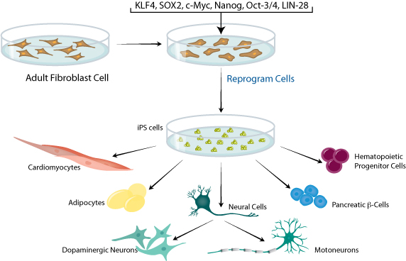
Marolt and de Peppo and their co-workers used skin grafts from their patients to isolate skin fibroblasts that were reprogrammed to induced pluripotent stem cells (iPSCs). Because iPSCs are made from the patient’s own cells, they have the same profile of cell surface proteins as the patient’s own tissues. Therefore, they are very unlikely to be rejected by the patient’s immune system. Also, iPSCs have the ability to differentiate into any cell type found in the adult body, and therefore, can be used to form bone cells.

iPS cells

After deriving iPSCs from patient skin cells, de Peppo, Marolt, and colleagues coaxed the cells to form osteoblasts (the cells that form bone), and seeded them onto a scaffold that mimicked three-dimensional bone structure. These structures were grown in a bioreactor that fed the cells oxygen and nutrients.

According to Marolt, “Bone is more than a hard mineral composite, it is an active organ that constantly remodels. Blood vessels shuttle important nutrients to healthy cells and remove waste; nerves provide connection to the brain; and, bone marrow cells form new blood and immune cells.”

Previous studies have demonstrated that cells from other sources also possess bone-forming potential. However, these same studies have revealed serious shortcomings of the clinical potential of such cells. A patient’s own bone marrow stem cells can form bone and cartilaginous tissue, but not the accompanying underlying vasculature and nerve compartments. Also, embryonic stem cell derived bone may prompt an immune rejection. Therefore, the use of iPSCs can overcome many of these limitations.

As de Peppo noted: “No other research group has published work on creating fully viable functional three-dimensional bone substitutes from humans iPS cells. These results bring us closer to achieving our ultimate goal, to develop the most promising treatments.”

Since bone injuries and defects are often treated with bone grafts that are taken from other parts of the body or a tissue bank. Alternatively, synthetic alternatives can also be used, but none of these possibilities provide the means for complex reconstruction and they may also be rejected by the immune system, or fail to integrate with surrounding connective tissue. n the case of trauma patients who suffer from shrapnel wounds or vehicular injuries , the traditional treatments provide only limited functional and cosmetic improvements.

To access the integrity of the bioreactor-made bone, the NYSCF team implanted them into animals. Implantation of undifferentiated iPSCs formed tumors, but transplantation of the iPSC-derived bone produced no tumors, but also produced grafts that effectively integrated into the bones, connective tissues and blood vessels of the animals.

Susan Solomon, CEO of NYSCF, said of this work, “Following from these findings, we will be able to create tailored bone grafts, on demand, for patients without any immune rejection issues. She continued: “it is the best approach to repair devastating damage or defects.”

The therapeutic relevance of this work aside, these adaptive bone substitutes can also serve as models for bone development and various bone pathologies. Such bone exemplars could serve as models for drug testing and drug development.

Isolating Mammary Gland Stem Cells


Female mammary glands are home to a remarkable population of stem cells that grow in culture as small balls of cells called “mammospheres.” Clayton and others were able to identify these stem cells in 2004 (Clayton, Titley, and Vivanco, Exp Cell Res 297 (2004): 444-60), and Max Wicha’s laboratory at the University of Michigan showed that a signaling molecule called Sonic Hedgehog and a Polycomb nuclear factor called Bmi-1 are necessary for the self-renewal of normal and cancerous mammary gland stem cells (Lui, et al., Cancer Res June 15, 2006 66; 606). The biggest problem with mammary gland stem cells is isolating them from the rest of the mammary tissue.

Mammary gland stem cells or MaSCs are very important for mammary gland development and during the induction of breast cancer. Getting cultures of MsSCs is really tough because the MaSCs share cell surface markers with normal cells and they are also quite few in number.

Gregory Hannon and his co-workers at Cold Spring Harbor Laboratory used a mouse model to identify a novel cell surface protein specific to MaSCs. By exploiting this unusual marker, Hannon and his team were able to isolate MaSCs from mouse mammary glands of rather high purity.

Camila Do Santos, the paper’s first author, said that “We are describing a marker called Cd1d.” Cd1d is also found on the surfaces of cells of the immune system, but is specific to MaSCs in mammary tissue. Additionally, MaSCs divide slower than the surrounding cells. Do Santos and her colleagues used this feature to visually isolate MaSCs from cultured mammary cells.

They used a mouse strain that expresses a green glowing protein in its cells and then made primary mammary cultures from these green glowing mice. After shutting of the expression of the green glowing protein with doxycycline, the cultured cells divided, and diluted the quantity of green glow protein in the cells. This caused them to glow less intensely. However, the slow-growing MaSCs divided much more slowly and glowed much more intensely. Selecting out the most intensely glowing cells allowed Dos Santos and her colleagues to enrich the culture for MaSCs.

“The beauty of this is that by stopping GFP expression, you can directly measure the number of cell divisions that have happened since the GFP was turned off,” said Dos Santos. She continued: “The cells that divide the least will carry GFP the longest and are the ones we characterized.”

Using this strategy, Dos Santos and others selected stem cells from the mammary glands in order to examine their gene expression signature. They also confirmed that by exploiting Cd1d expression in the MaSCS, in combination with other techniques, they could enhance the purity of the cultures several fold.

Hannon added, “With this advancement, we are now able to profile normal and cancer stem cells at a very high degree of purity, and perhaps point out which genes should be investigated as the next breast cancer drug targets.”

Will we be able to use these cell for therapeutic purposes some day?  Possibly, but at this time, more must be known about them and MaSCs must be better characterized.