Placental Stem Cell Provides Model System for Pregnancy Complications


Preeclampsia occurs during pregnancy, and is characterized by a gradual rise in blood pressure to dangerous levels. It usually presents after the 20th week of pregnancy, and can even persist after delivery.

How common is preeclampsia? In the United States, preeclampsia affects 5-8% of all births. Among the women of Canada, the United States, and Western Europe, the births affected by preeclampsia range from 2-5%. (5,6) In the developing world, the percentage of births affected by preeclampsia range from 4% of all deliveries to as high as 18% in parts of Africa. In Latin America, preeclampsia is the number one cause of maternal death.

Globally, ten million women develop preeclampsia each year, and 76,000 pregnant women die each year from preeclampsia and related disorders. The number of babies who die from these disorders is thought to be on the order of 500,000 per year.

In developing countries, a woman is seven times more likely to develop preeclampsia than a woman in a developed country, and between 10-25% of those cases will result in the death of the mother.

Now that I’ve hopefully convinced you that preeclampsia is a problem, how do we address it? Research in laboratory mice have told us a great deal about preeclampsia and other disorders that arise during pregnancy, but finding a sound model system that can be used to develop effective and safe treatments requires something closer to humans.

To that end, Hanna Mikkola and her research team and the University of California, Los Angeles Eli and Edythe Broad Center of Regenerative Medicine and Stem Cell Research (that’s a mouthful), have identified a type of progenitor cell that is key to the growth of a health placenta.

Work in laboratory mice has shown that preeclamsia often arises because of a malformed placenta. This poorly-formed placenta does not provide enough oxygen and nutrients for the growth needs of the baby at the fetal stages of development, and the mother’s body responds by increasing the mother’s blood pressure in order to increase blood flow through the placenta.

The work by Mikkola and her colleagues have provided physicians and developmental biologists with a new “tool box” for understanding the development of the placenta and the different cell types that compose it. Hopefully, various complications during pregnancy might be due to malfunctions of these particular cell types and the progenitor cells that produce them.

Mikkola and others started with laboratory mice, since it is possible to label single cells in mouse embryos and track exactly where those cells and their progeny go and what they do. The powerful genetic tools available in laboratory mice also allows scientists to identify the various biochemical signaling pathways that cells use to communicate with other cells during placental development. Also, if something goes wrong with particular cell signaling pathways, the mouse model allows scientists to precisely characterize the developmental consequences of much dysfunction.

Through their work in the mouse, Mikkola and her co-workers identified a placental progenitor cells called the Epcamhi labyrinth trophoblast progenitor or LaTP. The LaTP is like a multipotent adult of tissue-specific stem cell that can become many of the cells required to make the placenta.

Mikkola and her group also showed that the “c-Met” signaling pathway was required to sustain the growth of LaTPs during placental development and that this same signaling pathway was required to form a specific group of cells (syncytiotrophoblasts) that form the interface between the placenta and the mother’s endometrium. Elimination of c-Met signaling completely compromised the growth of the fetus and its development.

g abs7

This new cell type should provide a wealth of opportunities to examine complications during pregnancy like preeclampsia and others and design treatments that can save the lives of mothers and their babies.

A Stem Cell-Based Therapy for Colon Cancer


Colorectal cancer is the third leading cause of death in the Western World. Like many other types of cancer, colorectal cancer spreads and is propagated by cancer stem cells. Therefore, understanding how to inhibit the growth of cancer stem cells provides a key to treating the cancer itself.

By inactivating a gene that drives stem cell renewal in cancer stem cells, scientists and surgeons at the Princess Margaret Cancer Centre in Toronto, Canada, have discovered a promising new approach to treating colorectal cancer.

John Dick, a senior scientist at the Princess Margaret Cancer Centre, said, “This is the first step toward clinically applying the principles of cancer stem cell biology to control cancer growth and advance the development of durable cures.”

In preclinical experiments with laboratory rodents, Dick and his team identified a gene called BMI-1 as a pivotal regulator of colon cancer stem cell proliferation. With this knowledge in hand, Dick’s laboratory dedicated many hours to finding small molecules that disarm BMI-1. Then Dick and his co-workers replicated human colorectal cancer in mice, and used their BMI-1-inhibiting small molecules to treat these cancer-stricken mice.

According to lead author of this work, Antonija Kreso: “Inhibiting a recognized regulator of self-renewal is an effective approach to control tumor growth, providing strong evidence for the clinical relevance of self-renewal as a biological process for therapeutic targeting.”

Dr. Dick explained: “When we blocked the BMI-1 pathway, the stem cells were unable to self-renew, which resulted in long-term and irreversible impairment of tumor growth. In other words, the cancer was permanently shut down.”

The clinical potential of this approach is significant, since it provides a viable treatment that specifically targets colon cancer. About 65% of all colorectal cancers have an activated BMI-1 pathway. Since physicians now have techniques for identifying the presence of BMI-1 and the tools to inhibit it, this strategy could translate into a clinical treatment that might radically transform the treatment of aggressive, advanced colorectal cancers. Such a treatment would be specific, personal, and specific. May the phase 1 trials begin soon!!!

Heart Regeneration and the Heart’s Own Stem Cell Population


For years scientists were sure that the heart virtually never regenerated.

Today this view has changed, and researchers at the Max Plank Institute for Heart and Lung Research have identified a stem cell population that is responsible for heart regeneration. Human hearts, as it turns out, do constantly regenerate, but at a very slow rate.

This finding brings the possibility that it might be possible to stimulate and augment this self-healing process, especially in patients with diseases or disorders of the heart, with new treatments.

Some vertebrates have the ability to regenerate large portions of their heart. For example zebrafish and several species of amphibians have the ability to self-heal and constantly maintain the heart at maximum capacity. This situation is quite different for mammals that have a low capacity for heart regeneration. Heart muscle cells in mammals stop dividing soon after birth.

However, mammalian hearts do have a resident stem cell population these cells replace heart muscle cells throughout the life of the organism, In humans, between 1-4% of all heart muscle cells are replaced every year.

Experiments with laboratory mice have identified at heart stem cells called Sca-1 cells that replace adult heart muscle cells and are activated when the heart is damaged. Under such conditions, Sca-1 cells produce significantly more heart muscle.

Unfortunately, the proportion of Sca-1 cells in the heart is very low, and finding them has been likened to searching for a diamond at the bottom of the Pacific Ocean.

Shizuka Uchida, the project leader of this research, said, “We also faced the problem that Sca-1 is no longer available in the cells as a marker protein for stem cells after they have been changed into heart muscle cells. To prove this, we had to be inventive.”

This inventiveness came in the form of a visible protein that was made all the time in the Sca-1 cells that would continue being made even if the cells differentiated into heart muscle.

Uchida put it this way: “In this way, we were able to establish that the proportion of the heart muscle cells originating from Sca-1 stem cells increased continuously in healthy mice. Around five percent of the heart muscle cells regenerated themselves within 18 months.”

When the same measurements were taken in mice with heart disease, the number of heart muscle cells made from Sca-1 stem cells increased three-fold.

“The data show that in principle the mammalian heart is able to trigger regeneration and renewal processes. Under normal circumstances, however, these processes are not enough to ultimately repair cardiac damage,” said Thomas Braun, the principal investigator in whose laboratory this work was done.

The aim is to devise and test strategies to improve the activity and number of these stem cells and, ultimately, to strengthen and augment the heart’s self-healing powers.

Using Human Stem Cells to Predict the Efficacy of Alzheimer’s Drugs


Scientists who work in the pharmaceutical industry have seen this time and time again: A candidate drug that works brilliantly in laboratory animals fails to work in human trials. So what’s up with this?

Now a research consortium from the University of Bonn and the biomedical company Life & Brain GmbH has shown that animal models of Alzheimer’s disease fail to recapitulate the results observed with cultured human nerve cells made from stem cells. Thus, they conclude that candidate Alzheimer’s disease drugs should be tested in human nerve cells rather than laboratory animals.

In the brains of patients with Alzheimer’s disease beta-amyloid protein deposits form that are deleterious to nerve cells. Scientists who work for drug companies are trying to find compounds that prevent the formation of these deposits. In laboratory mice that have a form of Alzheimer’s disease, over-the-counter drugs called NSAIDs (non-steroidal anti-inflammatory drugs), which include such population agents as aspirin, Tylenol, Advil, Nuprin and so on prevent the formation of beta-amyloid deposits. However in clinical trials, the NSAIDs royally flopped (see Jaturapatporn DIsaac MGMcCleery JTabet N. Cochrane Database Syst Rev. 2012 Feb 15;2:CD006378).

Professor Oliver Brüstle, the director of the Institute for Reconstructive Neurobiology at the University of Bonn and Chief Executive Officer of Life and Brain GmbH, said, “The reasons for these negative results have remained unclear for a long time.”

Jerome Mertens, a former member of Professor Brüstle’s research, and the lead author on this work, said, “Remarkably, these compounds were never tested directly on the actual target cells – the human neuron.”

The reason for this disparity is not difficult to understand because purified human neurons were very difficult to acquire. However, advances in stem cell biology have largely solved this problem, since patient-specific induced pluripotent stem cells can be grow in large numbers and differentiated into neurons in large numbers.

Using this technology, Brüstle and his collaborators from the University of Leuven in Belgium have made nerve cells from human patients. These cells were then used to test the ability of NSAIDs to prevent the formation of beta-amyloid deposits.

According to Philipp Koch, who led this study, “To predict the efficacy of Alzheimer drugs, such tests have to be performed directly on the affected human nerve cells.”

Nerve cells made from human induced pluripotent stem cells were completely resistant to NSAIDs. These drugs showed no ability to alter the biochemical mechanisms in these cells that eventually lead to the production of beta-amyloid.

Why then did they work in laboratory animals? Koch and his colleagues think that biochemical differences between laboratory mice and human cells allow the drugs to work in one but not in the other. In Koch’s words, “The results are simply not transferable.”

In the future, scientists hope to screen potential Alzheimer’s disease drugs with human cells made from the patient’s own cells.

“The development of a single drug takes an average of ten years,” said Brüstle. “By using patient-specific nerve cells as a test system, investments by pharmaceutical companies and the tedious search for urgently needed Alzheimer’s medications could be greatly streamlined.”

Adult Stem Cells Suppress Cancerous Growth While Dormant


William Lowry and his postdoctoral fellow Andrew White at UCLA’s Eli and Edythe Broad Center of Regenerative Medicine and Stem Cell Research have discovered the means by which particular adult stem cells suppress their ability to trigger skin cancer during their dormant phase. A better understanding of this mechanism could provide the foundation to better cancer-prevention strategies.

This study was published online Dec. 15 in the journal Nature Cell Biology. William Lowry, Ph.D. is an associate professor of molecular, cell and developmental biology in the UCLA College of Letters and Science.

Hair follicle stem cells are those tissue-specific adult stem cells that generate the hair follicles. Unfortunately, they also are the cell population from which cutaneous squamous cell carcinoma, a common skin cancer, begins. However, these stem cells cycle between active periods, when they grow, and dormant periods, when they do not grow.

Diagram of the hair follicle and cell lineages supplied by epidermal stem cells. A compartment of multipotent stem cells is located in the bulge, which lies in the outer root sheath (ORS) just below the sebaceous gland. Contiguous with the basal layer of the epidermis, the ORS forms the external sheath of the hair follicle. The interior or the inner root sheath (IRS) forms the channel for the hair; as the hair shaft nears the skin surface, the IRS degenerates, liberating its attachments to the hair. The hair shaft and IRS are derived from the matrix, the transiently amplifying cells of the hair follicle. The matrix surrounds the dermal papilla, a cluster of specialized mesenchymal cells in the hair bulb. The multipotent stem cells found in the bulge are thought to contribute to the lineages of the hair follicle, sebaceous gland, and the epidermis (see red dashed lines). Transiently amplifying progeny of bulge stem cells in each of these regions differentiates as shown (see green dashed lines).

White and Lowry used transgenic mouse models for their work, and they inserted cancer-causing genes into these mice that were only expressed in their hair follicle stem cells. During the dormant phase, the hair follicle stem cells were not able to initiate skin cancer, but once they transitioned into their active period, they began growing cancer.

Dr. White explained it this way: “We found that this tumor suppression via adult stem cell quiescence was mediated by PTEN (phosphatase and tensin homolog), a gene important in regulating the cell’s response to signaling pathways. Therefore, stem cell quiescence is a novel form of tumor suppression in hair follicle stem cells, and PTEN must be present for the suppression to work.”

Retinoids are used to treat certain types of leukemias because they drive the cancer cells to differentiate and cease dividing. Likewise, understanding cancer suppression by inducing quiescence could, potentially, better inform preventative strategies for certain patients who are at higher risk for cancers. For example, organ transplant recipients are particularly susceptible to squamous cell carcinoma, as are those patients who are taking the drug vemurafenib (Zelboraf) for melanoma (another type of skin cancer). This study also might reveal parallels between squamous cell carcinoma and other cancers in which stem cells have a quiescent phase.

Reactivation of Hair Follicle Stem Cells Restarts Hair Growth


Sarah Millar and her team at the Perelman School of Medicine at the University of Pennsylvania have exploited a known property of hair follicle stem cells to restart hair growth in laboratory animals.

The Wnt signaling pathway is an important regulator of hair follicle proliferation, but does not seem to be required for hair follicle survival. Wnt signaling in cells culminated in the activation of a protein called beta-catenin, which goes to the nucleus of the cell and causes changes in gene expression.

wnt signaling

Millar and her colleagues disrupted Wnt signaling in laboratory animals by expressed an inhibitor called Dkk1 in hair follicles. Dkk1 expression prevented hair growth, and when the hair follicles were examined, they still had their stem cell populations, but these stem cells were dormant. Removal of Dkk1 resumed Wnt/beta-catenin signaling, and restored hair growth.

Dkk1 activity

Interestingly, Millar’s group found Wnt activity in non-hairy regions of the skin, such as palms, soles of feet, and so on. Therefore, in order for Wnt signaling to induce hair growth, it must occur within specific cell types.

This work also has additional applications: skin tumors often show over-active beta-catenin. Removing beta-catenin could prevent the growth of skin tumors, just as removing beta-catenin in the skin of these mice prevented proliferation of any hair follicles. However, agents that can activate beta-cateinin in hair follicles could reactivate dormant hair follicles and induce new hair growth.

Finding ways to safely reactivating the Wnt pathway in particular cells in the skin is a major focus of Millar’s research group.  Such work may lead to treatments for male pattern baldness.

Merry Christmas to All My Readers!!


Matthew 1:18-25
18 This is how the birth of Jesus the Messiah came about: His mother Mary was pledged to be married to Joseph, but before they came together, she was found to be pregnant through the Holy Spirit. 19 Because Joseph her husband was faithful to the law, and yet did not want to expose her to public disgrace, he had in mind to divorce her quietly.

20 But after he had considered this, an angel of the Lord appeared to him in a dream and said, “Joseph son of David, do not be afraid to take Mary home as your wife, because what is conceived in her is from the Holy Spirit. 21 She will give birth to a son, and you are to give him the name Jesus, because he will save his people from their sins.”

22 All this took place to fulfill what the Lord had said through the prophet: 23 “The virgin will conceive and give birth to a son, and they will call him Immanuel” (which means “God with us”).

24 When Joseph woke up, he did what the angel of the Lord had commanded him and took Mary home as his wife. 25 But he did not consummate their marriage until she gave birth to a son. And he gave him the name Jesus.

Human Neural Stem Cells Heal Damaged Limbs


The term “ischemia” refers to conditions under which a part of your body, organ, or tissue is deprived of oxygen. Without life-giving cells begin to die. Therefore, ischemia is usually a very bad thing.

Critical limb ischemia or CLI results when blood vessels to the legs, feet or arms are severely obstructed. The results of CLI are never pretty, and CLI remains a medical condition that presents few treatment options.

A study from a research team and the University of Bristol’s School of Clinical Sciences has used stem cells in a trial that uses laboratory mice to treat CLI. The success of this study provides a new direction and new hope for procedures that relieve symptoms and prolong the life of the limb.

Autologous stem cells treatments, or those stem treatments that utilize a patient’s own stem cells care subject to clear limitations. After collection from bone marrow, fat, or other source, the stem cells must be expanded in culture after stimulation with chemicals called cytokines. After growth in culture, the cells typically contain a collection of different types of stem cells of variable quality and potency. Also, if the patients has had a heart attack or has diabetes, then the quality and potency of their own stem cells are seriously compromised.

To circumvent this problem, Paulo Madeddu and his team at the Bristol Heart Institute have used an immortalized human neural stem cell line called CTX to treat animals who suffered from diabetes mellitus and CLI.

The CTX cell line comes from a biotechnology company called ReNeuron. This company is using this cell line in a clinical trial for stoke patients, and wants to use the CTX cell line in a clinical trial for CLI patients in the future.

When CTX cells are injected into the muscle of diabetic mice with CLI, the cells promote recovery from CLI. The CTX cells do so by promoting the growth of new blood vessels.

Madeddu said, “There are not effective drug interventions to treat CLI. The consequences are a very poor quality of life, possible major amputation and a life expectancy of less than one year from diagnosis in 50 percent of all CLI patients.”

Dr. Madeddu continued: “Our findings have shown a remarkable advancement towards more effective treatments for CLI and we have also demonstrated the importance of collaborations between universities and industry that can have a social and medical impact.”

New Approach for Corneal Stem Cell Treatments


More than 8 million people worldwide suffer from corneal blindness; a form of blindness that results from cloudiness of the outermost covering of the eye, the cornea.

Usually, the cornea copes quite well with minor injuries or scrapes and scratches. If the cornea is scratched, healthy cells slide over quickly and patch the injury before infection occurs and vision is not adversely affected. However, if the scratch penetrates the cornea more deeply, then the healing process takes longer and can result in greater pain, blurred vision, tearing, redness, and extreme sensitivity to light. Such scratches may require professional treatment. Even deeper scratches can also cause corneal scarring, which results in a haze on the cornea that can greatly impair vision, and the patient might require a corneal transplant.

Alternatively, corneal stem cells can help heal a damaged cornea; especially in those cases where the cornea has been damaged to the point where the native stem cell population has suffered irreparable damage (e.g., chemical burns, eye infections, or cases where the patient was born with a corneal stem cell deficiency).

A feasible treatment for such cases is a corneal stem cell transplant from another eye or from cultured corneal stem cells. Unfortunately, this procedure has not yet been standardized to date.

Fortunately, researchers at the Eye Program at the Cedar-Sinai Regenerative Medicine Institute have designed a fast, new procedure for preparing human amniotic membrane to use as a scaffold for corneal stem cells. The membrane provides a foundation that supports the growth of stem cells that can be grafted onto the cornea.

To date, a standardized method does not exist for the preparation of amniotic membranes for culturing corneal stem cells. Many methods use chemicals and may leave behind amniotic cells and membrane components.

This new procedure, however, takes less than one minute and ensures complete amniotic cell removal and preservation of amniotic membrane components, and, as an added bonus, supports the overall growth of various stem and tissue cells.

“We believe that this straightforward and relatively fast procedure would allow easier standardization of amniotic membrane as a valuable stem cell support and improve the current standard of care in corneal stem cell transplantation,” said the lead author of this work Alexander Ljubimov, the director of the Eye Program at the Cedar-Sinai Regenerative Medicine Institute. “This new method may provide a better method for researchers, transplant corneal surgeons, and manufacturing companies alike.”

The amniotic membrane has several beneficial properties for corneal stem cells culturing and use in corneal transplantations. For this reason it is an attractive framework for the growth and culture of corneal stem cells and for corneal transplantations.

The new method for amniotic membrane preparation will provide a fast way to create scaffolds for cell expansion and might potentially streamline clinical applications of cell therapies.

New Clinical Trial to Examine Stem Cell Treatment for Cerebral Palsy in Children


A new clinical trial that is probably one of the first of its kind will study two types of stem cell treatments for children who have cerebral palsy. The University of Texas Health Science Center at Houston (UTHealth) Medical School will host this trial.

This trial will be conducted in a blinded fashion and will test the efficacy of stem cells against a placebo. The types of stem cells investigated in this clinical trial include banked cord blood stem cells and bone marrow stem cells. Charles S. Cox Jr., M.D., professor of pediatric surgery at the UTHealth Medical School and director of the Pediatric Trauma Program at Children’s Memorial Hermann Hospital will lead this clinical trial, and Sean I. Savitz, M.D., chair of the UTHealth Department of Neurology will serve as the co-principal investigator.

This FDA-approved study builds on Dr. Cox’s previous work on traumatic brain injury and the use of stem cell therapy to treat it in children and adults. In particular, Cox has focuses on those patients who have been admitted to Children’s Memorial Hermann and Memorial Hermann-Texas Medical Center after having suffered a traumatic brain injury. Prior research by Cox and others have shown that stem cells derived from a patient’s own bone marrow can be used safely used in pediatric patients with traumatic brain injury. In this clinical trial, Cox is also studying cord blood stem cell treatment for these injuries in a separate clinical trial.

Cox’s trials will enroll a total of 30 children between the ages of 2 and 10 who have cerebral palsy. 15 of these subjects have will have their own cord blood banked at Cord Blood Registry (CBR), and 15 will not have banked any cord blood. In each of these groups, five subjects will be randomized to a placebo control group.

After treatment the children will be neurologically assessed at six, 12 and 24 months. None of the parents will be told if their child received stem cells or a placebo until the 12-month follow-up exam, and at this time, those parents whose children received the placebo may elect to have their child receive a stem cell treatment either by means of stem cells isolated from bone marrow harvest or with stem cells from cord blood banked with CBR.

Collaborators in the study include CBR, Let’s Cure CP, TIRR Foundation and Children’s Memorial Hermann Hospital.

Growing Intestinal Stem Cells


Researchers from MIT and Brigham and Women’s Hospital in Boston, MA have discovered a protocol that allows them to grow unlimited quantities of intestinal stem cells. These intestinal stem cells can then be induced to differentiate into pure populations of various types of mature intestinal cells. Scientists can used these cultured intestinal cells to develop new drugs and treat gastrointestinal diseases, such as Crohn’s disease or ulcerative colitis.,

The small intestine has a small repository of adult stem cells that differentiate into mature adult cells that have specialized functions. Until recently, there was no good way to grow large numbers of these intestinal stem cells in culture. Intestinal stem cells, you see, only retain their immature characteristics when they are in contact with supportive cells known as Paneth cells.

paneth cells

In order to grow intestinal stem cells in culture, researchers from the laboratories of Robert Langer at the MIT Koch Institute for Integrative Cancer Research and Jeffrey Karp from the Harvard Medical School and Brigham and Women’s Hospital, determined the specific molecules that Paneth cells make that keep the intestinal stem cells in their immature state. Then they designed small molecules that mimic the Paneth cell-specific molecules. When Langer and Karp’s groups grew the intestinal stem cells in culture with those small molecules, the cells remained immature and grew robustly in culture.

Langer said, “This opens the door to doing all kinds of thing, ranging from someday engineering a new gut for patients with intestinal diseases to doing drug screening for safety and efficacy. It’s really the first time this has been done.”

The inner mucosal layer of the intestine has several vital functions: the absorption of nutrients, the secretion of mucus of create a barrier between our own cells and the bacteria and viruses and habitually inhabit our bowels, and alerting the immune system to the presence of potential disease-causing agents in the bowel.

The intestinal mucosa is organized into a collection of folds with small indentations called “intestinal crypts.”  At the bottom of each crypt is a small pool of intestinal stem cells that divide to routinely replace the specialized cells of the intestinal epithelium.  Because the cells of the intestinal epithelium show a high rate of turnover (they only last for about five days), these stem cells must constantly divide to replenish the intestine.

INTESTINES COMPARED

Once these intestinal stem cells divide, they can differentiate into any type of mature intestinal cell type.  Therefore, these intestinal stem cells provide a marvelous example of a “multipotent stem cell.”

Obtaining large quantities of intestinal stem cells could certainly help gastroenterologists  treat gastrointestinal diseases that damage the epithelial layer of the gut.  Fortunately, recent studies in laboratory animals have demonstrated that the delivery of intestinal stem cells can promote the healing of ulcers and regeneration of new tissue, which offers a new way to treat inflammatory bowel diseases like ulcerative colitis.

This, however, is only one of the many uses for cultured intestinal stem cells.  Researchers are literally salivating over the potential of studying things like goblet cells, which control the immune response to proteins in foods to which many people are allergic.  Alternatively, scientists would like to investigate the properties of enteroendocrine cells, which secrete hunger hormones and play a role in obesity.  I think you can see, that large numbers of intestinal stem cells could be a boon to gastrointestinal research.

Karp said, “If we had ways of performing high-throughput screens of large numbers of these very specific cell types, we could potentially identify new targets and develop completely new drugs for diseases ranging from inflammatory bowel disease to diabetes.”

The laboratory of Hans Clevers in 2007 identified a molecule that is specifically made by intestinal stem cells called Lgr5.  Clevers is a professor at the Hubrecht Institute in the Netherlands and he and his co-workers have just identified particular molecules that enable intestinal stem cells to grow in synthetic culture.  In culture, these small clusters of intestinal stem cells differentiate and form small sphere-like structures called “organoids,” because they consist of a ball of intestinal cells that have many of the same organizational properties of our own intestines, but are made in culture.

Clevers and his colleagues tried to properly define the molecules that bind Paneth cells and intestinal stem cell together.  The purpose of this was to mimic the Paneth cells in culture so that the intestinal stem cells would grow robustly in culture.  Clevers’ team discovered that Paneth cells use two signal transduction pathways (biochemical pathways that cells use to talk to each other) to coordinate their “conversations” with the adjacent stem cells.  These two signal transduction pathways are the Notch and Wnt pathways.

Fortunately, two molecules could be used to induce intestinal stem cell proliferation and prevent their differentiation: valproic acid and CHIR-99021.  When Clevers and others grew mouse intestinal stem cells in the presence of these two compounds, they found that large clusters of cells grew that consisted of 70-90 percent pure stem cells.  When they used inhibitors of the Notch and Wnt pathway, they could drive the cells to form particular types of mature intestinal cells.

“We used different combinations of inhibitors and activators to drive stem cells to differentiate into specific populations of mature cells,” said Xiaolei Yin, first author of this paper.  Yin and others were able to get this strategy to work with mouse stomach and colon cells, and that these small molecules also drove the proliferation of human intestinal stem cells.

Presently, Clevers’ laboratory is trying to engineering intestinal tissues for potential transplantation in human patients and for rapidly testing the effects of drugs on intestinal cells.

Ramesh Shivdasani from Harvard Medical School and Dana-Farber Cancer Institute would like to use these cells to investigate what gives stem cells their ability to self-renew and differentiate into other cell types.  “There are a lot of things we don’t know about stem cells,” said Shivdasani.  “Without access to large quantities of these cells, it’s very difficult to do any experiments.  This opens the door to a systematic, incisive, reliable way of interrogating intestinal stem cell biology.”

X. Yi, et al. “Niche-independent high-purity cultures of Lgr5 intestinal stem cells and their progeny.” Nature Methods 2013; DOI:10.1038/nmeth.2737.

Human Stem Cells Converted into Functional Lung Cells


Scientists from the Columbia University Medical Center have succeeded in transforming human stem cells into functional lung and airway cells. This finding has significant potential for modeling lung disease, screening lung-specific drugs, and, hopefully, generating lung tissue for transplantation.

Study leader, Hans-Willem Snoeck, professor of medicine and affiliated with the Columbia Center for Translational Immunology and the Columbia Stem Cell Initiative, said, “Researchers have had relative success in turning human stem cells into heart cells, pancreatic beta cells, intestinal cells, liver cells, and nerve cells, raising all sorts of possibilities for regenerative medicine. Now, we are finally able to make lung and airway cells. This is important because lung transplants have a particularly poor prognosis. Although any clinical application is still many years away, we can begin thinking about making autologous lung transplants – that is, transplants that use a patient’s own skin cells to generate functional lung tissue.”

The research builds on Snoeck’s earlier discoveries in 2011 that a set of chemical factors could induce the differentiation of embryonic or induced pluripotent stem cells into “anterior foregut endoderm,” which is the embryo in the tissue from which the lungs form (Green MD, et al. Generation of anterior foregut endoderm from human embryonic and induced pluripotent stem cells. Nat Biotechnol. 2011 Mar;29(3):267-72).

Human Embryological Development - one month

In his new study, Snoeck and his colleagues found new factors that can transform anterior foregut endoderm cells into lung and airway cells. In particular, Snoeck and his co-workers were able to establish the presence of “type 2 alveolar epithelial cells,” which secrete the lung surfactant that maintains the lung alveoli (those tiny sacs in the lung where all the oxygen exchange takes place).

lung alveolus

With these techniques, lung researchers hope to study diseases like idiopathic pulmonary fibrosis (IPF), in which type 2 epithelial cells seem to divide and produce scarring in the lungs.

“No one knows what causes the disease, and there’s no way to treat it,” said Snoeck. “Using this technology, researchers will finally be able to create laboratory models of IPF, study the disease at the molecular level, and screen drugs for possible treatments or cures. In the longer term, we hope to use this technology to make an autologous lung graft. This would entail taking a lung from a donor, removing all the lung cells, leaving only the lung scaffold; and seeding the scaffold with new lung cells derived from the patient. In this way, rejection problems could be avoided.”

Snoeck is investigating this approach in collaboration with researchers in the Columbia University Department of Biomedical Engineering.

Priming Cocktail for Cardiac Stem Cell Grafts


Approximately 700,000 Americans suffer a heart attack every year and stem cells have the potential to heal the damage wrought by a heart attack. Stem cells therapy has tried to take stem cells cultured in the laboratory and apply them to damaged tissues.

In the case of the heart, transplanted stem cells do not always integrate into the heart tissue. In the words of Jeffrey Spees, Associate Professor of Medicine at the University of Vermont, “many grafts simply didn’t take. The cells would stick or would die.”

To solve this problem, Spees and his colleagues examined ways to increase the efficiency of stem cell engraftment. In his experiments, Spees and others used mesenchymal stem cells from bone marrow. Mesenchymal stem cells are also called stromal cells because they help compose the spider web-like filigree within the bone marrow known as “stroma.” Even though the stroma does not make blood cells, it supports the hematopoietic stem cells that do make all blood cells.  Here is a picture of bone marrow stroma to give you an idea of what it looks like:

Immunohistochemistry-Paraffin: Bone marrow stromal cell antigen 1 Antibody [NBP2-14363] Staining of human smooth muscle shows moderate cytoplasmic positivity in smooth muscle cells.
Stromal cells are known to secrete a host of molecules that protect injured tissue, promote tissue repair, and support the growth and proliferation of stem cells.

Spees suspected that some of the molecules made by bone marrow stromal cells could enhance the engraftment of stem cells patches in the heart. To test this idea, Spees and others isolated proteins from the culture medium of bone marrow stem cells grown in the laboratory and tested their ability to improve the survival and tissue integration of stem cell patches in the heart.

Spees tenacity paid off when he and his team discovered that a protein called “Connective tissue growth factor” or CTGF plus the hormone insulin were in the culture medium of these stem cells. Furthermore, when this culture medium was injected into the heart prior to treating them with stem cells, the stem cell patches engrafted at a higher rate.

“We broke the record for engraftment,” said Spees. Spees and his co-workers called their culture medium from the bone marrow stem cells “Cell-Kro.” Cell-Kro significantly increases cell adhesion, proliferation, survival, and migration.

Spees is convinced that the presence of CTGF and insulin in Cell-Kro have something to do with its ability to enhance stem cell engraftment. “Both CTGF and insulin are protective,” said Spees. “Together they have a synergistic effect.”

Spees is continuing to examine Cell-Kro in rats, but he wants to take his work into human trials next. His goal is to use cardiac stem cells (CSCs) from humans, which already have a documented ability to heal the heart after a heart attack. See here, here, and here.

“There are about 650,000 bypass surgeries annually,” said Spees. “These patients could have cells harvested at their first surgery and banked for future application. If they return for another procedure, they could then receive a graft of their own cardiac progenitor cells, primed in Cell-Kro, and potentially re-build part of their injured heart.”

The Therapeutic Potential of Fat-Based Stem Cells Decreases With Age


Fat is a rich source of stem cells for regenerative medicine.  Treating someone with their own stem cells from their own fat certainly sounds like an attractive option.  However, a new study shows that demonstrates that the therapeutic value of fat-based stem cells declines when those cells come from older patients.

“This could restrict the effectiveness of autologous cell therapy using fat, or adipose-derived mesenchymal stromal cells (ADSCs), and require that we test cell material before use and develop ways to pretreat ADSCs from aged patients to enhance their therapeutic potential,” said Anastasia Efimenko, M.D., Ph.D.  Dr Efimenko and Nina Dzhoyashvili, M.D., were first authors of the study, which was led by Yelena Parfyonova, M.D., D.Sc., at Lomonosov Moscow State University, Moscow.

Heart disease remains the most common cause of death in most countries.  Mesenchymal stromal cells (MSCs) collected from either bone marrow or fat are considered one of the most promising therapeutic agents for regenerating damaged tissue because of their ability to proliferate in culture and differentiate into different cell types.  Even more importantly they also have the ability to stimulate the growth of new blood vessels (angiogenesis).

In particular, fat is considered an ideal source for MSCs because it is largely dispensable and the stem cells are easily accessible in large amounts with a minimally invasive procedure.  ADSCs have been used in several clinical trials looking at cell therapy for heart conditions, but most of the studies used stem cells from relatively healthy young donors rather than sick, older ones, which are the typical patients who suffer from heart disease.

“We knew that aging and disease itself may negatively affect MSC activities,” Dr. Dzhoyashvili said. “So the aim of our study was to investigate how patient age affects the properties of ADSCs, with special emphasis on their ability to stimulate angiogenesis.”

The Russian team analyzed age-associated changes in ADSCs collected from patients of different age groups, including some patients who suffered from coronary artery disease and some without.  The results showed that ADSCs from the older patients in both groups showed some of the characteristics of aging, including shorter telomeres (the caps on the ends of chromosomes that protect them from deterioration), which confirms that ADSCs do age.

“We showed that ADSCs from older patients both with and without coronary artery disease produced significantly less amounts of angiogenesis-stimulating factors compared with the younger patients in the study and their angiogenic capabilities lessened,” Dr. Efimenko concluded. “The results provide new insight into molecular mechanisms underlying the age-related decline of stem cells’ therapeutic potential.”

“These findings are significant because the successful development of cell therapies depends on a thorough understanding of how age may affect the regenerative potential of autologous cells,” said Anthony Atala, M.D., director of the Wake Forest Institute for Regenerative Medicine, and editor of STEM CELLS Translational Medicine, where this research was published.

A Protein from Fat-Based Stem Cells Prevents Light-Induced Damage to the Retina


Japanese researchers from Gifu Pharmaceutical University and Gifu University have reported that a type of protein found in stem cells taken from adipose (fat) tissue can reverse and prevent age-related, light-induced retinal damage in mice. These results may lead to treatments for patients faced with permanent vision loss.

According to the work done by these two research teams led by Drs. Hideaki Hara and Kazuhiro Tsuruma, a single injection of fat-derived stem cells (ASCs) reduced the retinal damage induced by light exposure in mice. This study also discovered that when fat-derived stem cells were grown in culture with retinal cells, the stem cells prevented the retinal cells from suffering damage after exposure to hydrogen peroxide and visible light both in the culture and in the retinas of live mice.

Additionally, Hara and Tsuruma and their colleagues discovered a protein in fat-derived stem cells called “progranulin.” This protein, progranulin, seems to play a central role in protecting other cells from suffering light-induced eye damage.

In the retina, which lies at the back of the eye, excessive light exposure causes degeneration of the photoreceptor cells that respond to light. Several studies have suggested that a long-term history of exposure to light might be an important factor in the onset of age-related macular degeneration. Photoreceptor loss is the primary cause of blindness in particular eye-specific degenerative diseases such as age-related macular degeneration and retinitis pigmentosa.

“However, there are few effective therapeutic strategies for these diseases,” Hideaki Hara, Ph.D., R.Ph., and Kazuhiro Tsuruma, Ph.D., R.Ph.

“Recent studies have demonstrated that bone marrow-derived stem cells protect against central nervous system degeneration with limited results. Just like the bone marrow stem cells, ASCs also self-renew and have the ability to change, or differentiate, as they grow. But since they come from fat, they can be obtained more easily under local anesthesia and in large quantities.”

The fat tissue used in the study was taken from mice and processed in the laboratory to isolate the fat-based stem cells. Afterwards, those cells were tested with cultured mouse retinal cells, and they show a robust protective effect. These successes suggested to the team to test their theory on a live group of mice that had retinal damage after exposure to intense levels of light.

Five days after receiving injections of the fat-based stem cells, the animals were tested for photoreceptor degeneration and retinal dysfunction. The results showed the degeneration had been significantly inhibited.

“Progranulin was identified as a major secreted protein of ASCs, which showed protective effects against retinal damage in culture and in animal tests using mice,” Drs. Hara and Tsuruma said. “As such, it may be a potential target for the treatment of degenerative diseases of the retina such as age-related macular degeneration and retinitis pigmentosa. The ASCs reduced photoreceptor degeneration without engraftment, which is concordant with the results of previous studies using bone marrow stem cells.”

“This study, suggesting that the protein progranulin may play a pivotal role in protecting against retinal light-induced damage, points to the potential for new therapeutic approaches to degenerative diseases of the retina,” said, Anthony Atala, MD, editor of STEM CELLS Translational Medicine and director of the Wake Forest Institute for Regenerative Medicine, where this work was published.

Stem Cells from Bone Marrow Help Heal Hard-to-Heal Bone Fractures


A new study that has appeared in the journal STEM CELLS Translational Medicine demonstrates the potential of a subset of stem cells called CD34+ in treating stubborn bone fractures that prove hard to heal.

The body has mechanisms for the repair of broken bones. Consequently, most patients recover from broken bones with little or no complication. However, up to 10 percent of all fracture patients experience fractures that refuse to heal. Such heard to heal fractures can lead to several debilitating side effects that include infection and bone loss, and the healing of hard to heal fractures often requires extensive treatment that includes multiple operations and prolonged hospitalization as well as long-term disability.

Regenerating broken bones with stem cells could offer an answer to this medical conundrum. Adult human peripheral blood CD34+ cells have been shown to contain a robust population of endothelial progenitor cells (EPCs) and hematopoietic stem cells, which give rise to all types of blood cells. These two types of stem cells might be good candidates for this therapy.

However, while other types of stem cells have been tested for their bone regeneration potential, the ability of CD34+ stem cells to facilitate bone healing has not been examined; that is until now. A phase I/II clinical study that evaluated the capacity of CD34+ to stimulate bone regeneration was published in the current edition of STEM CELLS Translational Medicine. This study was conducted by researchers at Kobe University Graduate School of Medicine, led by Tomoyuki Matsumoto, M.D., and Ryosuke Kuroda, M.D., members of the university’s department of orthopedic surgery and its Institute of Biomedical Research and Innovation (IBRI).

Matsumoto’s and Kuroda’s study was designed to evaluate the safety, feasibility and efficacy of autologous and G-CSF-mobilized CD34+cells in patients with non-healing leg bone breaks that had not healed in nine months. Seven patients were treated with CD34+ stem cells after receiving bone grafts.

In case you were wondering, G-CSF is a drug that releases stem cells from the bone marrow into the blood. It is given by injection or intravenously, and works rather well to mobilize bone marrow stem cells into the peripheral circulation.  It has clinical uses for patients recovering from chemotherapy.  Filgrastim (Neupogen) and PEG-filgrastim (Neulasta) are two commercially-available forms of recombinant G-CSF.

“Bone union was successfully achieved in every case, confirmed as early as 16.4 weeks on average after treatment,” Dr. Kuroda said.

Dr. Matsumoto added, “Neither deaths nor life-threatening adverse events were observed during the one year follow-up after the cell therapy. These results suggest feasibility, safety and potential effectiveness of CD34+ cell therapy in patients with nonunion.”

Atsuhiko Kawamoto, MD, Ph.D., a collaborator in IBRI, said, “Our team has been conducting translational research of CD34+ cell-based vascular regeneration therapy mainly in cardiovascular diseases. This promising outcome in bone fracture opens a new gate of the bone marrow-derived stem cell application to other fields of medicine.”

Although the study documents a relatively small number of patients, the results suggest the feasibility, safety and potential effectiveness of CD34+ cell therapy in patients with non-healing breaks,” said Anthony Atala, M.D., editor of STEM CELLS Translational Medicine and director of the Wake Forest Institute for Regenerative Medicine.

A More Efficient Way to Grow Heart Muscle from Stem Cells Could Yield New Regenerative Therapies


An improved method to produce heart muscle from embryonic stem cells or induced pluripotent stem cells could potentially fulfill the demand for heart disease treatments and models of testing new heart drugs. The challenging part of making heart muscle in the laboratory is the production of cells that are all the same. Otherwise their response to drugs or their transplantation into a damaged heart will be unpredictable and unreliable. Fortunately a new study published in the journal STEM CELLS Translational Medicine may provide a way to make large, homogeneous batches of heart muscle cells.

By mixing some small molecules and growth factors together, an international research team led by investigators at the Cardiovascular Research Center at Icahn School of Medicine at Mount Sinai developed a two-step system that induced embryonic stem cells (ESCs) and induced pluripotent stem cells (iPSCs) to efficiently differentiate into ventricular heart muscle cells. This protocol was not only highly efficient but also very reproducible. It also seemed to nicely recapitulate the developmental steps of normal heart development.

“These chemically induced, ventricular-like cardiomyocytes (termed ciVCMs) exhibited the expected cardiac electrophysiological and calcium handling properties as well as the appropriate heart rate responses,” said lead investigator Ioannis Karakikes, Ph.D., of the Stanford University School Of Medicine, Cardiovascular Institute. Other members of this research team consisted of scientists from the Icahn School of Medicine at Mount Sinai, New York, and the Stem Cell & Regenerative Medicine Consortium at the University of Hong Kong.

One of the unusual aspects of this research project was the integrated approach it took. This research group combined computational and experimental systems and by using these techniques, they showed that the use of particular small molecules modulated the Wnt pathway. Signals from the Wnt pathway pass from cell to cell and play a key role in determining whether cells differentiate into an atrial or ventricular muscle cell.

“The further clarification of the molecular mechanism(s) that underlie this kind of subtype specification is essential to improving our understanding of cardiovascular development. We may be able to regulate the commitment, proliferation and differentiation of pluripotent stem cells into heart muscle cells and then harness them for therapeutic purposes,” Dr. Karakikes said.

“Most cases of heart failure are related to a deficiency of heart muscle cells in the lower chambers of the heart,” said Anthony Atala, MD, editor of STEM CELLS Translational Medicine and director of the Wake Forest Institute for Regenerative Medicine. “An efficient, cost-effective and reproducible system for generating ventricular cardiomyocytes would be a valuable resource for cell therapies as well as drug screening.”

Stem cells at a closer view


Larry H. Bernstein at Pharmaceutical Intelligence reblogged some of my posts. They are shown here. Thanks Larry!! If you want the straight skinny on new and cool things in the pharmaceutical research world, you can do no better than Pharmaceutical Intelligence. I highly recommend it!!

Leaders in Pharmaceutical Business Intelligence (LPBI) Group

Larry H. Bernstein, MD, FCAP, Reporter and Curator

http://pharmaceuticalintelligence.com/2013-12-15/larryhbern/Stem cells at a closer view/

There are two bloggers who have brought a clear vision to the growing importance of Pleuripotential stem cell research, applications, and noted risks.  They are M Buratov and David O’Connell.
I repost  some work that needs more attention.  The technology has improved, and there are a number of successful applications.  The treatment of the cells, and the ability to put them on a stable and nontoxic resorbable matrix is a bioengineering advance.

Growing Skeletal Muscle in the Laboratory

Skeletal muscle – that type of voluntary muscle that allows movement – has proven difficult to grow in the laboratory. While particular cells can be differentiated into skeletal muscle cells, forming a coherent, structurally sound skeletal muscle is a tough nut to crack from a research perspective. Another problem dogging muscle research is the difficulty growing new…

View original post 6,237 more words

Australian Researchers Make A Kidney in the Laboratory With Stem Cells


Stem cell researchers from the University of Queensland in Australia have successfully grown a kidney in the laboratory with stem cells. This new breakthrough will almost certainly open the door to improved treatments for patients with kidney disease, and bodes well for the future of organ bioengineering.

Mini-kidney in dish. (Source: University of Queensland)

The principal investigator of this research project, Professor Melissa Little, from University of Queensland’s Institute for Molecular Bioscience (IMB), said that new treatments for kidney disease were urgently needed.

“One in three Australians is at risk of developing chronic kidney disease and the only therapies currently available are kidney transplant and dialysis,” Little said. “Only one in four patients will receive a donated organ, and dialysis is an ongoing and restrictive treatment regime. We need to improve outcomes for patients with this debilitating condition, which costs Australia $1.8 billion a year.”

Little’s research team designed a new step-wise protocol to coax embryonic stem cells to gradually form all the required kidney-specific cell types and to induce them to “self-organize” into a mini-kidney in a dish.  The embryonic stem cell line HES3 was used in this work, which derived by Reubinoff and others in the laboratory of Alan Trounson in 2000.

“During self-organization, different types of cells arrange themselves with respect to each other to create the complex structures that exist within an organ, in this case, the kidney,” Little said. “The fact that such stem cell populations can undergo self-organization in the laboratory bodes well for the future of tissue bioengineering to replace damaged and diseased organs and tissues. It may also act as a powerful tool to identify drug candidates that may be harmful to the kidney before these reach clinical trial.”

Despite the success of this research, Little cautioned that she and other kidney researchers had a great deal of work to do to before this protocol might be ready for human trials. Regardless, it is a very exciting step forward.

The Queensland Minister for Science and Innovation Ian Walker congratulated Little and her co-workers for their advances, and added that biomedical research was crucial in ensuring a healthier future for Queenslanders.

“The work by the IMB research team is an important milestone in developing improved treatments for chronic kidney disease and will ensure those with the condition can continue to live fulfilling and productive lives,” Walker said.

Little’s research team included Dr. Minoru Takasato, Pei Er, Melissa Becroft, Dr. Jessica Vanslambrouck, from IMB, and her collaorators, Professors Andrew Elefanty and Ed Stanley, from the Murdoch Children’s Research Institute and Monash University.

The research is published in the scientific journal Nature Cell Biology and supported by the Queensland Government, the Australian Research Council, as part of the Stem Cells Australia Strategic Research Initiative, and the National Health and Medical Research Council of Australia.

New Model for Kidney Regeneration


Harvard Stem Cell Institute Kidney Diseases Program Leader Benjamin Humphreys has examined tissue regeneration in the kidney. His interest in kidney regeneration has occupied a major part of his career, but some of his more recent work resulted from his skepticism of a particular theory of kidney regeneration.

The kidney stem cell repair model postulates that scattered throughout the kidney are small stem cell populations and are activated after the kidney is injured to repair it. This theory, however, conflicts with another view of kidney regeneration. Namely that after injury, the cells of the kidney dedifferentiate into more primordial versions of themselves and proliferate, after which they differentiate into the various tissues of the kidney.

Humphreys and his colleagues now have evidence that strongly suggests that all the cells of the kidney have the capacity to divide after injury and contribute to kidney regeneration.

Their evidence comes in the form of experiments in mice in which the cells of the kidney were genetically tagged, and then the kidneys were injured to determine what cells contributed to the regeneration of the kidney.

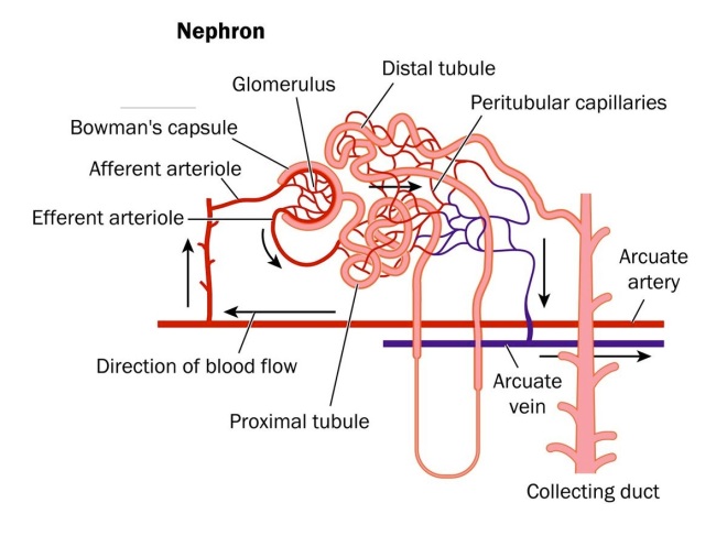
The tagging in this experiment is complicated, but quite technically brilliant. The kidney is composed of myriads of tiny functional units called nephrons. Each nephron is fed by a tiny knot of blood vessels called a glomerulus.  The structure of a nephron is shown below.  

Nephron-image

The blood supply to the kidney comes from branches off the descending aorta knows as renal arteries.  After entering the kidneys, the renal arteries branch multiple times until they become tiny vessels that feed into each nephron known as afferent arterioles.  The afferent arterioles forms a dense network of knot-like vessels that form the glomerulus and the portion of the nephron that interacts with the glomerulus is known as the Bowman’s capsule..  The blood vessels of the glomerulus are very special because they are exceptionally porous.  However, the Bowman’s capsule has a series of cells with foot-like extensions that coat the glomerulus called “podocytes.”  An especially beautiful picture of podocytes wrapped around a glomerular vessel is shown below.

normal-kidney-podocyte

The podocytes cover the pores of the glomerulus and only allows water and things dissolved in water through the pores.  Proteins do not make it through – they are too heavily charged.  Cells also do not make it through – they are too big.  But water, sodium ions, potassium ions, hydrogen ions, some drugs, metabolites, waste products, and things like that all make it through the podocyte-guarded pores.  For this reason, if you have excessive protein or some blood cells in your urine, it is usually an indication that something is wrong.  

Now, rest of the tubing attached to the nephron serve to reabsorb all the things you do not want to get rid of and not absorb all the things you do want to get rid of.  The amount of water you eliminate depends on your degree of hydration and is controlled by a hormone called antidiuretic hormone, which is release by the posterior lobe of your pituitary gland when you are dehydrated.  In the presence of ADH, the posterior tubing reabsorbs more water, and in lower concentrations of this hormone, it reabsorbs far less.  

Now that we know something about the kidney, here’s how Humphreys and others genetically marked the kidneys of their mice.  The sodium-dependent inorganic phosphate transporter (SLC34a1) is only expressed in mature proximal tubule cells.  Tetsuro Kusaba, the first author on this paper, and his colleagues inserted a CreERT2 cassette into this gene.  If you are lost at this point all you need to remember is this: the CreERT2 cassette is inserted into a gene that is ONLY expressed in specific kidney cells.  The Cre gene encodes a recombinase that clips out specific bits of DNA from a chromosome.  Kusaba and others crossed these engineered mice with another strain of mice that had the gene for a bright red dye inserted into another gene, but this dye could not be expressed because another piece of DNA was in the way.  When these hybrid mice were fed a drug called tamoxifen, it activated the expression of the Cre protein, but only in the proximal tubule cells of the kidney and this Cre protein clipped out the piece of DNA that was preventing the red dye gene from being expressed.  Therefore, these mice had a particular part of their nephrons, the proximal tubules glowing bright red.  This is a stroke of shear genius and it genetically marks these cells specifically and strongly.  

Next, Kusaba and colleagues used unilateral ischemia reperfusion injury (IRI) to damage the kidneys.  In IRI, the blood supply is stopped to one kidney but not the other for a short period of time (26 minutes).  This causes cell death and kidney damage.  The other kidney is not damaged and serves as a control for the experiment.  

Examination of the damaged kidneys showed that  red-glowing cells were found in areas other than the proximal tubules.  The only way these cells could have ended up in these places was if the differentiated cells divided and helped repair the damaged parts of the nephrons.  

Other research groups have seen similar results, but interpreted them as evidence of stem cell populations in the kidney.  However, Humphreys groups discovered something even more fascinating.  These “stem cell-markers” in the kidney are actually markers of kidney damage and regeneration and all cells in the kidney express them.  In Humphreys words, “What was really interesting is when we looked at the appearance and expression patterns of these differentiated cells, we found that they expressed the exact same ‘stem cell markers’ that these other groups claimed to find in their stem cell populations.  And so, if a differentiated cell is able to express a ‘stem cell marker’ after injury, then what our work shows is that that’s an injury marker – is doesn’t define a stem cell.”  

Indeed, several genes that have been taken to be signs of a kidney stem cell population (CD133, CD24, vimentin, and KIM-1) were expressed in red-glowing cells.  A stem cell population should not be fully differentiated and therefore, should not be able to express the red dye.  However, red-glowing cells clearly expressed these found genes after injury.  This rather definitely shows that it is the fully differentiated cells that are doing the regeneration in the kidney and not a resident stem cell population.  This does not prove that there is no resident stem cell population in the kidney, but only that the lion’s share of the regeneration is done by differentiated cells, and that under these conditions, no stem cell population was detected.  

This new interpretation of kidney repair suggests that cells can reprogram themselves in a way that resembles the way mature cells are chemically manipulated to revert to an induced pluripotent state.  

See Tetsuro Kusaba, Matthew Lalli, Rafael Kramann, Akio Kobayashi, and Benjamin D. Humphreys. Differentiated kidney epithelial cells repair injured proximal tubule. PNAS (October 14, 2013); doi:10.1073/pnas.1310653110.