Bone Marrow Mesenchymal Stem Cell Aggregates for Treating Deep Skin Wounds


Cuts, bruises, lacerations, abrasions, of the skin are some of the most common injuries. Deeper wounds that extend into the dermis are more susceptible to chronic inflammation and suffer greatly if they are bumped, knocked, or run into. Such wounds are also more difficult to heal, since a full-thickness cutaneous wound usually damages many different structures and cell lineages. Fortunately, the healing of these structures begins directly after production of the wound. Regeneration is largely orchestrated by growth factors such as Vascular Endothelial Growth Factor and Transforming Growth Factor-β. Since mesenchymal stem cells (MSCs) are a good source of these growth factors, they might be promising candidates for treating full-thickness wounds.

Yan Jin and his colleagues at the Research and Development Center for Tissue Engineering in Shaanxi, China have tested the ability of bone marrow-derived mesenchymal stem cells (BMMSCs) to accelerate the healing of deep skin wounds.

Jin and his colleagues, Yulin An, Wei Wei, Huan Jing, Leigo Ming, and Shiyu Lui used bone marrow from mice that had been genetically engineered to express Green Fluorescent Protein (GFP) in their bone marrow. After isolating mesenchymal stem cells from the bone marrow of these mice, they applied the cells to rats that had suffered full-layer skin cutaneous wounds. However, they tested several different ways of applying these cells to the wound.

In one group of rats, GFP+BMMSCs were grown in cell culture that used a growth medium that contained a steroid drug called dexamethasone and ascorbic acid phosphate. These chemicals caused the BMMSCs to grow in the form of clusters of cells called “cell aggregates” that could be scraped off mechanically and readily transplanted onto the wounds without the need for any scaffold.  These cell aggregates grow as sheets of cells that can act as a kind of patch made from healing MSCs that can be easily and readily applied to a wound or other lesion.  In a second group of rats, the same number of GFP+BMMSCs was topically administered around the wound. In the third group of rats, the BMMSCs were given intravenously through tail vein. All three groups of rats received the same number of BMMSCs. Samples from the bed of the wounds were taken at different time points, and the morphological, histological and molecular characteristics of the wounds were analyzed and compared.

(A) The rim of aggregate curled a little on dish bottom. (B) GFP+BMMSCs in the aggregate gave green fluorescence under 509 nm excitation light. (C) The whole aggregate was scratched off the dish. (D) HE staining revealed a certain thickness of the aggregate with cells in it (Bar = 20 nm). (E) RT-PCR showed that BMMSC aggregate presented significantly higher expression of TGF-β and collagen I but had a similar VEGFα expression with normal cultured cells. (N-C: normal cultured cells; A-C: Aggregate cells; **p < 0.05 is considered statistically different.)

According to the results, the BMMSCs administered in cell-aggregates produced the highest expression of pro-healing genes than the other methods. Also animals treated with the BMMSC cell aggregates also showed better vascularization and more regular dermal collagen deposition than the other two groups of rats.

(A,B) Wound bed size and vascularization state in BMMSC-transplanted rats with each control at 4-week post-operation. Yellow dashed circle outside and the dashed line inside showed the original wound size and the left wound bed respectively; (C) Quantification of wound bed size revealed that rats of C-ag group had the smallest wound bed left at 4W. p1 = 0.000, p2 = 0.001, n = 14; (D) Quantification of capillary number revealed that C-ag group models enjoyed the highest capillary density followed by models of Top-ad group and Int-ad group (p1 = 0.001, p2 = 0.000, p3 = 0.000, n = 15).
(A) HE staining (top, 10×) of 4-week samples showed epithelialization in three BMMSC transplanted groups while not in all their controls. Masson trichrome staining (bottom, 20×) of dermal layer showed a superior collagen deposition with certain direction and thicker bundle for C-ag group and Top-ad group to that of Int-ad group, while the collagen disposition of control groups samples was short without certain direction; (B) RT-PCR confirmed that the samples of C-ag group and the Top-ad group presented the highest collagen I expression among groups and followed by Int-ad group (p1 > 0.05, p2 < 0.05).

Detection of inflammatory cells as ascertained by immunofluorescence staining of inflammatory cells also revealed that the duration of inflammation in the cell-aggregate-treated group was significantly shorter than the other two groups. These results were corroborated by RT-PCR experiments that measured the expression of pro-inflammatory genes in the wound tissue.

RT-PCR showed the expression profile of inflammatory cytokines TNF-α (A, p1  0.05, p2 > 0.05, p3 < 0.05) and immune-regulating gene iNOS (C, p1 < 0.05, p2 < 0.05). Wound bed tissues of C-ag and Top-ad group expressed lower level of TNF-α and IL-1β, which were significantly higher in Control groups. Tissue of C-ag group expressed highest level of iNOS among groups.
RT-PCR showed the expression profile of inflammatory cytokines TNF-α (A, p1 < 0.05, p2 < 0.05, p3 < 0.01) and IL-1β (B, p1 > 0.05, p2 > 0.05, p3 < 0.05) and immune-regulating gene iNOS (C, p1 < 0.05, p2 < 0.05). Wound bed tissues of C-ag and Top-ad group expressed lower level of TNF-α and IL-1β, which were significantly higher in Control groups. Tissue of C-ag group expressed highest level of iNOS among groups.

In situ immunofluorescence staining also demonstrated higher rates of GFP+-cell engraftment in the rats treated with BMMSC cell-aggregates than the other groups.

(A) Immunofluorescence staining on CD45+ lymphocytes on wound bed samples at 4W (counterstained with Hoechst 33342) (Bar = 50); (B) Quantification of CD45+ cell among groups. CD45+ cell infiltration in Top-ad and Int-ad wound bed tissue was heavier than that that in C-ag ones. p1 = 0.003, p2 = 0.000, p3 = 0.405, n = 6; (C) Immunofluorescence staining on GFP+ cell on wound bed samples at 4W (counterstained with Hoechst 33342) (Bar = 50 nm); (D) Quantification of GFP+ cell among groups indicated better engraftment for C-ag group than the other two cell transplanted groups, difference being significant. p1 = 0.001, p2 = 0.000, p3 = 0.135, n = 6.

These data show that not only are BMMSC cell aggregates safe, but they might also stimulate greater cutaneous regeneration for full layer cutaneous wounds than BMMSCs administered by other means.  These successful studies will hopefully be followed by large animal studies to confirm the expandability and efficacy of this technology in larger animals.

Forcing Stem Cells to Make Bone


Researchers in the laboratory of Janet Rubin from the University of North Carolina School of Medicine have discovered that a compound from mold can drive mesenchymal stem cells (MSCs) to become osteoblasts, which are the cells that make bone.

MSCs are found in many different tissues without our bodies, ranging from bone marrow, to fat, to tendons, to liver, muscle, brain, and the heart. MSCs have the ability to readily differentiate into bone, fat, and cartilage, and with a little coaxing in the laboratory, they can also form smooth muscle, blood vessels, and even neurons. The key to using these cells is fine-tuning their differentiation in the clinic to efficiently make one cell type over and above another.

Rubin and her colleagues showed that by treating MSCs with cytochalasin D, the cells overwhelmingly became bone cells (osteoblasts). Furthermore, when cytochalasin D was injected into the bone cavity inside bones, it also triggered the formation of bone.

Rubin commented that the “bone forms quickly. The data and images are so clear; you don’t have to be a bone biologist to see what cytochalasin D does in one week in a mouse.”

Rubin continued: “This is not what we expected. This was not what we were trying to do in the lab. But what we’ve found could become an amazing way to jump-start local bone formation. However, this will not address osteoporosis, which involves bone loss throughout the skeleton.”

Cytochalasins are known to target the major cytoskeletal protein actin. Actin self-assembles to form long chain known as microfilaments, and are involved in such vital cell processes as movement, cell shape, cell extensions, vesicle trafficking, and other essential processes in the cell. Cytochalasins disassemble actin microfilaments, which increases the pool of actin monomers in the cell. Rubin and her team showed that these actin monomers went into the cell nucleus and regulated gene expression. Specifically, they turned on the genes responsible for osteoblasts differentiation.

This a novel use of actin by cells, since actin is not normally known to traffic to the nucleus and affect gene expression.

If Rubin and her group disassembled the actin cytoskeleton but prevented actin from trafficking to the nucleus, the MSCs nerve differentiated into osteoblasts.

Cytochalasin D also worked in live mice to drive the formation of bone.

Because bone formation is largely the same in humans and mice, this research is probably translatable. Even though clinicians may not want to use cytochalasin D in human patients, screening compounds that trigger the transport of monomeric actin into the nucleus might be a good way to induce MSCs to form bone cells.

This work was published in the journal Stem Cells 2015; 33(10:3065.

Fetal Stem Cell Therapy Trial for Brittle Bone Disease


Dr. Cecilia Gӧtherström works as a medical researcher at the Karolinska Institutet in Stockholm, Sweden. Earlier this month, Dr Gӧtherström announced the commencement of the first clinical trial that utilizes fetal stem cell transplants to treat the brittle bone disease, Osteogenesis Imperfecta.

Osteogenesis imperfect (OI) was made famous by the Bruce Willis/Samuel Jackson movie “Unbreakable.” In this movie, Samuel Jackson played a wheel-chair bound savant whose bones were incredibly fragile, but acted as a mentor to Bruce Willis’ character who had a tendency to not become injured despite being in accidents and other traumatic events. Willis becomes a kind of local protector of the weak and innocent in his community under Jackson’s tutelage. I will not give away the surprising ending, but the fact that Jackson’s character had OI and his bones broke so easily put OI in the public’s consciousness.

OI is actually a group of genetic disorders that affects an estimated 6 to 7 per 100,000 people worldwide and prevents the bones from forming properly. This disease results from mutations in the COL1A1, COL1A2, CRTAP, and P3H1 genes. More than 90 percent of all cases of OI result from mutations in the COL1A1 and COL1A2 genes. The COL1A1 and COL1A2 genes encode the type I collagen proteins. Collagen is the most abundant protein in bone, skin, and other connective tissues. Patients with OI have fragile bones that break easily, sometimes with no apparent cause. OI can also cause loose joints, fatigue, early hearing loss, and respiratory problems. Multiple fractures are common, and in severe cases, can occur even before birth. Milder cases may involve only a few fractures over a person’s lifetime.

The publication SelectScience interviewed Dr. Gӧtherström who is the coordinator of this clinical trial that will use stem cell therapy to treat babies diagnosed with OI before they are ever born. Dr Gӧtherström told SelectScience that she and her colleagues selected OI as a disease to attack with stem cell treatment because “no good treatment exists.” Dr. Gӧtherström continued: “OI is a chronic disorder that affects the patient throughout their lifetime with reduced quality of life.” Also, because OI causes poor bone mineralization, fractures and malformation of the bones commences by the time the baby is born. Therefore, physicians can diagnose OI during pregnancy, and once it has been diagnosed, it is crucial to initiate treatment as soon as possible.

Dr. Gӧtherström and her colleagues will infuse stem cells into the fetal bodies of babies afflicted with OI by employing the same protocol that is generally used for blood transfusions during pregnancy. This is a very well-tested technique that carries a very little risk to the mother and her baby. According to Dr. Gӧtherström, there is a theoretical risk of the donor cells acquiring mutations that causing cancer in the mother, but this is very unlikely.

Fetal stem cell therapy has some benefits over other types of stem cell therapy. According to Dr. Gӧtherström, “Fetuses do not have a fully developed immune system, so the donor cells may have a better engraftment potential.” Also, fetal Mesenchymal Stem Cells (MSC) have a far better ability to form bone tissue than adult stem cells.

“If this proves to be safe and efficient, we will explore other disorders that can be treated prenatally, such as other skeletal dysplasias, or metabolic disorders,” Dr Gӧtherström explained. The success of this trial could open up new avenues for prenatal therapies to become more common. Dr. Gӧtherström believes that prenatal diagnosis of similar chronic disorders will shift, from delaying or slowing down the onset of a condition to actually treating it.

Regenerating Whole Teeth With A Tissue-Engineered Scaffold


As tissues go, teeth are relatively simple. They only consist of a few cell types, arranged in a rather straight-forward manner. Therefore, regenerating teeth, while more difficult than it seems, should represent a tractable problem for stem cell biologists and tissue engineers. While some progress has been made, tooth regeneration procedures will require more fine-tuning before they will be hailed as successful.

Tzong-Fu Kuo and others from the School of Veterinary Medicine, National Taiwan University, Taipei, Taiwan have examined the feasibility of whole-tooth regeneration in minipigs. Kuo and his group used a tissue-engineered tooth germ-like construct.

To construct their tooth germ constructs, Kuo and his colleagues extracted dental pulp from upper incisors, canines, premolars, and molars from mature miniature pigs. They grew the dental pulp tissue in culture in order to expand the faster-growing dental pulp stem cells (DPSCs) that can outgrowth everything else from the pulp in culture. They differentiated the DPSCs into odontoblasts, which make the dentine of the tooth, and osteoblasts, which make bone. Kuo’s team also acquired gingival epithelial cells from the gums of the minipigs.

Next the gum epithelial cells, odontoblasts, and osteoblasts were implanted onto the surface (upper, and lower layers, respectively), of a bioactive scaffold. This scaffold had the odontoblasts inside, the osteoblasts outside, and the gum epithelial cells outside the osteoblasts. Then Kuo and his coworkers transplanted these seeded bioactive scaffolds into the tooth sockets of the lower jaw of a minipig whose lower first and second molar tooth germs were removed.

13.5 months after the scaffolds were implanted, seven of eight pigs had formed two new teeth that had crowns, roots, and pulp. When the newly-formed teeth were extracted and sectioned, they had enamel-like tissues, dentin, cementum, odontoblasts, and periodontal tissues.

A fascinating finding in this study was that all the pigs, without exception, had regenerated molar teeth regardless of the original tooth from which the DPSCs were isolated. As an important control, minipigs that had their tooth germs removed or received empty scaffolds did not develop teeth.

This study from Kuo’s laboratory showed that implantation of a tooth germ-like structure can produce a complete tooth can do so successfully and efficiently. This study also established that the location of the implant seemed to deeply influence the morphology of the regenerated tooth.

Partial Repair of Full-Thickness Rotator Cuff Tears By Guided Application of Umbilical Cord Blood Mesenchymal Stem Cells


Baseball players, weight lifters, tennis players, basketball players, and other athletes have experienced the pain and frustration of a rotator cuff injury. The rotator cuff is the capsule that surrounds the shoulder joint, in combination with the fused tendons that support the arm at the shoulder joint. A tear in any of these tendons constitute a rotator cuff tear, and it is painful, and debilitating. Furthermore, rotator cuff tears are notoriously slow healing, if they heal at all.

The main option for a rotator cuff tear is microsurgical repair of the tendon. However, as Christopher Centeno at the Regenexx blog points out, sewing together atrophied tissue does not make a lot of sense, and consequently, rotator cuff repairs by means of microsurgery can have a high percentage of re-tearing. Is there a better way?

In the journal Stem Cells and Translational Medicine, Dong Rak Kwon and his two colleagues, Gi-Young Park and Sang Chui Lee, from the Catholic University of Daugu School of Medicine in Daegu, Korea have reported the results of treating whole-thickness rotator cuff tears in rabbits with human umbilical cord blood mesenchymal stem cells (UCB-MSCs). The results are quite interesting.

Kwon and his colleagues broke a colony of New Zealand White rabbits into three groups and surgically subjected all animals to full-thickness tears in the subscapularis tendon. Because rabbits are four-legged creatures, such tears severely compromise their ability to walk, and Kwon and his team measured the ability of these rabbits to walk and the speed at which they walked. All three groups of rabbits showed about the same ability to walk: they walked at about the same speed at for the same distance before giving up.

Human umbilical cord blood-derived mesenchymal stem cell (MSC) and ultrasound images. (A): Human umbilical cord blood-derived MSCs. (B): Injection was made in the left shoulder subscapularis (SCC) full-thickness tears under ultrasound guidance. (C): Longitudinal ultrasound image showed the needle (arrows) in the left shoulder SCC of the rabbit. Abbreviations: S, mesenchymal stem cell; T, tendon.

The first group of rabbits received injections of UCB-MSCs into their rotator cuffs. These injections were guided by ultrasound so that Kwon and his colleagues were able to place the stem cells directly on the damaged tendons. The second group of rabbits received injections of hyaluronic acid (HA), which is a component of connective tissue and the synovial fluid within bursal sacs that surround and lubricated some our joints. The third group received injections of sterile saline into their joints. The animals were then examined four weeks later.

shoulder-joint

The HA- and saline-injected animals showed few changes, but the UCB-MSC-injected animals were able to walk almost twice as far as the other rabbits and almost twice as fast. When the joint tissue of these animals was examined in detail, the HA and saline-injected animals still had full-thickness rotator cuff tears, although the HA-injected animals showed more healing that then the saline-injected rabbits. When the UCB-MSC-injected animals were examined, seven of the ten animals have rotator cuffs that had healed so that the tears could be classified as partial-thickness tears rather than full-thickness tears. Furthermore, a more detailed examination of these joint revealed that they showed regeneration of the tendon and the production of tough, high-quality collagen I.

Gross morphological (A–F) and histological (G–I) findings of the subscapularis tendons in groups 1, 2, and 3. The polygon in each of the first six images depicts the area of the full-thickness subscapularis tendon tear. (A–C): Pretreatment images. (D–F): Posttreatment images. (G): Parallel arrangement of hypercellular fibroblastic bundles (arrow) was noted in group 1. (H, I): Histological findings in groups 2 and 3 showed absence of fiber bundles. Group 1 received a 0.1-ml injection of MSCs; group 2, 0.1 ml of HA; group 3, 0.1 ml of saline. Hematoxylin-and-eosin stain, ×40. Abbreviations: MSC, human umbilical cord blood-derived mesenchymal stem cell; HA, hyaluronic acid; SSC, subscapularis muscle.

Collagen I is the tough material that makes tendon. When rotator cuff surgeries fail, it can be for a variety of reasons, such as poor blood supply, intrinsic tendon degeneration, fatty infiltration, or muscle atrophy (see UG Longo, et al., British Medical Bulletin 2011, 98:31-59).

Histological micrographs of tissue from group 1 rabbits. (A): Newly regenerated tendons are shown in the blue-stained fibers (black arrow; Masson’s trichrome stain; magnification, ×12.5). (B): Regenerated tendon fibers (yellow arrowhead; Masson’s trichrome stain; magnification, ×250) are connected to adjacent M fibers. (C): The regenerated tendon fibers (black arrow) stained with anti-type 1 collagen antibody. The defect was reconstructed with human umbilical cord blood-derived mesenchymal stem cells (magnification, ×100). Abbreviation: M, muscle.

However, tendon failures after surgery usually result from the production of collagen III, which is mechanically weaker than collagen I, instead of collagen I (see MF Pittenger, et al., Science 1999, 284: 143-147; V Rocha, et al., New England Journal of Medicine 2000, 342: 1846-1854). None of the animals in the other groups showed any sign of collagen I production.

This experiment shows that full thickness tears in the subscapularis tendon of the rotator cuff of rabbits, which is functionally similar to the supraspinatus in humans (see figure below), can be partially healed by the ultrasound-guided infusion of UCB-MSCs.

th48RY4PHI

If larger numbers of UCB-MSCs were implanted, it is possible that the tears would have been completely repaired. Also, it is possible that partial tears can be completely repaired by this procedure, but clearly more work is required.

Other questions also remain besides the optimal dose of the cells. What sized tears can be regenerated by this procedure? What immobilization procedures are appropriate after the stem cell injections and for how long? What are the most effective rehabilitation techniques after the surgery? These are all questions that are amenable to research so take heart athletes; a better cure is slowly, but surely on its way.

Combining Umbilical Cord Cells with Hyaluronic Acid Improves Heart Repair After a Heart Attack


Umbilical cord blood cells have an advantage over bone marrow or peripheral blood cells in that aging, systemic inflammation, and stress or damage caused by cell processing procedures can potentially compromise and diminish the regenerative capability of these cells. This problem is particularly acute in the case of treating patients who have recently suffered a heart attack, since transplanted cells experience a rather hostile environment that kills off most cells. Additionally, blood flow through the heart tends to wash out infused cells, which further decreases any regenerative activities the cells might have otherwise exerted.

With this in mind, Patrick Hsieh and his colleagues at the Academia Sinica, in Taipei, Taiwan tested if ability of human cord blood mononuclear cells (CB-MNCs) injected into the heart in combination with a hyaluronan (HA) hydrogel could extend the regenerative abilities of these cells in a pig model. HA is a common component of connective tissue, and, in general, it is very well tolerated by patients and implanted cells. Furthermore, it has the added bonus of shielding cells from a hostile environment and preventing them from being washed out of the heart.

Hsieh used a total of 34 minipigs and divided them into five different groups. One group was the sham operation group in which minipigs received surgical incisions but no heart attack was induced. The second group had heart attacks surgically induced and received infusions of normal saline solutions. The third group of minipigs also experienced heart attacks, and had HA injected into the heart walls. The fourth group also suffered heart attacks and received injections of human umbilical cord stem cells into their heart walls. The fifth group experienced heart attacks and received injections of both HA and human umbilical cord blood cells. The animals were kept and examined two months after surgery.

Two months after the surgery, the minipigs that received injections of human umbilical cord blood cells plus HA showed the highest left ventricle ejection fraction (51.32% ± 0.81%). This is significant when compared to 42.87% ± 0.97%, for the group that received injections of normal saline, 44.2% ± 0.63% for the group that received injections of HA alone, and 46.17% ± 0.39% for the group that received injections of umbilical cord blood cells only. Additionally, hearts from minipigs that received cord blood cells plus HA improved the systolic and diastolic function significantly better than the other experimental groups. Injections of either cord blood cells alone or in combination with HA significantly decreased the scar area and promoted the formation of new blood vessels in the infarcted region. In general, this study suggests that combined infusion of umbilical cord blood cells and HA improves the function of the heart after a heart attack and might prove to be a promising treatment option of heart attack patients.

This is a preclinical study, but it is a preclinical study in a larger animal model system. Umbilical cord blood cells have a demonstrated ability to induce healing in the heart after a heart attack. However, the combination of these cells with HA almost certainly significantly increases cell retention in the heart, thereby significantly improving cardiac performance, and preventing cardiac remodeling. Therefore, using healthy cells donated from another source to replace damaged or moribund cells may be a better option to treat a heart patient and repair their sick heart.

This work appeared in Stem Cells Trans Med November 2015, doi: 10.5966/sctm.2015-0092

Muscular Dystrophy is a Stem Cell-Based Disease


Michael Rudnicki, who has done pioneering work in muscle stem cell biology and muscle regeneration, and whose work has been featured several times on this blog, has struck again. Rudnicki, who serves as director of the Regenerative Medicine Program at The Ottawa Hospital and a professor at the University of Ottawa and holds the prestigious Canada Research Chair in Molecular Genetics, teamed up with workers from the Sprott Centre for Stem Cell Research and the Sinclair Centre for Regenerative Medicine to investigate the role of muscle-specific stem cells in patients who suffer from Duchenne muscular dystrophy. This new earth-shaking study, which was published in the journal Nature Medicine (November 16, 2015), has changed the way we think about muscular dystrophy and will almost certainly force people to rethink the treatments and cures for this dreadful disease.

According to this new study, Duchenne muscular dystrophy directly affects muscle stem cells, and is, largely a disease of muscle stem cells.

Rudicki said: “For nearly 20 years, we’ve thought that the muscle weakness observed in patients with Duchenne muscular dystrophy is primarily due to problems in their muscle fibers, but our research shows that it is also due to intrinsic defects in the function of their muscle stem cells. This completely changes our understanding of Duchenne muscular dystrophy and could eventually lead to far more effective treatments.”

Muscular dystrophy comes in several different forms, but the predominant sign of muscular dystrophy is progressive muscle weakness. Altogether, muscular dystrophy refers to a group of more than 30 genetic diseases, all of which cause progressive weakness and degeneration of skeletal muscles used during voluntary movement. Approximately half of all who suffer from muscular dystrophy have Duchenne muscular dystrophy (DMD). Because muscular dystrophy results from mutations in the dystrophin gene, which is on the X chromosome, the vast majority of muscular dystrophy patients are male. Girls can be carriers of muscular dystrophy and can be mildly affected.

Interestingly, somewhere around one-third of boys who suffer from DMD have no family history of the disease. Because the dystrophin gene is so large, spontaneous mutations in it are probably relatively common.

The signs and symptoms typically appear between the ages of 2 and 3, and may include frequent falls, difficulty getting up from a lying or sitting position, trouble running and jumping, a strange, shuffling way of walking or having a tendency to walk on their toes, calf muscles that are abnormally large, muscle pain and stiffness, and some learning disabilities.

Becker muscular dystrophy (BMD) has signs and symptoms that are largely similar to those of DMD, but BMD tends to be a milder form of the disease that progresses more slowly. Symptoms typically begin in the teens but, some patients may not experience symptoms until their mid-20s and some may not experience symptoms until later.

There are also several different types of muscular dystrophy-type diseases. Steinert’s disease or myotonic muscular dystrophy, which is characterized by an inability to relax muscles at after contractions, is the most common form of adult-onset muscular dystrophy. The first muscles to be affected are the muscles of the face and neck. Facioscapulohumeral muscular dystrophy affects the muscles of the face and shoulders, where symptoms first begin. When patients with facioscapulohumeral raise their arms, their shoulder blades noticeably protrude. This disease may first manifest itself in children, teenagers as late as age 40. This disease tends to affect one side more than the other.

Limb-girdle muscular dystrophy affects the muscles of the shoulders and hips. There are over 20 inherited forms of this disease, and because this condition is not due to mutations in dystrophin, but to mutations in genes that encode proteins that interact with dystrophin, the inheritance of limb-girdle muscular dystrophy is not sex-linked. Some forms of this disease are recessive and some are dominant. Patients with this type of muscular dystrophy usually trip more often because they have trouble raising the front part of their feet. Some autosomal recessive forms of the disorder are now known to be due to a deficits in proteins called sarcoglycans or dystroglycan.

Congenital muscular dystrophy is extremely varible and is probably a cluster of several different diseases caused by mutations in different genes. Some of types of congenital muscular dystrophy show sex-linked inheritance while others do not. Most cases of congenital muscular dystrophy result from the absence of a muscle protein called merosin, which is found in the connective tissue that surrounds muscle fibers. Other types of congenital muscular dystrophy have normal merosin and still others result from abnormal motor neuron migration. Clinically, this disease is also extremely variable and can manifest itself at birth or before age 2, progress slowly or rapidly, and cause mild disability or severe impairment.

Muscular dystrophy affects all ethnic groups and occurs globally. It affects around 1 in every 3,500 to 6,000 male births each year in the United States.  DMD affects approximately one in 3,600 boys.

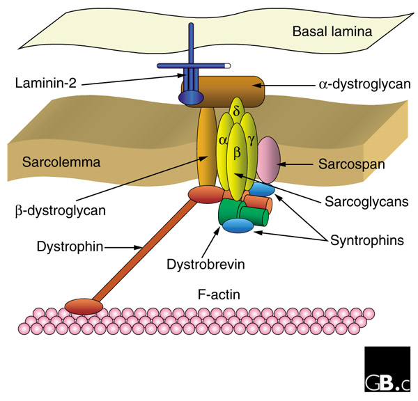
Because DMD results from mutations in the dystrophin gene, the vast majority of muscular dystrophy research was based on a simple model in which the Dystrophin protein played a structural role in the structural integrity of muscle fibers. Abnormal versions of the Dystrophin protein caused the muscle fibers to become damaged and die as a result of contraction.  Dystrophin anchors the cytoskeleton of the muscle fibers, which are essential for muscle contraction, to the muscle cell membrane, and then to the extracellular matrix outside the cell that serves as a foundation upon which the muscle cells are built.

gb-2001-2-4-reviews3006-3

However in this current study, Rudnicki and his team discovered that muscle stem cells also express the dystrophin protein. This is a revelation because Dystrophin was thought to be protein that ONLY appeared in mature muscle. However, in this study, it became exceedingly clear that in the absence of Dystrophin, muscle stem cells generated ten-fold fewer muscle precursor cells, and, consequently, far fewer functional muscle fibers. Dystrophin is also a component of a signal transduction pathway that allows muscle stem cells to properly ascertain if they need to replace dead or dying muscle.  Muscle stem cells repair the muscle in response to injury or exercise by dividing to generate precursor cells that differentiate into muscle fibers.

“Muscle stem cells that lack dystrophin cannot tell which way is up and which way is down,” said Dr. Rudnicki. “This is crucial because muscle stem cells need to sense their environment to decide whether to produce more stem cells or to form new muscle fibres. Without this information, muscle stem cells cannot divide properly and cannot properly repair damaged muscle.”

Even though Rudnicki used mice as a model system in these experiments, the Dystrophin protein is highly conserved in most vertebrate animals. Therefore, it is highly likely that these results will also apply to human muscle stem cells.

Treatment for DMD patients is limited to steroids to decrease muscle inflammation and muscle cell death, and physical therapy to increase muscle use and prevent muscle atrophy. These approaches only delay the progression of the disease and alleviate symptoms. Gene therapy experiments and trials are in progress and even show some promise, but Rudnicki’s work tell us that gene therapy approaches must target muscle stem cells as well as muscle fibers if they are to work properly.

“We’re already looking at approaches to correct this problem in muscle stem cells,” said Dr. Rudnicki. “I’m not sure if we will ever cure Duchenne muscular dystrophy, but I’m very hopeful that someday in the future, we will have new therapies that correct the ability of muscle stem cells to repair the muscles of afflicted patients and turn this devastating, lethal disease into a chronic but manageable condition.”

This paper has received high praise from the likes of Ronald Worton, who was one of the co-discovers of the dystrophin gene with Louis Kunkel in 1987.  Worton later served as Vice-President of Research at The Ottawa Hospital from 1996 to 2007.

“When we discovered the gene for Duchenne muscular dystrophy, there was great hope that we would be able to develop a new treatment fairly quickly,” said Dr. Worton, who is now retired. “This has been much more difficult than we initially thought, but Dr. Rudnicki’s research is a major breakthrough that should renew hope for researchers, patients and families.”

Blocking Differentiation is Enough to Turn Mature Cells into Stem Cells


Hiroshi Kawamoto led a collaboration between the RIKEN Center for Integrative Medical Science and other institutions in Japan and Europe that examined the possibility that adult cells can be maintained in a stem cell-like state where they can proliferate without undergoing differentiation. They discovered that in immune cells, blocking the activity of one transcription factor can maintain the cells in a stem cell-like state where they continue to proliferate and still have the capacity to differentiate into different mature cell types.

Kawamoto and his team genetically engineered hematopoietic progenitor cells from mice to overexpress the Id3 protein. Id3, or inhibitor of DNA binding 3, is an inhibitory protein that forms nonfunctional complexes with other transcription factors. In particular, Id3 inhibits so-called “E-proteins,” (such as TCF3) which drive the progenitor cells to differentiate into immune cells.

Overexpression of Id3, in addition to soaking the cells in a cocktail of cytokines, cause the cells to continue to divide as stem cells. However, when the cytokines were withdrawn, the cells differentiated into various types of immune cells.

Next, Kawamoto and his collaborators infused these engineered hematopoietic progenitors into mice that had been depleted of white blood cells. They discovered that their Id3-overexpressing cells could expand and replenish the white blood cell population of these.

In a follow-up experiment, Kawamoto and his crew recapitulated this experiment using human umbilical cord blood hematopoietic progenitors. Just like their mouse counterparts, these umbilical cord cells could be maintained in culture, and then, upon change of culture conditions, could differentiate into blood cells.

Because these cells can be kept in an undifferentiated state and can extensively proliferate, this culture system provides a model for studying the genetic and epigenetic basis of stem cell self-renewal. And it might also allow scientists to inexpensively grow large quantities of immune cells for regenerative medicine or immune therapies.

This work was published in Stem Cell Reports, October 2015 DOI: 10.1016/j.stemcr.2015.09.012.

Skin Cells Converted into Placenta-Generating Cells


Yosef Buganim and his colleagues from Hebrew University of Jerusalem have successfully reprogrammed skin fibroblasts in placenta-generating cells.

The placenta is a marvelously complex, but it is also a vital organ for the unborn baby. It supplies oxygen and nutrients to the growing baby and removes waste products from the baby’s blood. The placenta firmly attaches to the wall of the uterus and the umbilical cord arises from it.

The placenta forms from a population of cells in the blastocyst-stage embryo known as trophoblast cells. These flat, outer cells interact with the endometrial layer of the mother’s uterus to gradually form the placenta, which firmly anchors the embryo to the side of the uterus and produce a structure that serves as an embryonic kidney, endocrine gland, lung, gastrointestinal tract, immune system, and cardiovascular organ.

Trophoblast form after an embryonic event known as “compaction,” which occurs at about the 12-cell stage (around day 3). Compaction binds the cells of the embryo tightly together and distinguishes inner cells from outer cells. The outer cells will express the transcription factor Cdx2 and become trophoblast cells. The inner cells will express the transcription factor Oct4 (among others too), and will become the cells of the inner cell mass, which make the embryo proper.

Fetal growth restriction, which is also known as intrauterine growth restriction, refers to a condition in which a fetus is unable to achieve its genetically determined potential size. It occurs when gas exchange and nutrient delivery to the fetus are not sufficient to allow it to thrive in utero. Fetal growth restriction can lead to mild mental retardation or even fetal death. This disease also cause complications for the mother.

Modeling a disease like fetal growth restriction has proven to be very difficult largely because attempts to isolate and propagate trophoblast cells in culture have been unsuccessful. However, these new findings by Buganim and his colleagues may change that.

Buganim and his coworkers screened mouse embryos for genes that support the development of the placenta. They identified three genes – Gata3, Eomes, and Tfap2c – that, when transfected into skin fibroblasts, could drive the cells to differentiate into stable, fully-functional trophoblast cells. Buganim called these cells “induced trophoblast stem cells” or iTSCs.

In further tests, Hana Benchetrit in Buganim’s laboratory and her colleagues showed that these iTSCs could integrate into a developing placenta and contribute to it.

Buganim and his team are using the same technology to generate fully functional human placenta-generating cells.

If this project succeeds, it might give women who suffer from the curse of recurrent miscarriages or other placenta dysfunctions diseases the chance to have healthy babies. Also, since these iTSCs integrate into the placenta and not the embryo, they pose little risk to the developing baby.

This work was published in Cell Stem Cell 2015; DOI: 10.1016/j.stem.2015.08.006.

A Cell Delivery Methods that Uses Magnetic Fields


Injecting stem cells into the brain has serious risks. In the a case of patients who have experienced traumatic brain injury (TBI), intracranial injections of stem cells can cause intracranial hemorrhage. Also, the injected stem cells often fail to find their way to the injured parts of the brain. Therefore, you have a high-risk procedure that may yield few benefits.

However a new technique for getting stem cells into the brain has been designed and tested by Paul Yarowsky and his colleagues at the University of Maryland and the Veterans Administration Maryland Healthcare System.

Yarowsky and his coworkers labeled human neural progenitor cells with iron oxide “superparamagnetic nanoparticles” and directed team to the site of a brain injury by means of a magnetic field.

They tested this technique in rats that had suffered TBI and discovered that the delivery methods has no deleterious effects on the viability of the stem cells and not only increases stem cell homing to the site of injury, but also increased stem cell retention.

“Magnetic cell targeting is ideally suited to augmenting cell therapies. The external magnetic field and field gradient can guide cells to sites of injury and, using MRI, the iron-oxide superparamagnetic nanoparticles can be visualized as they travel to the site of injury. The goal when employing this method is not only guiding the particles to the site of injury, but also enhancing entry into the brain and the subsequent retention of the transplanted cells,” said Yarowsky.

The intensity of the magnetic field neither affects the viability of the cells in culture, nor their proliferation nor differentiation. This is also the case when the cells are loaded with iron oxide nanoparticles. These results suggest that this is indeed a promising technique for cell delivery in TBI patients and might also be useful for treating other neurological injuries and neurodegenerative diseases as well.

A critical question is, “what happens to the cells when the magnetic field is no longer present?”. Also, the patient must wear a magnetic hat in order to subject the cells to a magnetic field, but what is the minimum time the patient must wear it in order for the procedure to be successful?  All of these questions must be addressed to some degree if they this technique is to be properly understood. For now, Yarowsky and his colleagues assume that the optimized magnetic intensity observed in experiments with rodents must be extrapolated to larger animals, which may or may not be a legitimate extrapolation. Until larger animal experiments are conducted, this will remain a speculation.

Even though a good deal of work remains to be done, Yarowsky and his colleagues are still optimistic that their ingenious iron oxide nanoparticle procedure has promise and might, some day, be translated to human clinical trials.

This work was published in the journal Cell Transplantation, 21 September 2015.

Cord Blood Cells As a Potential Treatment for Alzheimer’s Disease


Jared Ehrhart from the University of South Florida, who also serves as the Director of Research and Development at Saneron CCEL Therapeutics Inc, and his coworkers have shown that cells from umbilical cord blood can not only improve the health of mice that have an experimental form of Alzheimer’s disease (AD), but these can also be administered intravenously, which is safer and easier than other more invasive procedures.

Laboratory mice can be engineered to harbor mutations that can cause a neurodegenerative disease that greatly resembles human AD. One such mouse is the PSAPP mouse that harbors two mutations that are known to cause an inherited, early-onset form of AD in humans. By placing both mutations in the same mouse, the animal forms the characteristic protein plaques more rapidly and shows significant AD symptoms and brain pathology.

Ehrhart used PSAPP mice to test the ability of human umbilical cord blood to ameliorate the symptoms of AD. He injected one million Human Umbilical Cord Blood Cells (HUCBCs) into the tail veins of PSAPP mice and 2.2 million into the tail veins of Sprague-Dawley rats. Then he harvested their tissues at 24 hours, 7 days, and 30 days after injection. Then Ehrhart and his team used a variety of techniques to detect the presence of the HUCBCs.

Interestingly, the HUCBCs were able to cross the blood-brain barrier and take up residence in the brain. The cells remained in the brain and survived there for up to 30 days and did not promote the growth of any tumors.

Several studies have shown that the administration of HUCBCs to mice with a laboratory form of AD can improve the cognitive abilities of those mice (see Darlington D, et al., Cell Transplant. 2015;24(11):2237-50; Banik A, et al., Behav Brain Res. 2015 Sep 15;291:46-59; Darlington D, et al., Stem Cells Dev. 2013 Feb 1;22(3):412-21). However, in such cases it is essential to establish that the administered cells actually found their way to the site of damage and exerted a regenerative response.

Even though Ehrhart and his troop found that the intravenously administered HUCBCs were widely distributed throughout the bodies of the animals, they persisted in the central nervous system for up to one month after they were injected. In the words of this publication, which appeared in Cell Transplantation, the HUCBCs were “broadly detected in both in the brain and several peripheral organs, including the liver, kidneys, and bone marrow.”. The fact that such a minimally invasive procedure like intravenous injection can effectively introduce these cells into the bodies of the PSAPP mice and still produce a significant therapeutic effect is a significant discovery.

Ehrhart and his colleagues concluded that HUCBCs might provide therapeutic effects by modulating the inflammation that tends to accompany the onset of AD. Furthermore, these cells do not need to be delivered by means of an invasive procedure like intracerebroventricular injection. Furthermore, even though HUCBCs were detected in other organs, their numbers in those places was not excessive and the ability of the HUCBCs to cross the blood-brain barrier suggests that these cells might serve as safe, effective therapeutic agents for AD patients some day.

Drugs that Increase Bone Marrow Stem Cell Mobilization Improves Heart Healing After a Heart Attack in Laboratory Mice


The laboratory of Ahmed Abdel-Latif at the University of Kentucky has used an acute heart attack model in laboratory mice to examine if fatty signaling molecules have the ability to improve the healing of the heart after a heart attack.

A host of studies have examined the ability of transplanted stem cells to help heal the heart after a heart attack. Many laboratories have examined the efficacy of stem cells from bone marrow (Afzal MR, et al., Circ Res. 2015 Aug 28;117(6):558-75), fat (Suzuki E, et al., World J Cardiol. 2015 Aug 26;7(8):454-65), and umbilical cord blood (Xing Y, et al., Cell Mol Biol (Noisy-le-grand). 2014 Jun 15;60(2):6-12) to improve heart function, prevent remodeling, help heart muscle cells survive, and promote the growth of new blood vessels. Unfortunately, while these studies have produced largely positive results, such stem cell treatments lack consistency in their activity and efficacy.

Heart attacks result from oxygen deprivation of the heart. The lack of oxygen causes heart muscle cells to die off. Heart muscle cells are not like skeletal muscle cells, which can work at an oxygen deficit. Instead, the oxygen-deprived heart muscle cells will die even after a show period of ischemia. This cell death causes the release of a host of molecules into the vicinity of the heart muscle that induces a sizable inflammatory response, which kills off even more cells. This inflammatory response, however, has a positive side too, since it can send signals to the rest of the body, in particular the bone marrow, and mobilize stem cells into the blood steam that eventually home to the damaged heart tissue. Once in the heart, these cells can mediate repair of the damaged heart (see Hsieh PC, et al., Nature Medicine 2007;13:970-974; Abdel-Latif A, et al., Exp Hematol 2010;38:1131-1142; Finan A and Richard S, Frontiers in Cell and Developmental Biology 2015; 3: 57).

The nature of the signals that bring bone marrow stem cells to the doorstep of the damaged heart have been the subject of some interest to several laboratories. Work from several different laboratories have shown that bone marrow stem cells are held in the bone marrow by means of a molecule called stromal-derived growth factor-1 (SDF-1), which is made by the bone marrow cells that surround the stem cell that binds to a receptor on the surface of the bone marrow stem cell called-CXCR4. This SDF-1/CXCR4 interaction keeps the bone marrow stem cell happy with its location. And additional binding between a stem cell surface protein called Very Late Antigen-4 (VLA-4; α4β1 integrin) and a receptor for VLA-4 called Vascular Adhesion Molecule-1 (VCAM-1; CD106), which is found on the surfaces bone marrow cells, tethers the bone marrow stem cells to the bone marrow and bone marrow niches (see Lapidot T, Dar A, Kollet O. Blood. 2005;106(6):1901–1910; Peled A, et al., J Clin Invest. 1999;104(9):1199–1211;Lévesque JP, et al., Blood. 2001;98(5):1289–1297; Lévesque JP, et al., J Clin Invest. 2003;111(2):187–196).

Bone marrow stem cells can be mobilized into the peripheral blood by infection, tissue injury, or after the administration of particular pharmacological agents such as granulocyte colony stimulating factor (G-CSF) or some polysaccharides such as Zymosan. Earlier thinking focused on the protein SDF-1, because several papers seemed to suggest a role for SDF-1 in stem cell recruitment of tissue repair after injury (Bobadilla M, et al., Stem Cells Dev. 2014 Jun 15;23(12):1417-27; Wen J, Am J Cardiovasc Dis. 2012;2(1):20-8; Yang JX, et al., J Biol Chem. 2015 Jan 23;290(4):1994-2006). However, SDF-1 does not seem to be the major signaling molecule that mobilizes bone marrow stem cells after a heart attack, because stem cell mobilization is not blocked if an antagonist for CXCR4 called AMD3100 is administered (See Ratajczak and others below). Instead, a group of lipids that are precursors for the synthesis of a group of membrane lipids known as “sphingolipids” seem to be the main signaling molecules for this event (see Ratajczak MZ, et al., Leukemia 2010;24:976-985).

sphingo

In particular, two molecules, sphingosine-1-phosphate (S1P) and ceramide-1-phosphate (C1P) are probably the main players for this response. Thus, several stem cell scientists have predicted that giving people drugs that increase the concentrations of S1P and C1P might enhance healing of the heart after a heart attack through improved stem cell mobilization.

This is the point at which Ahmed Abdel-Latif and his colleagues com into the story, because Abdel-Latif’s lab used a drug called tetrahydroxybutylimidazole (THI) to do exactly that. THI inhibits an enzyme called S1P lyase (SPL), which degrades S1P. Therefore, THI raises the concentrations of S1p in the peripheral blood. Abdel-Latif and his coworkers administered THI to mice 4 days after they had suffered a heart attack. This time lag is essential because the first few days after the heart attack, the heart is a very hostile place, and any recruited or injected cells will die. However, 4 days is also well before scarring and scar formation occur in the heart.

Abdel-Latif and others observed that THI treatment lengthens the time period during which stem cells from the bone marrow are recruited and sent to the blood stream. The greater number of stem cells sent to the heart resulted in enhanced heart regeneration. The hearts of the THI-treated animals showed significantly better ejection fractions (average percentage of blood ejected from the ventricles per heart beat), increased heart wall thickness, and reductions in the size of the heart scar 5 weeks after their heart attacks.

When the mobilized bone marrow stem cells were isolated from peripheral blood and screened for gene expression, it was clear that these cells expressed a gaggle of stem cell homing, mobilization, cell survival, and blood vessel making genes. Thus, these mobilized stem cells were not only ready to go to the heart, but they were fully primed for to stimulate tissue healing.

Labeling studies also showed that bone marrow stem cells and progenitor cells flocked to the damaged hearts. The THI-treated mice had more than twice the number of labeled cells in their hearts at the edge of the infarct zone than the control animals 5 weeks after their heart attack. The THI-treated animals also showed significant increases in capillary densities in the THI-treated animals. As expected, there was no evidence that the mobilized bone marrow stem cells that differentiated into heart muscle cells. Thus, whatever benefits these cells convey to the heart is probably mostly by means of secreted exosomes, growth factors, and other mechanisms or so-called paracrine mechanisms.

This procedure worked rather well in laboratory mice. Can it work in human patients? That’s the $64,000 question. We have hints that increase bone marrow stem cells mobilization after a heart attack might improve recovery. However, this hint comes from a small clinical study in which levels of mobilized stem cells in the bloodstream after a cardiac event was correlated with clinical outcomes one year after the episode (Wyderka R, et al. Mediators Inflamm 2012;2012:564027). Such a study is at odds with studies that have pharmacologically mobilized stem cells from the bone marrow with intravenous G-CSF in patients after a heart attack with little benefit (Hilbert B, et al., CMAJ. 2014 Aug 5;186(11):E427-34; Archilli F., et al., Heart. 2014 Apr;100(7):574-81; Moazzami K, et al., Cochrane Database Syst Rev. 2013 May 31;5:CD008844. doi: 10.1002/14651858.CD008844.pub2). However, as noted in this paper, a drug called LX2931 is a THI analog and is already given as a treatment for rheumatoid arthritis, LX2931 is a safe drug and also inhibits SPL. Possibly future clinical trials that use either LX2931 or something akin to it will be tested in heart attack patients.

Gene Therapy for Stroke Applied with Eye Drops


Administering growth factors to the brains of patients with neurodegenerative diseases can prevent neurons from dying and maintain the structure of their brains. For example, a recently published clinical trial by Nagahara and others from the Department of Neuroscience and the University of California, San Diego examined 10 Alzheimer’s disease (AD) patients and showed that these patients responded to Nerve Growth Factor gene therapy. When they compared treated and nontreated sides of the brain in 3 patients who underwent gene transfer, expansion of cholinergic neurons was observed on the NGF-treated side. Both neurons exhibiting the typical pathology of AD and neurons free of such pathology expressed NGF, which indicates that degenerating cells can be infected with therapeutic genes. No adverse pathological effects related to NGF were observed. In the words of this study, “These findings indicate that neurons of the degenerating brain retain the ability to respond to growth factors with axonal sprouting, cell hypertrophy, and activation of functional markers. [Neuronal s]prouting induced by NGF persists for 10 years after gene transfer. Growth factor therapy appears safe over extended periods and merits continued testing as a means of treating neurodegenerative disorders.” See JAMA Neurol. 2015 Oct 1;72(10):1139-47.

Another study that also shows that the brains of AD patients can respond to growth factors comes from a paper by Ferreira and others from the Journal of Alzheimers Disease. These authors hail from the Karolinska Institutet, Stockholm, Sweden, and they implanted encapsulated NGF-delivery systems into the brains of AD patients. Six AD patients received the treatment during twelve months. These patients were classified as responders and non-responders according to their twelve-month change in the Mini-Mental State Examination (MMSE), which is a standard. In order to set a proper standard of MMSE decline and brain atrophy in AD patients, Ferreira and other examined 131 AD patients for longitudinal changes in MMSE and brain atrophy. When these results provided a baseline, the NGF-treated were then compared with these baseline data. Those patients who did not respond to the implanted NGF showed more brain atrophy, and neuronal degeneration as evidenced by higher CSF levels of T-tau and neurofilaments than responding patients. The responders showed better clinical status and less pathological levels of cerebrospinal fluid (CSF) Aβ1-42, and less brain shrinkage and better progression in the clinical variables and CSF biomarkers. In particular, two responders showed less brain shrinkage than what was normally experienced in the baseline data. From these experiments, Ferreira and others concluded that encapsulated biodelivery of NGF might have the potential to become a new treatment strategy for AD.

Now new, even simpler treatment strategy has been developed by a research team funded by the National Institute of Biomedical Imaging and Bioengineering for delivering gene therapy to the brains of AD patients. This team invented an eye drop cocktail that can deliver the gene for a growth factor called granulocyte colony stimulating factor (G-CSF) to the brain. They have tested these eye drops on mice with stroke-like injuries.

When treated with these eye drops, the mice experienced a significant reduction in shrinkage of the brain, neurological defects, and death. Ingeniously, this research group also devised a way to use Magnetic Imaging Systems to monitor how well the gene delivery worked. This one-two punch of an inexpensive and noninvasive delivery system combined with a monitoring technique that is equally noninvasive might have the ability to improve gene therapy studies in laboratory animals. Such a strategy might also be transferable to human patients. Imagine that acute brain injury might be treatable in the near future by emergency medical workers by means of eye drops that carry a therapeutic gene.

The growth factor G-CSF (granulocyte-colony stimulating factor) has more than proven itself in several animal studies. In model systems for stroke, AD, and Parkinson’s disease, G-CSF promotes neuronal survival and decreases inflammation (See McCollum M, et al., Mol Neurobiol. 2010 Jun;41(2-3):410-9; Frank T, et al., Brain. 2012 Jun;135(Pt 6):1914-25; Prakash A, Medhi B, Chopra K. Pharmacol Biochem Behav. 2013 Sep;110:46-57; Theoret JK, et al., Eur J Neurosci. 2015 Oct 16. doi: 10.1111/ejn.13105). Unfortunately, when G-CSF was when tested in a human trial in more than 400 stroke patients, it failed to improve neurological outcomes in stroke patients. Therefore, it is fair to say that the excitement this growth factor once generated is not what is used to be. A caveat with this clinical trial, however, is that G-CSF expression in the brains of these patients might have been rather poor in comparison to the expression achieved in mice. To properly establish the efficacy or lack of efficacy of gene therapies in human patients, scientists MUST convincingly determine that the gene is expressed in the target tissue of test subjects. This has been a perennial problem that has dogged many gene therapy trials.

Philip K. Liu, Ph.D., of the Martinos Center for Biomedical Imaging at Harvard Medical School, and his collaborators, H. Prentice and J. Wu of Florida Atlantic University, developed the novel MRI-based techniques for monitoring G-CSF treatment and the eye drop-based delivery system as well. MRI can efficiently confirm successful administration and expression of G-CSF in the brain after gene therapy delivery. This work was published in the July issue of the journal Gene Therapy.

“This new, rapid, non-invasive administration and evaluation of gene therapy has the potential to be successfully translated to humans,” says Richard Conroy, Ph.D., Director of the NIBIB Division of Applied Science and Technology. “The use of MRI to specifically image and verify gene expression, now gives us a clearer picture of how effective the gene therapy is. The dramatic reduction in brain atrophy in mice, if verified in humans, could lead to highly effective emergency treatments for stroke and other diseases that often cause brain damage such as heart attack.”

Liu’s motivation for this project was to develop a gene delivery method that was simple, and could rapidly and effectively deliver the genes to the brain. A simple gene delivery technique would obviate the need for highly trained staff and expensive, sophisticated equipment. They also sought to successfully demonstrate the efficacy of their technology in laboratory animals so that it could be translated to humans.

To test their system, they deprived mice of blood flow to their brains, and then administered a genetically-engineered adenovirus that had the G-CSF gene inserted into its genome. This particular adenovirus is known to be quite safe in humans and can also efficiently infect brain cells. The adenovirus was also safely and effectively administered through eye drops. The simplicity of the eye drops means that it is easy to give multiple gene therapy treatments. By delivering the G-CSF gene at multiple time points after the induced blockage, Liu and others found that the treated mice showed significant reductions in deaths, brain atrophy, and neurological deficits as measured by behavioral testing of these mice.

MRI examinations also confirmed that G-CSF was expressed in treated mouse brains. Liu and his group used an MRI contrast agent tethered to a segment of DNA that targets the G-CSF gene. This inventive strategy enabled MRI imaging of G-CSF gene expression in mouse brains. The brains of mice treated with the recombinant adenovirus showed significant expression of the G-CSF gene. Control mice treated with the same adenovirus carrying the contrast agent bound to a different piece of DNA produced no MRI signal in the brain.

Control mice that did not receive G-CSF in eye drops, MRI scan identified areas of the brain with reduced metabolic activity and shrinkage as a result of the stroke. Mice treated with the G-CSF gene therapy, however, kept their usual levels of metabolic activity and did not have any evidence of brain atrophy. On average, after a stroke, mouse brain striatum size decreased more than 3-fold, from 15 square millimeters in normal mice to less than 5 square millimeters. But in contrast, G-CSF-treated mice retained an average striatum volume of more than 13 square millimeters, which is close to normal brain volume.

“We are very excited about the potential of this system for eventual use in the clinic,” says Liu, “The eye drop administration allows us to do additional treatments with ease when necessary. The MRI allows us to track gene expression and treatment success over time. The fact that both methods are non-invasive increases the ability to develop, and successfully test gene therapy treatments in humans.”

Liu and his collaborators are now jumping through the multitudes of hoops to take this work to a clinical trial. They are trying to secure FDA approval for the use of the G-CSF gene therapy in human patients. Finally, they also need to invite collaborating with physicians to develop their clinical trial protocol.

3D Printing of Stem Cells on Bioceramic Molds to Reconstruct Skulls


Skull defects or injuries can be very difficult to repair. However, an Australian research team has pioneered a new technique that can regrow skulls by applying stem cells to a premade scaffold with a 3D printer.

This research team consists of a surgeon, a neurosurgeon, two engineers, and a chief scientist. This five-person team is collaborating with a 3D printing firm that is based in Vienna in order to manufacture exact replicas of bone taken from the skulls of patients.

The protocol for this procedure utilizes stem cells and 3D printers, and is funded by a $1.5 million research grant that is aimed at reducing costs and improving efficiency of the Australian public health service.

The first subjects for this procedure will include patients whose skulls were severely damaged, or had a piece of their skull removed for brain surgery, and require cranial reconstruction. The skull reconstructions will take place at the Royal Perth Hospital. The first trial will commence next year. If this procedure proves to be successful it could reduce the risk of complications and surgical time, and provide massive cost savings.

If a patient has a skull injury or some other skull issue, pieces of skull bone were removed bone and stored it in a freezer for later implantation into the skull. Unfortunately, this procedure often resulted in infection or resorption of the bone. Alternatively, titanium plates can be used but these eventually they degrade, and therefore, are not ideal.

Neurosurgeon Marc Coughlan, who is a member of the five-person research team that developed this procedure, said this protocol represents the first time stem cells have been used on a 3D printed scaffold to regrow bone. “What we’re trying to do is take it one step further and have the ceramic resorb and then be only left with the patient’s bone, which would be exactly the same as having the skull back,” Coughlan told The Australian.

If this procedure proves successful, it could revolutionize cranial reconstruction surgeries. According to health minister Kim Hames, “This project highlights some of the innovative and groundbreaking research that is under way in WA’s public health system, and the commitment of the government to supporting this crucial work.”

We will keep tabs on this clinical trial to determine if it works as well as reported.

Combination of Mesenchymal Stem Cells and Schwann Cells Used to Treat Spinal Cord Injury


Spinal cord injuries represent an immensely difficult problem for regenerative medicine. The extensive nature of the damage to the spinal cord is difficult to repair, and the transformation that the injury wrecks in the spinal cord makes the spinal cord inhospitable to cellular repair.

Fortunately some headway is being made, and several clinical trials have shown some success with particular stem cells. Neural stem cells can differentiate into new neurons and glial cells and replace dead or damaged cells (see Tsukamoto A., et Al., Stem Cell Res Ther 4,102, 2013 ). Oligodendrocyte progenitor cells (OPCs) derived from embryonic stem cells or other sources can replace the myelin sheath that died off as a result of the injury (Alsanie WF, Niclis JC, Petratos S. Stem Cells Dev. 2013 Sep 15;22(18):2459-76).  Olfactory ensheathing cells can move across the glial scar and facilitate the regrowth of severed axons across the scar (Tabakow P, et al., Cell Transplant. 2014;23(12):1631-55). Mesenchymal stem cells can mitigate the inflammation in the damaged spinal cord, and, maybe, stimulate endogenous stem cell populations to repair the spinal cord (Geffner L.F., et al., Cell Transplant 17,1277, 2008). Therefore, several cell types seem to have some ability to heal the damaged spinal cord.

A new clinical trial from the Zali laboratory at Shahid Beheshti University of Medical Sciences, in Tehran, Iran, has examined the used of two different stem cells to treat spinal cord injury patients. This trial was a small, Phase I trial that only tested the safety of these treatments.

Zali and his colleagues assessed the safety and feasibility of transplanting a combination of bone marrow mesenchymal stem cells (MSCs) and Schwann cells (SCs) into the cerebral spinal fluid (CSF) of patients with chronic spinal cord injury. SCs are cells that insulate peripheral nerves with a myelin sheath. Even though SCs are not found in the central nervous system, they do the same job as oligodendrocytes, and several experiments have shown that when transplanted into the central nervous system, SCs can do the job of oligodendrocytes in the central nervous system.

In this trial, six subjects with complete spinal cord injury according to International Standard of Neurological Classification for Spinal Cord Injury (ISNCSCI) developed by the American Spinal Injury Association were treated with co-transplantation of their own MSCs and SCs by means of a lumbar puncture. The neurological status of these patients was ascertained by the ISNCSCI and by assessment of each patient’s functional status according to the Spinal Cord Independent Measure. Before and after cell transplantation, the spinal cord of each patient was imaged by means of magnetic resonance imaging (MRI). All patients also underwent electromyography, urodynamic study (UDS) and MRI tractograghy before the procedure and after the procedure if patients reported any changes in motor function or any changes in urinary sensation.

In a span of 30 months following the procedure, radiological findings were unchanged for each patients. There were no signs or indications of neoplastic tissue overgrowth in any patient. In one patients, their American Spinal Injury Association class was downgraded from A to B. This same patients had increased bladder compliance, which correlated quite well with the axonal regeneration detected in MRI tractography. None of these patients showed any improvement in motor function.

To summarize, there were no adverse effects detected around 30 months after the transplantations. These results suggest that this stem cell combination is safe as a treatment for spinal cord injury. While improvement of observed in one patients, because the trial was not designed to investigate the efficacy of the treatment, it is difficult to make any hard-and-fast conclusions about the efficacy of this treatment at this time. However, the fact that one patient did improve is at least encouraging.

These data were reported in the journal Spinal Cord (Spinal Cord. 2015 Nov 3. doi: 10.1038/sc.2015.142).

Skin Cells Converted into Placenta-Generating Cells


Researchers from the laboratory of Yosef (Yossi) Buganim at Hebrew University of Jerusalem have used genetic engineering techniques to directly reprogram mouse skin cells into stable, and fully functional placenta-generating cells called induced trophoblast stem cells (iTSCs).

The placenta forms a vital link between a mother and her baby. When the placenta does not work as well as it should, the baby will receive less oxygen and nutrients from the mother. Consequently, the baby might show signs of fetal stress (that is the baby’s heart does not work properly), not grow nearly as well, and have a more difficult time during labor. Such a condition is called “placental insufficiency” and it can cause recurrent miscarriages, low birth weight, and birth defects.

Placental dysfunction has also been linked to a condition called fetal growth restriction (AKA Intrauterine growth restriction). Intrauterine growth restriction or IUGR is a condition characterized by poor growth of a baby while in the mother’s womb during pregnancy.

How can scientists study the placenta? Virtually all attempts to grow placental cells in culture have been largely unsuccessful.

Buganim and his colleagues have solved this problem. A screen for genes that support the development of the placenta yielded three genes: GATA3, Eomes, and Tfap2c. Next the Buganim team took mouse skin fibroblasts and forced the expression of these three placenta-specific genes in them. This initiated a cascade of events in the cells that converted them into stable and fully functional placenta-generating cells.

These skin-derived TSCs behave and look like native TSCs and they also function and contribute to developing placenta. The Bugamin laboratory used mouse cells for these experiments, but they want to expand their experiments to include human cells to make human iTSCs.

The success of this study could potentially give women who suffer from recurrent miscarriage and placental dysfunction diseases the ability to have healthy babies. The embryo is not at risk from such cells, since iTSCs integrate into the placenta and not into the embryos itself.

See Cell Stem Cell. 2015 Sep 22. pii: S1934-5909(15)00361-6. doi: 10.1016/j.stem.2015.08.006.

Cardiac Muscle Cells Work as Well as Cardiac Progenitor Cells to Repair the Heart


Cell therapies for the heart after a heart attack provide some healing, but the success of these treatments in inconsistent and the majority of the improvements are modest. Whole bone marrow or even bone marrow stem cells can promote the growth of new blood vessels in the heart after a heart attack (Zhou Y, et al., Ann Thorac Surg. 2011 Apr;91(4):1206-12). The treatment of the heart after a heart attack, can also stimulate the regeneration of new heart muscle, but such new muscle comes from endogenous stem cells populations that are induced by the implanted stem cells (Hatzistergos KE, et al., Circ Res. 2010 Oct 1;107(7):913-22).

Nevertheless, the clinical trials with bone marrow cells have produced mixed results. Bone marrow implants work well in some patients and hardly at all in others. The quality of the patient’s bone marrow might be part of the reason for the disparate findings of these trials, but the fact remains, that using cells that can replace dead heart muscle can potentially treat a damaged heart better than bone marrow stem cells.

Pluripotent stem cells, either embryonic stem cells or induced pluripotent stem cells (iPSCs) can efficiently differentiate into heart muscle cells, but a debate remains as to which cell does a better job for healing the heart: Should young heart muscle cells called progenitor cells be used, or can mature heart muscle cells do the job just as well?

Charles Murray from the University of Washington, who has pioneered the use of stem cells to treat the hearts of laboratory animals, and his colleagues tested the ability of heart progenitor cells to repair the heart versus mature heart muscle cells. Both of these cell types were tested against bone marrow stem cells as a control.

Murray and his colleagues used heart muscle cells made from human embryonic stem cells and heart progenitor cells made from the same human embryonic stem cell line to treat the hearts of laboratory rats. These rats were given heart attacks and then the cells were injected directly into the walls of the heart. Injections were given four days after the heart attacks were induced. Each treatment group contained ten rats, including a control group that received injections of cells that are known to possess no healing capabilities.

Measurements of heart function four weeks after treatment showed that both heart progenitor cells and mature heart muscle cells improved the heart equally well and both cells improved heart significantly better than bone marrow stem cells.

Murray said, “There’s no reason to go back to more primitive cells, because they don’t seem to have a practical advantage over more definitive cells types in which the risk for tumor formation is lower.”

In the future, Murry would like to determine if these same cells work in a larger animal model system and then, eventually start clinical trials in human heart attack patients.

Fernandes and Chong et al., Stem Cell Reports, October 2015 DOI: 10.1016/jstemcr.2015.09.011.