The STAP paper sage continues – the Knoepfler post


Stem cell scientist and blogger Paul Knoepfler (from my alma mater, UC Davis), has written a nice summary of the STAP situation as it sits.  See his very useful post here.  He points out the ironic truth that Obokata and her co-authors agreed to retract the Nature STAP cell letter, but not the Nature article even though the Nature letter is not the one found by the RIKEN Center to contain figures that were manipulated.   Dr. Knoepfler wrote an editorial to the journal Nature in which he called for the journal to retract BOTH papers.  This is pretty much the view of the scientific community in general, at least from where I sit.  

Once the STAP papers came out, a host of labs tried to recapitulate the experiments described in the papers.  These are some very successful stem cell labs with very talented people.  They pretty universally had trouble recapitulating the results of Obokata and others.  Now that’s not definitive proof that something’s wrong.  Some experiments are really hard to do and it takes time to learn how to do them even if you are really good.  However, even after the detailed STAP protocol was made available, people still had trouble getting it to work.  Now things started to look hinkey.  Further mining of the papers began to show some really deep problems – things that did not make sense.  When clarification was asked for, the problems began to look even bigger.  This is the point at which the RIKEN Center became involved.

I think we should give the RIKEN Center some credit.  After all, looking into a signature publication from your own institute and the workings of one of your own is not easy.  But investigate they did, and the results were not pretty.  They did not sugar-coat their findings, but reported them forthrightly.  According to Dr. Knoepfler, RIKEN is currently determining a punishment for what it called “Dr. Obokata’s misconduct.”  If misconduct produced the Nature article then it should be retracted.  If there is some good science in that paper, then let the authors re-do it and resubmit it.  But as it stands, I think Dr. Knoepfler is completely correct when he writes, “the whole STAP story is fundamentally flawed.”

Nature should request and then demand a retraction from the authors.  If they do not get this approval, it seems to me that they are well within their rights to either retract the papers on their own pending further review or take legal action to get the papers retracted.  Most of the stem cell community, bloggers included, just want to put this whole affair behind us.

Polymer Nanoflower Encapsulates Two Cancer Drugs to Hit Tumors with More Punch


Get a load of this!!

Lyra Nara Blog

Many existing anti-cancer drugs can be disappointingly ineffective in clinical practice, but often it is the delivery method and not the medication itself that limits effectiveness. Being able to deliver multiple drugs together, each with a different mechanism of action, to their target can be considerably more powerful than separate administrations. Researchers at North Carolina State University and the University of North Carolina at Chapel Hill have developed a “nanoflower” made out of a hydrophilic polymer that carries camptothecin and doxorubicin directly into cancer cells.

nanoflower Polymer Nanoflower Encapsulates Two Cancer Drugs to Hit Tumors with More Punch

The hydrophobic drugs are encapsulated within the polyethylene glycol structure similarly to how proteins fold in on themselves. At about 50 nanometers in diameter, the nanoflowers can be injected into the bloodstream to seek out cancer cells. In an animal study, the structures stayed together until they penetrated lung cancer cells by taking advantage of “lipid raft and clathrin-mediated endocytotic pathway without premature leakage,” according…

View original post 39 more words

Human Infra-patellar Fat Pad-Derived Stromal Cells Show Great Cartilage-Making Potential, Which is Enhanced By Connective Tissue Components


With age and overuse, our knees wear out and we sometimes need an artificial one. The cartilage shock absorber at the ends of our bones simply does not regenerate very well, and this results in large problems when we get older.

Is there an effective way to regenerate cartilage? Stem cells do have the ability to make cartilage, but finding the right stem cell and delivering enough of them to make a difference remains a challenge.

To that end, Tang-Yuan Chu and his colleagues from Tzu Chi University and the Buddhist Tzu Chi General Hospital in Hualien, Taiwan have discovered that stem cells from the fat pad that surrounds the knee appear to be one of the best sources of cartilage-making cells for the knee.

The infra-patellar fat pad or IFP contains a stem cell population called infra-patellar fat pad-derived stromal cells or IFPSCs. These IFPSCs were isolated by Chu and his colleagues from patients who were undergoing arthroscopic surgery. When Chu and others grew these cells in culture, the IFPSCs grew robustly for two weeks. The culture protocol was a standard one and no special requirements were required. In fact, after two weeks, the IFPSCs grew to more than 10 million cells on the third passage.

When the ability of IFPSCs to form cartilage-making cells (chondrocytes) were compared with mesenchymal stem cells from bone marrow, fat and umbilical cord connective tissue (Wharton’s jelly), the IFPSCs showed a clear superiority to these other cells types, and differentiated into chondrocytes quite effectively.

Next, Chu and his crew cultured the IFPSCs on a material called hyaluronic acid (HA). HA is a common component of the synovial fluid that helps lubricate our larger joints and in connective tissue, and basement membranes upon which epithelial cells sit.

Hyaluronic Acid

When grown on 25% HA, the IFPSCs were better at making bone or fat than IFPSCs grown on no HA. Furthermore, when grown on 25% HA, IFPSCs showed a four-fold increase in their ability to form chondrocytes. The HA also did not affect the ability of the cells to divide.

In conclusions, these IFPSCs seem to possess a strong potential to differentiate into chondrocytes and regenerate cartilage. Also, this ability is augmented in a growth environment of 25% HA. Certainly some preclinical trials with laboratory animal are due. Wouldn’t you say?

Source: Dah-Ching Ding; Kun-Chi Wu; Hsiang-Lan Chou; Wei-Ting Hung; Hwan-Wun Liu; Tang-Yuan Chu. Human infra-patellar fat pad-derived stromal cells have more potent differentiation capacity than other mesenchymal cells and can be enhanced by hyaluronan.  Cell Transplantation, http://dx.doi.org/10.3727/096368914X681937.

Using Patient Stem Cells to Make “Heart-Disease-on-a-Chip”


Personalized medicine is a new but quickly advancing innovation in medicine that tailors the diagnosis and treatment of a particular patient according to their specific genetic and physiological idiosyncrasies. As an example, if a patient has high blood pressure, which treatment would work the best? If the patient is an African-American, it is unlikely that a group of drugs called ACE inhibitor or another group called ARBs would work terribly well because African-American patients tend to lack sufficient quantities of the targets of these two type of drugs for them to work properly. Therefore, diuretics or beta blockers are better drugs to lower the blood pressure of such patients.

Other examples include the enzymes that chemically modify drugs are they circulate throughout our bodies. Some patients have excessive amounts of a liver enzyme called CYP2D6, and this enzyme modifies the painkiller codeine.  Codeine, you see, is not given in an active form.  It only becomes active after the liver enzyme CYP2D6 modifies it.  People with large amounts of CYP2D6, which includes about 10% of Arabs, over-activate codeine, which causes side effects like profound sedation and stomach cramps (codeine or hydrocodone is a form of morphine).  Therefore, before the patient is prescribed codeine, which is present in several different types of prescription painkillers (e.g., Norco, Lortab, Tusnel-HC, Canges-HC, Drocon-CS, Excof-SF, TriVent-HC, etc.), it would be immensely useful to know if your patient had this condition in order to cut their codeine dose or prescribe an altogether different pain-killer.

Now that you have, hopefully been convinced that personalized medicine can potentially save lives, I hope to tell you about a new advance that brings stem cells into the personalized medicine arena.  Kevin Kit Parker and William Pu have used stem cell and “organ-on-a-chip” technologies to grow functioning heart tissue that carries an inherited cardiovascular disease.  This research appears to be a big step forward for personalized medicine.

Parker and Pu modeled a cardiovascular disease called Barth Syndrome, which is caused by mutations on a gene that resides on the X chromosome called TAZ, which encodes the Tafazzin protein.  Barth Syndrome is also affects heart and skeletal muscle function.  Skin biopsies were taken from two male patients who suffer from Barth Syndrome.  These cells were de-differentiated into induced pluripotent stem cells (iPSCs) that were further differentiated into heart muscle cells.  To differentiate the iPSCs into heart muscle cells , the cells were grown on small slides known as chips lined with human extracellular matrix proteins that mimicked the environment of the human heart.  The cells were tricked into thinking that they were in a heart and they differentiated into heart tissue.  Not surprisingly, the heart tissue made on a chip contracted very weakly compared to normal heart tissue.

To confirm that they were not barking up the wrong tree, Parker and Pu used normal cells that had been genetically engineered to possess mutations in the TAZ gene.  When these engineered cells were used to make heart tissue on a chip, they too contracted very weakly.  This told Parker and Pu that they were definitely on the right track.

“You don’t relay understand the meaning of a single cell’s genetic mutation until you build a huge function,” said Parker, who has spent over a decade working on “organs-on-a-chip” technology.  “In the case of the cells grown out of patients with Barth Syndrome, we saw much weaker contractions and irregular tissue assembly.  Being able to model the disease from a single cell all the way up to heart tissue, I think that’s a big advance.”.

The TAZ mutation disrupts the activity of the powerhouse of the cell, a small structure called the mitochondrion.  Even though the TAZ mutation did not affect the over all energy supply of the cells, it seems to affect the way the heart muscle constructs itself so that it can properly contract.

Since mitochondria use a process known as oxidative phosphorylation to make the lion share of their chemical energy in the form of the molecule ATP (adenosine triphosphate), mitochondria also generate toxic byproducts called reactive oxygen species or ROS.  Cells have mechanisms to squelch ROS, but these mechanisms can be overwhelmed if cells make excessive quantities of ROS.  Heart muscle that contains the TAZ mutation seems to make excessive quantities of ROS, and this affects the integrity of the heart muscle.

Can drugs that quench ROS be used to retreat patients with Barth Syndrome?  It is difficult to say, but this chip-on-a-dish is surely an excellent model system to determine if such an approach can work.  Already, Pu and his colleagues are testing drugs to treat this disorder by testing those drugs on heart tissue grown on chips.

“We tried to thread multiple needles at once and it certainly paid off,” said Parker.  “I feel that the technology that we’ve got arms industry and university-based researchers with the tools they need to go after this disease.”.

Authors Agree to Retract One STAP Paper


Embattled stem cell scientist Haruko Obokata from the Riken Center for Developmental Biology in Japan has agreed, albeit reluctantly, to retract one of the two Nature papers that describes a controversial technique for generating pluripotent stem cells by stressing adult cells with acid or pressure.

Obokata and her colleagues pioneered the STAP protocol that generates Stimulus-Triggered Acquisition of Pluripotency or STAP cells in two papers that were published in the international journal Nature in January, 2014. When these papers appeared, they were regarded as a revolutionary finding in the field of stem cells. Nevertheless, these papers also generated more than a fair share of suspicions, and rightly so. After all, these papers challenged many previous observations. Therefore, many laboratories tried to repeat Obokata’s results, without any success. While in and of itself this was not a definitive refutation of these papers, further mining of the data in these papers revealed discrepancies and inconsistencies. Again, while this is not a definitive refutation of the results in the paper, it was enough to implement further investigation. Therefore an internal investigation by the Riken Center was conducted.

In their investigation, Riken found evidence of misconduct.  According to the Riken report, two pictures of electrophroresis gels were spliced together, and that data from Obokata’s doctoral thesis was reused in two images despite that fact that these data came from experiments that had been conducted under different conditions.

Obokata apologized for her errors, but insisted that these mistakes were unintentional and that they did not detract from the validity of her work in general. She also said she would be appealing the findings. That appeal, however, was rejected earlier this month.

Now, Obokata has agreed to retract one, but not both, of two Nature papers. According to the Nature News Blog, which is editorially independent of the research editorial team, the “Bidirectional developmental potential in reprogrammed cells with acquired pluripotency” paper is to be retracted. Riken told the Nature News Blog that each co-author either agreed to the retraction or did not oppose it.  According to the Japan Times:

Of the three researchers, her lawyer said University of Yamanashi professor Teruhiko Wakayama is responsible for the paper Obokata has agreed to retract. He was engaged in all experiments, and Obokata wrote the paper under his guidance, lawyer Hideo Miki said.

She e-mailed the other main co-author, Yoshiki Sasai, deputy director of the Riken Center for Developmental Biology in Kobe, that she would have no problem if Wakayama wants to retract it, Miki said.

Both papers were published in the Jan. 30 edition of the journal, one as a “letter” and the other as an “article.”

However, the journal Nature couldn’t confirm the request. “Nature does not comment on corrections or retractions that may or may not be under consideration, nor does it comment on correspondence with authors, which is confidential,” a spokesperson tells the Nature News Blog. “We are currently conducting our own evaluation and we hope that we are close to reaching a conclusion and taking action.”

According to the Japan Times, Obokata has said that she will not retract the other paper.

Expanding Functional Cord Blood Stem Cells for Transplantation


Patients who suffer from blood-based diseases such as leukemia, lymphoma, and other blood-related diseases sometimes require bone marrow transplants in order to live. The paucity of available bone marrow necessitates the use of umbilical cord blood for these patients, but cord blood suffers from one flaw and that is small volumes of blood and low numbers of stem cells. Scientists have tried to grow cord blood stem cells in culture in order to beef up the numbers of stem cells, but cord blood stem cells sometimes lose their ability to repopulate the bone marrow while in culture.

To solve this problem, researchers at the Icahn School of Medicine at Mount Sinai have designed a new technique to expand the number of cord blood stem cells without causing any loss of potency.

“Cord blood stem cells have always posed limitations for adult patients because of the small number of stem cells present in a single collection,” said Partita Chaurasia of the Tisch Cancer Institute at Mount Sinai. “These limitations have resulted in a high rate of graft failure and delayed engraftment in adult patients.”.

Chaurasia and coworkers used a technique called “epigenetic reprogramming” to reshape the structure of the genome of the stem cells. They used a combination of a drug called valproic acid and histone deacetylase inhibitors (HDACIs). The valproic acid-treated cells produced greater numbers of marrow repopulating stem cells in culture. These expanded cord blood stem cells were also able to reconstitute the bone marrow of immune-deficient mice, and when the reconstituted bone marrow of that mouse could be used to reconstitute the bone marrow of another immune-deficient mouse. Bone marrow from this second mouse could also reconstitute the bone marrow of a third immune deficient mouse.

These results have extremely important implications for patients who are in the midst of a battle with blood cancers, and might mean the difference between a successful cord blood transplant and one that fails.

Stem Cells Inc. Reports Additional Spinal Cord Injury Patients Transplanted with Neural Stem Cell Line Show Functional Improvements


StemCells, Inc. has developed a proprietary stem cell line called HuCNS-SC.  This stem cell line is a neural stem cell line, and neural stem cells can readily form neurons (the conducting cells of the nervous system), or glial cells (the support cells of the nervous system). In order to determine if these cells can regenerate spinal nerves in patients who have suffered a spinal cord injury, StemCells Inc. has commissioned a clinical trial to test their cells in human spinal cord injured patients.

Early indications showed that the HuCNS-SC cells were safe, but some patients have shows improvements in sensation.  Now StemCells Inc has issued an announcement that these initially reported improvements in only a few patients have also been confirmed in other patients.

According to Armin Curt, M.D., Professor and Chairman of the Spinal Cord Injury Center at Balgrist University Hospital, University of Zurich, and the principal investigator of their Phase I/II trial, the initial improvements that were observed in the first two patients treated with their HuCNS-SC neural stem cells have now been observed in two additional patients who have also been treated with these stem cells. These results come from an interim analysis of recent clinical data.

In a presentation to the Annual Meeting of the American Spinal Injury Association in San Antonio, Texas, Dr. Curt showed data on AIS B subjects who were transplanted with HuCNS-SC neural stem cells in the Phase I/II chronic spinal cord injury trial. This trial is different from the AIS A patients who have no mobility or sensory perception below the point of injury, since AIS B subjects are less severely injured, and are paralyzed but retain sensory perception below the point of injury. Two of the three AIS B patients who are participating in the study showed significant gains in sensory perception. The third patient remained stable.  These interim results confirm the favorable safety profile of these stem cells and the surgical implant procedure used to transplant them into the spinal cords of spinal cord injury patients.

Also included in Dr. Curt’s presentation was data from a total of five new subjects with a minimum six-month follow-up. In total, Stem Cells Inc. has now reported clinical updates on a total of eight of the twelve patients enrolled in its Phase I/II clinical trial that is testing this Company’s proprietary HuCNS-SC (purified human neural stem cells) platform technology for treating chronic thoracic spinal cord injury.

“Thoracic spinal cord injury was chosen as the indication in this first trial primarily to demonstrate safety. This patient population represents a form of spinal cord injury that has historically defied responses to experimental therapies and is associated with a very high hurdle to demonstrate any measurable clinical change. Because of the severity associated with thoracic injury, gains in multiple sensory modalities and segments are unexpected, and changes in motor function are even more unlikely,” said Dr. Curt. “In contrast, the cervical cord, which controls more motor function, may represent a patient population in which motor responses to transplant may be more readily anticipated.”

“We are seeing multi-segmental gains and a return of function in the cord in multiple patients. This indicates something that was not working in the spinal cord, now appears to be working following transplantation. This is even more significant because of the time that has elapsed from the date of injury, which ranges from 4 months to 24 months across the subjects with sensory gains,” said Stephen Huhn, M.D., FACS, FAAP, vice president, CNS clinical research at StemCells, Inc. “These results are exciting with respect to the expansion of this trial into patients with cervical injury because even a gain of one to two segments in cervical spinal cord injury patients can allow for additional function in the upper extremities.”

Caduceus Clinical Trial One-Year Update


The CADUCEUS clinical trial, which stands for CArdiosphere-Derived aUtologous stem CElls, to reverse ventricUlar dySfunction) was the brainchild of Cedar-Sinai cardiologist Eduardo Marbán and his colleagues. 

This CADUCEUS trial used a heart-specific stem cell called CDCs or cardiosphere-derived cells to treat patients who had recently suffered a heart attack.  CDCs are extracted from the patient’s own heart and they can be grown in culture, expanded, and then implanted back into the patient’s heart. The initial assessments of those patients who had received the stem cell treatments was published in 2012 in the Journal Lancet (R.R. Makkar, R.R. Smith, K. Cheng et al. Intracoronary cardiosphere-derived cells for heart regeneration after myocardial infarction (CADUCEUS): a prospective, randomised phase 1 trial. Lancet, 379 (2012), pp. 895–904). The initial assessments of these patients showed shrinkage of their heart scars.  However, these patients showed regional improvements in heart function but no significant differences in global heart function.  Despite these caveats, the initial results were hopeful. 

Now the one-year follow-up of these patients has been published in the Journal of the American College of Cardiology.  The results of this examination are even more exciting.

CDCs were extracted from patients by means of heart biopsies of the inner part of the heart muscle (myocardium). After the cells were grown in culture to larger numbers, they were reintroduced to the hearts of the patients by means of “stop-flow” technique. This procedure utilizes the same technology as stents in that an over-the-wire balloon angioplasty catheter that was positioned in the blood vessels on the heart that were blocked. The figure below shows the cultured cardiospheres.

Specimen processing for human cardiosphere growth and CDC expansion. a, Schematic depicts the steps involved in specimen processing. b, Endomyocardial biopsy fragment on day 1. c, Explant 3 days after plating. d, Edge of explant 13 days after plating showing stromal-like and phase-bright cells. e, Cardiosphere-forming cells collected from the explant after 13 days and plated on poly-d-lysine for 2 days. f, Fully formed cardiospheres on day 25, 12 days after collection of cardiosphere-forming cells. g, CDCs during passage 2, plated on fibronectin for expansion. h and i, Cell growth is expressed as number of population doublings from the time of the first harvest for specimens from nontransplant patients (h) and specimens from transplant patients (i).

The initial assessment of these patients showed shrinkage of the heart scar and regional improvements in heart function. However in the one-year follow-up the scar showed even more drastic shrinkage (-11.9 grams or -11.1% of the left ventricle). Also, several of the indicators of global heart function showed substantial improvements (end-diastolic volume – -12.7 mls and end-systolic volume – -13.2 mls).

When it come to the all-important ejection fraction, which is the percentage of blood pumped from the left ventricle, the results are a little more complicated. When the ejection factions of each patient was compared with the size of their heart scars, there was a tight correlation between the increase in ejection fraction and the shrinkage of the heart scar. See the figure below for a scatter plot of ejection fraction versus heart scar size.

(A) Scatterplot showing the natural relationship between scar size and left ventricular ejection fraction ∼5 months post-myocardial infarction (circles). Each cross symbol represents the mean values (at the intersection of the vertical and horizontal bars [obtained from all patients with magnetic resonance imaging measurements]), whereas the width of each bar equals ±SEM of scar size and left ventricular ejection fraction of CADUCEUS patients at baseline, 6 months, and 1 year; the crosses are superimposed onto the scatterplot showing prior data from post-myocardial infarction patients with variable scar sizes. The changes in left ventricular ejection fraction in CDC-treated subjects are consistent with the natural relationship between scar size and ejection fraction in convalescent myocardial infarction, whereas the changes in left ventricular ejection fraction in controls fall within the margins of variability. (B) Changes in end-diastolic volume from baseline to 1 year. (C) Changes in end-systolic volume from baseline to 1 year. CDCs = cardiosphere-derived cells; EDV = end-diastolic volume; EF = ejection fraction; ESV = end-systolic volume; LV = left ventricle.

Other observations included safety assessments. When the number of adverse events between the control group and CDC-receiving group were measured, there were no differences between the two groups. The patients in the CDC-receiving group were more likely to be hospitalized and had transient cases of fast heartbeats, and there was also one death in this group. However the incidence of these events were not statistically different from the control group.

From these assessments, it is clear that the CDC treatments are safe, and decreased the scar size and regional function of infarcted heart muscle. From these results, the researchers state that “These findings motivate the further exploration of CDCs in future clinical studies.

Anti-Diabetes Drug Acts With Stem Cells to Repair Heart After Heart Attack


Exenatide is the generic name of a antidiabetes drug whose market name is Byetta. Made by Eli Lilly and Company, Exenatide binds to a receptor on the surface of insulin-secreting pancreatic beta cells and stimulates the insulin response of those cells. Insulin is the main hormone that tells the cells in our bodies to take up sugar and use it. Type 2 diabetics, however, have cells that have become de-sensitized to insulin and they require more insulin to signal to the cells to metabolize sugar, Once a type 2 diabetic injects himself with Exenatide, the body secretes more insulin than it might normally secrete, Thus, type 2 diabetics are able to more effectively control their blood sugar levels with this drug.

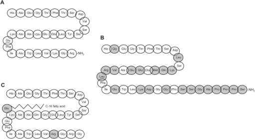
A) The molecular structure of human GLP-1. B) The molecular structure of exenatide (gray colors indicate differences in structure from human GLP-1. C) The molecular structure of liraglutide (gray colors indicate changes in structure from human GLP-1).

Normally, these receptors are bound by a small protein that is made by cells in the small intestine called GLP-1 (glucagon-like peptide-1). The small intestine makes GLP-1 when it is exposed to sugar, and it is the combination of high levels of sugar in the blood, plus the presence of GLP-1 that causes the pancreatic beta cells to release insulin. In type 2 diabetics, the beta cells release insulin, but the body is de-sensitized to it. Therefore, more insulin is needed to control the blood sugar levels. Exenatide does just that by acting like GLP-1.

What does this have to do with stem cells and regenerative medicine? It turns out that the heart also has GLP-1 receptors on the surfaces of its cells and the binding of GLP-1 to these receptors decreases inflammation in the heart, prevents the death of heart muscle cells, protects blood vessels, and protects against damage from reactive oxygen species (also known as free radicals). A fascinating paper has appeared in the Journal of Cellular and Molecular Medicine from the Chinese PLA Hospital in Beijing, China.  In this publication, Chinese cardiologists used a compound that is closely related to Exenatide called Exendin-4 in combination with stem cells to determine if the activation of the GLP-1 receptor influences the healing qualities of stem cells after a heart attack.

In this paper, Yundai Chen and colleagues extracted stem cells from the fat tissue of rats.  This fat tissue was minced and then the stem cells were isolated on the basis of cell surface proteins that are common only to stem cells in fat.  Then Chen and his co-workers gave heart attacks to 120 rats and divided the rats into five groups, with 30 animals each.  The first group received injections of buffer into their hearts immediately after the induction of a heart attack.  The second group received injections of Extendin-4 three days prior to the heart attack and seven days after.  The third group received injections of fat-derived stem cells into the heart tissue bordering the infarcted tissue.  The fourth group received the stem cell injections plus the treatments with Extendin-4.

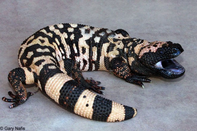
Exendin-4 is one of the compounds extracted from the salivary glands of the Gila monster, a colorful lizard (shown below)  found in the deserts of California.  Exenatide is the acetate salt of Extendin-4, and both compounds bind the GLP-1 receptor and elicits a biological response.

Gila Monster


The results of these experiments were as follows:  the animals that received buffer injections showed respectable amounts of cell death and oxidative damage in their hearts.  However, those animals that received Extendin-4 injections showed less oxidative damage and significantly less cell death.  The same could be said for those animals that received the fat-derived stem cell treatments.  However, those animals that were treated with Extendin-4 plus the stem cells showed substantially less oxidative damage and cell death than all the other groups.

The heart function tests show similar trends.  The ejection fraction, which measures the percentage of the blood that comes into that heart that is pumped out, was in the cellar in the buffer-injected animals, about 10% higher in the Extendin-4 and stem cell-treated animals, and almost 20% higher in the animals treated with both Extendin-4 and the stem cells.   The degree to which the heart muscle contracted (reported as % of shortening or fractional shortening) was over double that in the dually treated animals.  Also the size of the heart scar in the dually treated animals was half the size observed in the animals treated with buffer.

Further examinations of the heart of the dually treated animals showed that the fat-derived stem cells expressed genes normally found in blood vessels cells and heart muscle cells.  This is not definitive evidence that these cells differentiated into heart-specific cell types, but they clearly are surviving and doing something beneficial to the heart.

In culture, the fat-derived stem cells made a whole host of healing molecules when they are treated with Extendin-4.  Also, Extendin-4 treatment protected with stem cells from being damaged by noxious chemicals (e.g., hydrogen peroxide).  Biochemical studies showed that the stem cells that had been treated with Extendin-4 had activated the STAT3 pathway.  Why is this significant?  Because the STAT3 is normally activated by cells when they are stressed.  It is a “I want to survive” kind of pathway.  Extendin-4 seems to cause the stem cells to kick into high gear, survive better, and heal better.

A scheme illustrating the potential cardioprotective signalling pathways through which exenatide may reduce myocardial infarct size and protect the heart against lethal myocardial reperfusion injury. The actual mechanism underlying the cardioprotective effects elicited by exenatide remains to be elucidated, although it is assumed that many of the beneficial effects are mediated through the activation of the glucagon-like peptide-1 (GLP-1) receptor on the cardiomyocytes. The activation of this receptor then recruits pro-survival signalling cascades such as the phosphatidylinositol 3-kinase (PI3K)–Akt and adenylate cyclase (AC)–cAMP–protein kinase A (PKA) pathways which protect the heart against acute ischaemia–reperfusion injury through a number of potential mechanisms including: the inhibition of the mitochondrial permeability transition pore (mPTP), the activation of AKAPs (protein kinase A-anchoring proteins), increased myocardial glucose uptake (possibly via p38 mitogen-activated protein kinase and iNOS), reduced apoptotic cell death, and the transcription of cardioprotective factors (such as PPAR-β/δ, Nrf-2, and HO-1). eNOS, endothelial nitric oxide synthase; GSK, glycogen synthase kinase; HO-1, haem oxygenase 1; iNOS, inducible nitric oxide synthase; NO, nitric oxide; Nrf-2, nuclear respiratory factor 2; PKC, protein kinase; PKG, protein kinase G; PPAR, peroxisome proliferator-activated receptor; ROS, reactive oxygen species.

Now, these experiments were performed in rodents.  Therefore, it remains to be seen if the FDA-approved Exenatide will improve stem cell survival as well as the unapproved Extendin-4.  While they should have the same biological activity, different preparations of the similar molecules can elicit different responses.  Also, it is unclear if this strategy would work in humans.  Do human fat-derived or even bone marrow-derived stem cells respond in a similar fashion to Extendin-4 or Exenatide?  Would such an experiment work in humans?  Do the risks associated with Exenatide administration outweigh the potential benefits of administering it?  Could the stem cells simply be pre-treated with Exenatide before being administered?

This paper has truly opened up a can of worms that should keep scientists busy for many years to come.

Heart Muscle in Young Children May Be Capable of Regeneration


The heart of young children might possess untapped potential for regeneration, according to new research. For decades, scientists believed that after a child’s first few days of life, cardiac muscle cells did not divide. Heart growth was thought to occur by means of enlargement of muscle cells.

This view, however, has been seriously challenged in the last few years. New findings in mice that have recently been published in the journal Cell seriously question this dogma. These results have serious implications for the treatment of congenital heart disorders in humans.

Researchers at Emory University School of Medicine have discovered that in 15-day old mice, cardiac muscle cells undergo a precisely timed spurt of cell division that lasts about a day. The total number of cardiac muscle cells in the heart increases by about 40% during this time when the child’s body is growing rapidly. To give you some perspective, a 15-day-old mouse is roughly comparable to a child in kindergarten, and puberty occurs at day 30-35 in mice.

This burst of cell division is driven by a surge of thyroid hormone. This suggests that thyroid hormone might be able to aid in the treatment of children with congenital heart defects. Small trials have even tested thyroid hormone in children with congenital heart defects.

These findings also have broader hints for researchers who are developing regenerative heart therapies. Activating the regenerative potential of the muscle cells themselves is a strategy that is an alternative to focusing on the heart’s stem cells, according to senior author Ahsan Husain, PhD, professor of medicine (cardiology) at Emory University School of Medicine.

“It’s not as dramatic as in fish or amphibians, but we can show that in young mice, the entire heart is capable of regeneration, not just the stem cells,” he says.

This Emory group collaborated with Robert Graham, MD, executive director of the Victor Change Cardiac Research Institute in Australia.

One test conducted by these groups was to determine how well 15-day old mice can recover from the blockage of a coronary artery. Consistent with previous research, newborn (day 2) mice showed a high level of repair after such an injury, but at day 21, endogenous heart repair was quite poor. The 15-day old mice recovered better than the day 21 mice, indicating that some repair is still possible at day 15.

This discovery was an almost accidental finding while Naqvi and Husain were investigating the role of the c-kit gene. The c-kit gene is an important marker for stem cells in cardiac muscle growth. Adult mice that lack a functional c-kit gene in the heart have more cardiac muscle cells. When do these differences appear?

“We started counting the cardiomyocyte cell numbers from birth until puberty,” Naqvi says. “It was a fascinating thing, to see the numbers increasing so sharply on one day.” According to Naqvi, c-kit-deficient and wild-type mice both have a spurt of proliferation early in life but the differences in cardiac muscle cells between the c-kit+ and c-kit- mice appear later.

“Probably, previous investigators did not see this burst of growth because they were not looking for it,” Husain says. “It occurs during a very limited time period.” Even if in humans, the proliferation of cardiac muscle cells does not take place in such a tight time period as it does in mice, the finding is still relevant for human medicine, he says. “Cardiomyocyte proliferation is happening long after the immediate postnatal period,” Husain says. “And cells that were once thought incapable of dividing are the ones doing it.”

Naqvi and Husain plan to continue to investigate the relationships between thyroid hormone, nutrition during early life, and cardiac muscle growth.

Reference: N. Naqvi et al. A Proliferative Burst during Preadolescence Establishes the Final Cardiomyocyte Number. Cell 157, 795-807, 2014.

Modified RNA Induces Vascular Regeneration After a Heart Attack


Regenerating the heart after a heart attack remains one of the Holy Grails of regenerative medicine. It is a daunting task. Even though text books may say, “the heart is just a pump,” this pump has a lot of tricks up its sleeve.

Stem cell treatments can certainly improve the structure and function of the heart after a heart attack, but getting the heart back to where it was before the heart attack is a whole different ball game. To truly regenerate, the heart, the organ or parts of it need to be reprogrammed to a time when the heart could regenerate itself. If that sounds difficult, it’s because it is. But some recent work suggests that it might at least partially possible.

Kenneth Chien and his colleagues from the Department of Stem Cell Biology and Regenerative Medicine at Harvard University have published a terrific paper in the journal Nature Biotechnology that tries to turn back to clock of the heart to augment its regenerative capabilities.

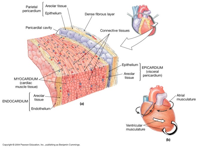
The outermost layer of the heart that surrounds the heart muscle is a layer called the “epicardium.”

epicardiumIn the epicardium are epicardial heart progenitors and these cells are activated within 48 hours after a heart attack in the mouse.  In the fetal heart, epicardial heart progenitors migrate into the heart and differentiate into heart muscle, blood vessels and smooth muscles.  In adults, these cells remain on the surface of the heart and differentiate largely into fibroblasts.  When it comes to regenerative medicine, can we take adult epicardial cells and reprogram them to act like fetal epicardial heart progenitors?

A few experiments have suggested that we can.  In 2011, Smart and others used a small peptide called thymosin β4 to reprogram epicardial cells in mice to form heart muscle and other heart-specific tissues.  Even though the reprogramming was not terribly robust, Smart and others convincingly showed that it was real (Nature 474,640–644).

The Chien group used modified RNA molecules made with unusual nucleotides that encoded the protein vascular endothelial growth factor-A (VEGF-A) to reprogram the epicardium of mice.  VEGF-A is very good and reprogramming the epicardium, and this modified RNA technique does not induce and immune response the way injecting DNA does and the RNA causes bursts of VEGF-A activity that efficiently reprograms the epicardium.

After giving mice heart attacks, Chien and others injected the VEGF-A modified RNAs into the border of the infarcted area of the heart. The modified RNAs induced new gene expression that is normally seen during the establishment of blood vessels.  VEGF-A expression was elevated for up to 6 days after the injections, and animals that had their hearts injected with modified VEGF-A RNA had smaller scars in their hearts, less cell death, and greater tissue volume in their hearts than animals that received either injections of VEGF-A DNA, buffer, or modified RNA that expressed a glowing protein.  Also, the effects of the modified VEGF-A RNA could be abrogated with co-administrating the drug Avastin, which is an antagonist of VEGF-A

Tests with cultured heart cells showed that VEGF-A modified RNA induced blood vessel-specific genes.  These inductions were sensitive to drugs that blocked the VEGF-A receptor, which shows that it is indeed the VEGF-A protein that is inducing these trends.  Finally, a heart muscle gene, Tnnt2 is also induced by the modified VEGF-A RNA.  When the efficacy of the modified VEGF-A RNA was tested in living animals, if was clear that the most numerous cells induced by the modified VEGF-A RNA was endothelial cells, which line blood vessels, followed by smooth muscle cells, and then by heart muscle cells.

Thus, the growth factor VEGF-A can signal to epicardial heart progenitor cells to heal the heart after a heart attack in mice.  It works through the VEGF-A receptor (KDR), and it induces epicardial derived cells (EPDCs) to differentiate into blood vessels, heart muscle cells, and smooth muscle cells, all of which are required to heal the heart.  If VEGF-A signaling can be used to augment heart healing after a heart attack, it might provide a new strategy for healing the heart after a heart attack in a manner that helps the heart heal itself from the inside rather than placing something from the outside into it.

Induced Pluripotent Stem Cells Slow to Grow Tumors in Monkeys


One of the major concerns that dogs the use of pluripotent stem cells in human clinical trials is the risk of tumor formation. Embryonic stem cells and induced pluripotent stem cells have an inherent ability to form special tumors known as teratomas. Teratomas are a rather strange group of tumors that develop from cells early in the developmental program of cells, before they have become committed to mature, adult cell types. Therefore, they contain a mixture of cell types organized to a greater or lesser extent into recognizable structures such as muscles or nerve tissue. In bizarre cases partial teeth may be found.

Embryonic stem cells and their derivatives have a distinct disadvantage in that they are rejected by the immune system of the patient. However, induced pluripotent stem cells (iPSCs), which are made from the patient’s own mature, adult cells, possess the same array of cell surface proteins as the patient’s own cells. Therefore, they are not rejected by the patient’s immune systems. Unfortunately, iPSCs can harbor cancer-causing mutations that were induced during the reprogramming process, and these mutations can seriously compromise their clinical usefulness and safety. Having, not all iPSC lines are the same. Some appear to be safer than others and screening methods that have been developed by stem cell scientists seem to be able to detect unsafe iPSC lines over others.

Now, a new study has shown that it takes a lot of effort to get iPSCs to form tumors after transplantation into a monkey. These findings will bolster the prospects of one day using iPSCs human patients.

Making iPSCs from an animal’s own skin cells and then transplanting them back into the creature also does not trigger an inflammatory response as long as the cells have first been differentiated into a more mature, specialized cell type.

“It’s important because the field is very controversial right now,” says Ashleigh Boyd, a stem-cell researcher at University College London, who was not involved in the work. “It is showing that the weight of evidence is pointing towards the fact that the cells won’t be rejected.”

Pluripotent stem cells have the ability to differentiate into many specialized cell types in culture. Therefore, they have been held out as potential sources of treatments for regenerative therapies for diseases such as Parkinson’s and some forms of diabetes and blindness. iPSCs, which are made by reprogramming adult cells, have an extra advantage because transplants made from them could be genetically matched to the recipient. Also, iPSC derivation is cheaper than cloning procedures and does not destroy a young embryo.

Globally, stem cells researchers are pursuing a variety of iPSCs-based therapies. For example, a group in Japan began enrolling patients for an iPSC-based human clinical trial last year. Experiments in mice from 2011 suggested that even genetically matched iPSCs can elicit an immune response, and pluripotent stem cells can also form slow-growing tumors. Both of these results have elicited deep safety concerns.

A stem-cell scientist from the National Institutes of Health in Bethesda, Maryland, named Cynthia Dunbar led this new study. She decided to evaluate both of these above-mentioned concerns in healthy rhesus macaques. The ability of pluripotent stem cells to form teratomas in laboratory mice is normally a test of their pluripotency. However, to prevent the immune systems of the mice from attacking and destroying these implanted stem cells, mice that lack the cell-mediated arm of the immune response are used. Such mice are called “nude” mice because they do have any hair.

Dunbar said, “We really wanted to set up a model that was closer to human. It was somewhat reassuring that in a normal monkey with a normal immune system you had to give a whole lot of immature cells to get any kind of tumor to grow, and they were very slow-growing.”

Dunbar and her team made iPSCs from skin and white blood cells from two rhesus macaques, and transplanted them back into the monkeys. She and her coworkers were careful to make sure that each monkey was injected with those iPSCs that had been derived from their own cells. For example, if monkey A provided cells that were used to derive iPSC cell line A1, then monkey A was only injected with iPSC line A1 cells and so on. Dunbar and others found that tumor formation required 20 times as many iPSCs as those needed for form a tumor in a nude mouse. These data are invaluable for safety assessments of potential iPSC-dependent therapies. Additionally, even though the injected iPSCs did trigger a mild immune response (white blood cells were attracted to the site of injection, which caused local but not systemic inflammation), when iPSCs were differentiated to a more mature cell types caused no such response.

Dunbar’s study is the first to examine the effects of transplanting undifferentiated iPSCs into the monkey they came from. However it is not the first primate study what happens when cells differentiated from iPSCs are transplanted into non-human primates. Scientists at Kyoto University in Japan transplanted monkey iPSCs that had been differentiated into dopaminergic neurons (the type of neuron that dies in Parkinson’s disease) into the brains of other monkeys and notes that these cells survived for months without forming tumors. Researchers at RIKEN in Kobe, Japan, observed similar results when they transplanted iPSCs that had been differentiated into retinal pigment epithelial cells, which support the photoreceptors at the back of the eye. In neither study did the implanted cells form tumors nor were they immunologically rejected when animals received their own cells. However, in both cases, the transplantation sites that were chosen tend to have a weak capacity to trigger immune responses.

In contrast, Dunbar differentiated iPSCs into bone precursor cells and placed them into small scaffolds just under the skin. Such a location can potentially elicit a robust immune response. However, the transplants did not cause irritation or inflammation, since the differentiated cells do not express embryonic proteins that are normally absent in mature tissues. By eight weeks, new bone had formed, and almost a year later no tumors had formed, and bone formation persisted.

The caveat to these studies is that some work has suggested that bone precursor cells can suppress the immune response against them. To circumvent this problem, Dunbar hopes to repeat these studies using iPSCs that have been differentiated into heart and liver cells.

Doubts About Cardiac Stem Cells


Within the heart resides a cell population called “c-kit cells,” which have the ability to proliferate when the heart is damaged. Several experiments and clinical trials from several labs have provided some evidence that these cells are the resident stem cell population in the heart that can repair the heart after an episode of cardiac injury.

Unfortunately, a few new studies, and in particular, one that was recently published in the journal Nature, seem to cast doubt on these results. Jeff Molkentin of Cincinnati Children’s Hospital Medical Center and his co-workers have used rather precise cell lineage tracing studies in mice to follow c-kit cells and their behavior after a heart attack. His results strongly suggest that c-kit cells rarely produce heart muscle cells, but they do readily differentiate into cardiac endothelium, which lines blood vessels.

“The conclusion I am led to from this is that the c-kit cell is not a cardiac stem cell, at least in term of its normal, in vivo role,” said Charles Murry, a heart regeneration researcher at the University of Washington who was not involved in this study.

Molkentin’s study is what some stem cells researchers are calling the nail in the coffin for c-kit cells. In fact the Molkentin paper is simply the latest in a series of papers that were unable to reproduce the results of others when it comes to c-kit cells. Worse still, one of the leading laboratories in the c-kit work, Piero Anversa at Harvard Medical School, has had to retract on of this papers and there is also some concern about his publication regarding the SCIPIO trial. Eduardo Marbán, an author of the new study and a cardiologist at the Cedars-Sinai Heart Institute in Los Angeles, said, “There’s been a tidal wave in the last few weeks of rising skepticism,” Nevertheless, the present dispute is not yet settled, and many scientists still regard the regenerative powers of c-kit cells as a firmly established fact.

In his laboratory, Piero Anversa and his colleagues and collaborators have shown that c-kit cells—cardiac progenitor cells expressing the cell surface protein c-kit—can produce new heart muscle cells (cardiomyocytes). Anversa and others also helped usher these cells, which are also known as CPCs or cardiac progenitor cells, into clinical trials to test whether they might help repair damaged cardiac tissue. This culminated in the SCIPIO trial, which showed that patients treated with their own CPCs showed long-lasting and remarkable improves in heart function.

Follow-up work by other research teams, however, has not been able to confirm these studies, and their work has raised doubts about the potential of c-kit cells to actually build new heart muscle. In his contribution to the c-kit controversy, Molkentin and his colleagues genetically engineered mouse strains in which any c-kit-expressing cells and their progeny would glow green. To do this, they inserted a green fluorescent protein gene next to the c-Kit locus. Therefore any c-kit-expressing cells in the heart would not only glow green, but whatever cell type they differentiated into would also glow green. After inducing heart attacks in these mice, Molkentin and others discovered that only 0.027 percent of the heart muscle cells in the mouse heart originated from c-kit cells. “C-kit cells in the heart don’t like to make myocytes,” Molkentin told The Scientist. “We’re not saying anything that’s different” from groups that have not had success with c-kit cells in the past,” Molkentin continued, “we’re just saying we did it in a way that’s unequivocal.”

Molkentin’s study did not address why there’s a discrepancy between his results and those of Anversa and another leader in the c-kit field, Bernardo Nadal-Ginard, an honorary professor at King’s College London. Last year in a paper published in the journal Cell, Nadal-Ginard and his colleagues showed that heart regeneration in rodents relies on c-kit positive cells and that depleting these cells abolishes the regenerative capacity of the heart.

In an email to a popular science news publication known as The Scientist, Nadal-Ginard suggested that technical issues with Molkentin’s mouse model could have affected his results, causing too few c-kit cells to be labeled. Additionally, “the work presented by Molkentin used none of our experimental approaches; therefore, it is not possible to compare the results,” Nadal-Ginard said in an e-mail.

Anversa said his lab is working with the same mouse model Molkentin used, “but our data are too preliminary to make any specific comment. Time will tell.”

Molkentin’s paper seems point to further problems with Aversa’s work with c-kit cells.  Last month, one of the papers Anvera and his group had published in the journal Circulation had to be retracted because the data used the write that paper were “sufficiently compromised.”  Then a few days later, the paper describing the results of the SCIPIO study that appeared the journal The Lancet expressed concern about supplemental data that was included with the published results.  These data came from the human clinical trial that treated heart patients with their own c-kit cells.  Harvard Medical School and Brigham and Women’s Hospital are investigating what went wrong with this study and the publication itself.

Regardless of Anversa’s present tribulations, Marbán is advancing another type of heart-specific stem cell, called cardiosphere-derived cells or CDCs.  Marbán and his colleagues have already used CDCs in a human clinical trial known as the CADUCEUS trial.  In this trial, heart attack patients treated with CDCs saw their heart scars shrink.  Marbán said he had been a true believer in c-kit cells, until the data started mounting against them. “The totality of the evidence now says the c-kit cell is no longer a cardiomyocyte progenitor,” he said.

Now even if c-kit cells do not make new heart muscle, it is possible that they heal the heart through other means.  The patients in the SCIPIO trial saw real, genuine improvements in their heart function and these results cannot be so cavalierly dismissed.  In fact, Murry said that just because the mechanistic basis for the human study remains in doubt, promising clinical results should not be dismissed. “Those results can be considered independent,” he said.  Molkentin also added that it’s possible that c-kit cells work in unknown ways to repair heart tissue.  Since clinical treatments involves high levels of c-kit cells that have been immersed in culture conditions, “Perhaps these cells act a little different,” Molkentin said.

Nadal-Ginard also noted that discrepancies do exist between his data and those of others, and that these discrepancies should not be papered over, but should be robustly debated and addressed.  He said he’d be willing to work with Molkentin to get to the bottom of it. “The concept under dispute is too important for the field of regenerative medicine—and regenerative cardiology, in particular—to turn into a philosophical/dogmatic argument instead of settling it in a proper scientific manner.”  Here here.

Stem Cell Therapy Replaces Dead Heart Muscle in Primates


The laboratory of Charles Murry at the University of Washington has used embryonic stem cells to make heart muscle cells that were then used to regenerate damaged hearts in non-human primates. This experiment demonstrates the possibility of using heart muscle cells derived from pluripotent stem cells, but it also underscores the many challenges that still must be overcome.

When the heart undergoes a heart attack or other types of damage, heart muscle cells begin to die off and these cells are not easy to replace. Heart muscle cells, also known as cardiomyocytes, do not readily replace themselves. Even though the heart has a resident stem cell population, (cardiac progenitor cells or CPCs) these heart-specific stem cells have a limited capacity to regenerate the heart. After a heart attack, as many as one billion cardiomyocytes or more die. The loss of so many beating heart muscle cells compromises heart function and can also lead to chronic heart failure and even death.

Physicians, cardiologists, and researchers have been on the lookout for new and improved procedures and technologies to replenish damaged heart tissue. Several different types of stem cells have shown promise in animal models and in human clinical trials. Stem cells from bone marrow have the ability to secrete a cocktail of molecules that stimulate heart regeneration. Whole bone marrow or isolated stem cell populations have shown variable, but statistically significant in patients who have had a recent heart attack. Unfortunately, stem cells from bone marrow do not have the ability to differentiate into heart muscle cells, and to maximize regeneration of the heart, damaged heart muscle must be replaced.

Human embryonic stem cells have proven promising in small animal models, but the long-term effects of embryonic stem cell-mediated improvements in some cases have proven to be transient. An additional problem with embryonic stem cell-derived heart muscle cells is their tendency to cause abnormal heart rates, otherwise known as arrhythmias.

Scientists in Murry’s laboratory tried to scale-up the production of cardiomyocytes from human embryonic stem cells in order to test the regenerative ability of these cells in a large animal model – non-human primates. These experiments were published online on April 30, 2014, in the journal Nature.

Murry’s team derived cardiomyocytes from genetically-engineered human embryonic stem cells that made a fluorescent calcium indicator that glowed in the presence of high calcium ion concentrations. With this fluorescent calcium indicator, Murray and his coworkers could track the calcium waves that mark the electrical activity of a beating heart. The animal subjects for this experiment were pigtail macaques (Macaca nemestrina) that had suffered heart damage and had been treated with drugs to suppress their immune systems. Five days later, the embryonic stem cell-derived cardiomyocytes were delivered in a surgical procedure to the damaged regions and surrounding border zones of the heart.

Over a 3-month period, the implanted cells infiltrated damaged heart muscle, matured, and organized themselves into muscle fibers in all the monkeys who received the treatment. An average of 40% of the damaged tissue was replaced by these grafts. Three-dimensional imaging showed that arteries and veins integrated into the grafts. Because sick hearts often contain clogged blood vessels, oxygen delivery to the damaged heart tissue was minimal. However, because these grafts contained integrated blood vessels, they would potentially be long-lasting.

Calcium activity studies showed that the heart muscle tissue within the grafts were electrically active and coupled to activity of the host heart. The grafts beat along with host muscle at rates of up to 240 beats per minute, the highest rate tested.

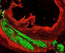
Cardiac cells derived from human stem cells (green) meshed and beat along with primates’ heart cells (red). Credit: Murry Lab/University of Washington.

All the macaques that received the grafts showed transient arrhythmias or irregular heart rates. However, these subsided by 4 weeks post-transplantation. The animals remained conscious and in no apparent distress during periods of arrhythmia. However, this problem will need to be addressed before this approach can be tested in humans.

“Before this study, it was not known if it is possible to produce sufficient numbers of these cells and successfully use them to remuscularize damaged hearts in a large animal whose heart size and physiology is similar to that of the human heart,” Murry says.

This article shows that despite the obstacles that remain, transplantation of human cardiomyocytes derived from pluripotent stem cells may be feasible for heart patients.

There are a few caveats I would like to mention.  First of all, these animals underwent immunosuppression.  If this procedure were to be used in a human patient, the human patient would need life-long immunosuppression, which has a wide range of side effects and tends to stop working over time.  Therefore, induced pluripotent stem cells are a better choice.  Secondly, the paper admits that the implanted cells underwent “progressive but incomplete maturation over a 3-month period.”  If the implanted cells are not maturing completely, then the risk of arrhythmias still exists, even though they may have subsided in these animals after 4 weeks.  This leads me to my third point.  These animals were watched for 3 months.  How do we know that these results were not transient?  Longer-term experiments are needed to establish that this treatment actually is long-term and not transient.  It is, however, gratifying to see an experiment that was extended to 12 weeks rather than the usual 4 weeks that is usually seen in mice.

Finally, tucked away in the extended data is the statement: “The cell-treated animals showed variable responses, with some having increased function and some having decreased function. Because of small group size, no statistical effects of hESC-CM therapy can be discerned.”  In other words, the treatments worked swimmingly in some animals and not at all in others.  This was a small animal trial and better numbers will be needed if this technology is to come to the clinic.

Compound from Sully Putty Might Advance Neural Stem Cell Therapies


According to a University of Michigan engineering team, human pluripotent stem cells differentiate differently in response to the sponginess of the surface upon which they grow.

University of Michigan assistant professor of mechanical engineering, Jianping Fu, and his colleagues, efficiently directed human embryonic stem cells to differentiate into working spinal cord cells by growing the cells on a carpet of poly(dimethylsiloxane), which is one of the main ingredients in the toy known as “Silly Putty.” This study established the importance of physical signals in the control of stem cell differentiation.

According to Fu, these data could be the beginning of a series of investigations that uncovers the most efficient way to guide pluripotent stem cells to differentiate into nervous tissues that can be used to replace diseased cells in patients with Alzheimer’s disease, Huntington’s disease or amyotrophic lateral sclerosis (Lou Gehring’s disease).

In Fu’s system, he and his co-workers engineered the poly(dimethylsiloxane) carpets by using this compound to form fine threads that were strung between microscopic posts. By varying the height of the posts, Fu discovered that he could vary the stiffness of the surface. Shorter posts gave a more rigid, stiff carpet and longer posts gave softer more plush carpets.

When embryonic stem cells were grown on poly(dimethylsiloxane) carpet strung between tall posts, they differentiated into neurons much more quickly and at a higher percentage than when they were grown on the more rigid and stiffer poly(dimethylsiloxane) carpets.  After 23 days, colonies of spinal cord motor neurons that control how muscles move grew on the softer micropost carpets.  These cell assemblages were four times more pure and 10 times larger than those growing on either traditional plates or rigid carpets.

“To realize promising clinical applications of human embryonic stem cells, we need a better culture system that can reliably produce more target cells that function well,” said Fu.  He added: “Our approach is a big step in that direction, by using synthetic micro-engineered surfaces to control mechanical environmental signals.”

Fu is presently collaborating with U-M Medical School professor of neurology, Eva Feldman.  Dr. Feldman is an expert in amyotrophic lateral sclerosis (ALS), and firmly believes in the power of stem cells to help ALS patients grow new stem cells that can replace the diseased, death or damaged nerve cells.  Feldman is also applying Fu’s ingenious technique to make neurons from a patient’s own cells.  Mind you, these results are purely exploratory at this point, since Feldman simply wants to determine the feasibility of this procedure.

Even if this technique does not pan out for regenerative treatments, it provides a very workable model system to study the electrical behavior of neurons from ALS patients in comparison to neurons from non-ALS individuals.

Fu’s system also has identified a cell signaling pathway that is involved in the regulation of mechanically sensitive behaviors.  This signaling pathway – the Hippo/Yap pathway – is also involved in controlling organ size and suppression of tumor formation.

Corresponding proteins in Drosophila and mammals are shown in the same colours. When organs are growing (Hippo pathway OFF), nuclear Yki/Yap binds to unknown DNA-binding factor(s) X and regulates the transcription of growth targets. When organs have reached the correct size (ON), the Hippo signalling pathway is activated (unknown ligand Y–Fat– Merlin–Expanded–Hippo interactions, in the Drosophila case; ligand Y–FatJ–NF2–FDM6–Mst½–Lats½ in mammals), and Yki and YAP is inactivated by localizing to the cytoplasm in response to Wts phosphorylation and 14-3-3 binding. ? indicates regulatory relationships that still need to be investigated. Figure adapted from reference 2.
Corresponding proteins in Drosophila and mammals are shown in the same colors. When organs are growing (Hippo pathway OFF), nuclear Yki/Yap binds to unknown DNA-binding factor(s) X and regulates the transcription of growth targets. When organs have reached the correct size (ON), the Hippo signalling pathway is activated (unknown ligand Y–Fat– Merlin–Expanded–Hippo interactions, in the Drosophila case; ligand Y–FatJ–NF2–FDM6–Mst½–Lats½ in mammals), and Yki and YAP is inactivated by localizing to the cytoplasm in response to Wts phosphorylation and 14-3-3 binding. ? indicates regulatory relationships that still need to be investigated. Figure adapted from reference 2.

The work of Fu and Feldman could certainly provide significant advances in our understanding of how pluripotent stem cells differentiate in the body.  This work also suggests that physical signals are important in patterning the nervous system, especially since the cells of the nervous system become specialized for specific tasks according to their physical location within the body and nervous system in general.

Stem Cell Treatments Cure Mice With MS-Like Disease


University of California researchers have discovered that human stem cells can reverse a multiple sclerosis-type condition in mice. Soon to be published in the journal Stem Cell Reports, this work could potentially lead to new treatments for multiple sclerosis (MS).

When this team first transplanted the stem cells into severely disabled MS mice, they were quite sure that the immune systems of the mice would attack these transplanted stem cells, which were from humans, would attack and reject them. However, the experiment had surprising results.

“My postdoctoral fellow Dr. Lu Chen came to me and said, ‘The mice are walking.’ I didn’t believe her,” said co-senior author, Tom Lane, PhD., who is presently a professor of pathology at the University of Utah, who began this study at University of California, Irvine.

Within next 10 to 14 days, the mice regained their lost motor skills, and six months later, they still show no signs of slowing down.

“This result opens up a whole new area of research for us,” said co-senior author Jeanne Loring, PhD, a professor at The Scripps Research Institute in La Jolla, Calif.

A chronic disease, MS results from the body’s own immune system attacks the body’s central nervous system. In particular, the insulating layer that surrounds many nerve fibers – a fatty substance called myelin – is slowly destroyed and this exposes nerves and slows or interrupts the transmission of nerve impulses. The symptoms of MS may be mild or severe, and includes numbness in the limbs, difficulty walking, paralysis, loss of vision, fatigue and pain.

Researchers say the MS mice treated with human stem cells experienced a dramatic reversal of symptoms within days. Immune attacks were blunted and damaged myelin was repaired.

Chen, Loring and their colleagues were further surprised that not only did the mice not initially reject these implanted stem cells, but they never showed any signs of rejection, even after one week.

Now Lane and others are eager to test this therapy in human clinical trials.

“Rather than having to engraft stem cells into a patient, which can be challenging, we might be able to put those chemical signals into a drug that can be used to deliver the therapy much more easily,” said Lane. He continued: “I would love to see something that could promote repair and ease the burden that patients with MS have.”

Previous work with stem cells has shown some promise in the treatment of MS. For example, in 2013, the US Food and Drug Administration (FDA) approved a small clinical trial of genetically modified mesenchymal stem cells initially harvested from bone marrow of MS patients. These stem cells were injected into the cerebrospinal fluid that surrounds the spinal cords of MS patients. Previous small studies conducted between 2005 and 2007 of this therapy in humans found that implantation of these modified stem cells reduced brain inflammation and repaired damaged layers of myelin. This small study only treated and tested seven patients.

There is no known cure for MS and drugs to treat it have limited effectiveness. An estimated 400,000 Americans have the disease and more than 2 million worldwide.

Making Safer Induced Pluripotent Stem Cells


Induced Pluripotent Stem Cells or iPSCs are made from mature, adult cells by means of genetic engineering techniques that introduce pluripotency genes into the adult cells. These introduced genes drive the adult cells to de-differentiate into an embryonic stem cell-like cells that have the ability to differentiate into almost all of the cells of the adult human body.

Despite the attractiveness of these cells for regenerative medicine, there is a dark side to iPSCs, since the production of iPSCs introduces new mutations into them. While not all iPSC lines are created equal and the methods by which they are derived also influences the degree of genetic damage to them, there are serious questions about the safety of iPSCs for clinical use.

A new paper from a Spanish group has discovered that a protein called SIRT1 is required to protect the chromosomal integrity of iPSCs during reprogramming.

Linear chromosomes are capped at their ends by special structures called telomeres. These telomeres shorten over time, but during reprogramming, the telomeres lengthen. This lengthening requires the SIRT1 protein, but SIRT1 is also required to maintain the telomeres at this elongated length. In this way, SIRT1 helps safeguard the chromosomes during reprogramming. The SIRT1 protein is also up-regulated in embryonic stem cells.

Using a mouse model system, researchers from the Spanish National Cancer Research Center’s Telomeres and Telomerase Group made cells that completely lacked any functional SIRT1 protein. Maria Luigia De Bonis, Sagrario Ortega, and Maria A. Blasco from CNIO discovered that SIRT1-deficient mouse cells could be reprogrammed, but the telomeres of these cells lengthened much less efficiently, and eventually experienced chromosome abnormalities and DNA damage. SIRT1-deficient iPSCs also formed larger, poorly differentiated tumors when transplanted into nude mice.  Thus SIRT1 seems to keep the chromosomes of iPSCs healthy.

Interestingly, the c-MYC protein, which is encoded by the c-myc gene – one of the four genes required to reprogram adult cells – is stabilized by SIRT1. Normally, the c-MYC protein has a very short half-life, but SIRT1 protects c-MYC from degradation, and c-MYC increases the production of the enzyme that replicates and elongates the telomeres; telomerase.

This work could very possibly lead to protocols that will stabilize the chromosomes of iPSCs during reprogramming. This will make iPSCs safer for possible use in the clinic.

Toxic Gas Prompts Mesenchymal Stem Cells to Become Bone Cells


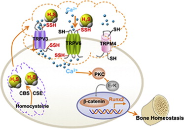
Hydrogen sulfide smells like rotten eggs and is toxic to human life at moderate concentrations. Therefore, imagine the surprise of researchers when they discovered that low concentrations of this poisonous gas actually stimulate mesenchymal stem cells from bone marrow to differentiate into bone-making cells.

In a paper published in the journal Cell Stem Cell, Yi Liu from the Ostrow School of Dentistry at the University of Southern California and colleagues have discovered that hydrogen sulfide (H2S), acts as a “gaseous signaling molecule” that mesenchymal stem cells actually produce at sub-lethal concentrations.

H2S acts as a “gasotransmitter” that regulates multiple signaling pathways. To determine the extent of these pathways, Liu and his colleagues made mice that were unable to synthesize any H2S. The H2S-deficient mice showed distinct abnormalities in bone marrow mesenchymal stem cells. Namely, mesenchymal stem cells (MSCs) from H2S-deficient mice were unable to properly self=-renew or differentiate into bone-making cells (osteoblasts).

When Liu and others dug a little deeper, they found that H2S deficiency results in aberrant influx of intracellular Ca2+. Problems with calcium handling arose because calcium channels have amino acids that actually react with the H2S. This reaction between the calcium channels and H2S opens the channels and allows entry of calcium into the cell. Now cells contain a host of enzymes that need calcium to operate properly.  Without the reaction of the calcium channels with H2S, calcium does not influx into the cell and the differentiation of mesenchymal stem cells into bone-making cells stops.

schematic diagram R1.ppt

Why is this important? Consider some of the diseases of bone, such as osteoporosis, in which the bones thin and become fragile. Restoring mesenchymal stem cell function in osteoporotic patients with treatments of H2S levels at nontoxic levels may provide treatments for diseases such as osteoporosis that might arise from H2S deficiencies.

Thus by understanding stem cell biology better, we can potentially treat a disease like osteoporosis with small amounts of a stinky gas. Incredible, isn’t it?

Mesenchymal Stem Cells Treating Fecal Incontinence in Animals


A new study published by the journal STEM CELLS Translational Medicine has demonstrated the regenerative effects of mesenchymal stem cells (MSC) on the anal sphincter. This work could have implications for the 11 percent of the population who suffer from fecal incontinence as a result of injury or disease.

Massarat Zutshi, M.D., and Levilester Salcedo, M.D., led the research team made up of their colleagues at the Cleveland Clinic (Cleveland, Ohio) and Summa Cardiovascular Institute and Northeast Ohio Medical University (Akron, Ohio).

Unfortunately none of the present treatments for fecal incontinence “are efficacious in the long-term or without complications related to the surgery or the device,” explained Dr. Zutshi. Additionally, she noted that mesenchymal stem cells (MSCs) from adipose tissue and skeletal muscle have improved heart function and urinary sphincter in animal models, On the strength of these successes, Zutshi and her colleagues decided to use these cells to treat damaged anal sphincters.

In this study. Zutshi and Salcedo and their research teams used a single intramuscular (IM) injection of MSCs compared to a series of intravenous (IV) treatments. For these experiments, they employed rats that had undergone an excision of 25 percent of their anal sphincter complex. 24 hours after anal sphincter injury, one group received a single IM injection of stem cells directly into their anal sphincters. A second group began a series of six consecutive daily treatments delivered by IV through their tail veins, as did a group of non-injured animals. A third group of injured animals received no stem cells.

The scientists measured anal pressures in these animals. Accordingly, they measured anal pressures were recorded prior to injury, then again at 10 days and five weeks after treatment. Ten days after the IM treatment, resting and peak pressures were significantly increased in the injured groups compared to the control group that received no treatment. At five weeks, the anal pressures of the two groups of injured rats receiving treatments were almost on par with the non-injured group.

“Both IM and IV MSC treatment after injury cause increase in anal pressures sustained at five weeks even though fewer cells were injected IM,” Dr. Zutshi concluded. “The MSC-treated groups showed less scarring than PBS treatment, with the IV infusion group showing the least scarring.

“Since MSC delivered IM or IV both resulted in functional recovery, the IM route may be preferable as fewer cells seem to be needed.”

This research demonstrates the regenerative effects of mesenchymal stem cells on the injured anal sphincter. Since fewer cells were needed for intramuscular injections, this mode of administration might be used in future clinical trials, said Anthony Atala, M.D., editor of STEM CELLS Translational Medicine and director of the Wake Forest Institute for Regenerative Medicine.

Genetically Modified As a Potential Treatment of Alzheimer’s Disease


A neurobiology team from UC Irvine (full disclosure, my alma mater) has used genetically engineered neural stem cells to treat mice with a form of Alzheimer’s disease (AD). Such implanted neural stem cells ameliorated some of the symptoms and pathological consequences of this disease in affected mice.

Patients with AD show accumulation of the protein amyloid-beta in their brains. These amyloid-beta clusters form clear plaques in the brain that are also quite toxic to nearby neurons.

Amyloid beta plaques can be cleared with the protein in them is degraded. Fortunately, the enzyme neprilysin can degrade these plaques, but the brains of AD patients show low levels of this enzyme. Neprilysin levels decrease with age and this is probably one of the reasons AD tends to be a disease of the aged.

The UC Irvine group, under the direction of Mathew Blurton-Jones, tried to deliver neprilysin to the brains of afflicted mice and used neural stem cells to do it. The goal of this work was to determine if increased degradation of the amyloid plaques abated the pathological effects of AD.

In this work, two different AD model systems were used. Thy1-APP and 3xTg-AD mice both exhibit many of the pathological effects of AD, and both were used in this study. Neural stem cells were transfected in express 25 times more neprilysin that normal. Then these genetically modified neural stem cells were transplanted into two areas of the brain known to be affected by AD: the hippocampus and the subiculum, which lies just below the hippocampus. Other AD mice were transplanted with neural stem cells that had not been transformed with neprilysin.

Post-mortem examination of both groups of mice even up to three months after transfection of the neural stem cells showed that those mice that received injections of neprilysin-expressing neural stem cells had significant reductions in amyloid-beta plaques within their brains compared to control mice. The neprilysin-expressing cells even seemed to promote the growth of neurons and the establishment of connections between them.

A truly remarkable finding of this work was that numbers of amyloid-beta plaques were also reduced in area of the brain that were some distance from the areas where the stem cells were injected. This suggests that the injected stem cells migrates across the brain, reducing plaque formation as they went.

Future experiments will seek to see if the reduction in amyloid-beta plaques also leads to improvements in cognition. Also, before this protocol can make its transition from animal models of human trials, the UC Irvine group will need to determine if the neprilysin also degrades soluble forms of amyloid-beta.

Every AD mouse model varies as to the types of pathologies observed in the brains of the affected mice. For this reason, this group tested their treatment strategy in two distinct AD mouse models, and in both cases, the neprilysin-expressing neural stem cells reduced the incidence of amyloid beta plaques. This strengthens the conclusion and neprilysin-expressing neural stem cells can indeed degrade amyloid-beta plaques.

More work needs to be done before this work can be used to support a human trial, but this is certainly an encouraging start to something great.