How Our Own Immune Systems Aid the Spread of Breast Cancer


Our immune systems help us fight off diseases and invasions of our bodies by foreign organisms. How surprising might it be to learn that our immune systems actually help tumors spread through our bodies?

Dr. Karin de Visser and her team at the Netherlands Cancer Institute have discovered that breast tumors cells induce certain immune cells to enable the spread of cancer cells. They published their findings online on March 30 in the journal Nature.

About one in eight women will develop breast cancer in Western countries. Of those women who die of this disease, 90 percent of them die because the cancer has spread to other parts of their body and formed metastases. Given these grim facts, cancer researchers are spending a good deal of time, treasure and energy to understand how metastasis occurs. A few years ago, several cancer biologists reported that breast cancer patients who showed high numbers of immune cells called neutrophils in their blood show an increased risk of developing metastatic breast cancers. Immune cells like neutrophils are supposed to protect our body. Why then are high neutrophil levels linked to worse outcome in women with breast cancer?

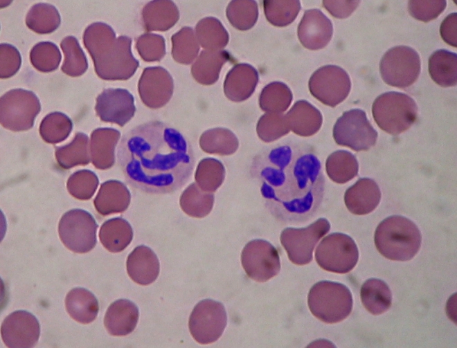
Neutrophils
Neutrophils in a blood smear amidst red blood cells.


Dr. Karin de Visser, group leader at the Netherlands Cancer Institute, and her team discovered that certain types of breast tumors use a signaling molecule called Interleukin-17 to initiate a domino effect of reactions within the immune system. The tumor cells stimulate the body to produce lots of neutrophils, which typically occurs during an inflammatory reaction. However, these tumor-induced neutrophils behave differently from normal neutrophils. These tumor-induced neutrophils block the actions of other immune cells, known as T cells. T cells are the cells that can (sometimes) recognize and kill cancer cells.

De Visser and her team went on to define the role of the signaling protein called interleukin-17 (or IL-17) in this process. “We saw in our experiments that IL-17 is crucial for the increased production of neutrophils”, says De Visser. “And not only that, it turns out that this is also the molecule that changes the behavior of the neutrophils, causing them to become T cell inhibitory.”

The first author of the Nature paper, postdoctoral researcher Seth Coffelt, showed the importance of the IL-17-neutrophil pathway when he inhibited the IL-17 pathway in a mouse model that mimics human breast cancer metastasis. When these neutrophils were inhibited, the animals developed much less metastases than animals from the control group, in which the IL-17-neutrophil route was not inhibited. “What’s notable is that blocking the IL-17-neutrophil route prevented the development of metastases, but did not affect the primary tumor,” De Visser comments. “So this could be a promising strategy to prevent the tumor from spreading.”

Inhibiting neutrophils would not be a prudent clinical strategy, since drugs that inhibit neutrophils would make patients susceptible to all kinds of infections. However, Inhibition of IL-17 might be a safer strategy. Fortunately, drugs that inhibit IL-17 already exist.  Presently, anti-IL-17 drugs are being tested in clinical trials as a treatment for inflammatory diseases, like psoriasis and rheumatism. Last month, the first anti-IL-17 based therapy for psoriasis patients was approved by the U.S. Federal Drug Administration (FDA). “It would be very interesting to investigate whether these already existing drugs are beneficial for breast cancer patients. It may be possible to turn these traitors of the immune system back towards the good side and prevent their ability to promote breast cancer metastasis,” De Visser says.

REALISTIC Trial to Test Efficacy of Bone Marrow Stem Cells on Liver Disease


Chronic liver disease is the fifth leading cause of death in the United Kingdom. With the long-standing shortage of donated, transplantable livers, the prognosis of such patients seems grim.

Several preclinical studies in animals have established that mobilization of bone marrow stem cells or direct injection of bone marrow stems into a damaged liver can augment healing and improve survival (Sukaida I, and others, Hepatology 2004;40:1304–11; and Yannaki E, and others, Exp Hematol 2005;33:108–19). Some small clinical trials have examined the use of a patient’s own bone marrow stem cells to prime the liver and stimulate its own internal healing mechanisms. These studies were small and varied in the manner in which the stem cells were delivered, but they di show that the stem cell treatments were safe and even improved the health of the liver significantly (Gordon MY, and others, Stem Cells 2006;24:1822–30; Terai S, and others, Stem Cells 2006;24:2292–8). Also, in patients with liver cancer who had to have portions of their livers removed, bone marrow stem cell treatments accelerated liver healing (am Esch JS, and others, Ann Surg 2012;255:79–85; am Esch JS, and others, Stem Cells 2005;23:463–70; and Furst G, and others, Radiology 2007;243:171–9).

Clearly there is a need for a larger, more systematic study of the efficacy of bone marrow stem cells as a therapeutic agent in patients with liver failure. To that end, Philip Newsome and his colleagues at the University of Birmingham, in collaboration with colleagues from Scotland, Newcastle, and Nottingham have initiated the REALISTIC trial, which stands for REpeated AutoLogous Infusions of STem cells In Cirrhosis.

This is a multi-center clinical trial and it will examine patients with Cirrhosis (fatty liver disease), regardless of the cause of that liver disease. Patients whose livers were damaged by excessive alcohol use, hepatitis B or C infections, or genetic conditions are all eligible for this study, but anyone who liver is too far-gone to be helped by a treatment like this or has had a liver transplant is not eligible.

Patients will receive injections of a drug called lenograstim (G-CSF) to mobilize bone marrow stem cells into the blood. These blood-based stem cells will then be collected and concentrated, and then implanted into the liver. Patients will be assessed at 3 months after the treatment and then followed-up for 1 year. Liver health will be assessed by means of medical imaging of the liver and various blood tests.

Patients will be evaluated using the Model for End-Liver Disease or MELD scoring system. Secondary tests will measure the degree of liver scarring, the degree of liver stiffness, blood tests, survival, and liver function.

Patients will also be placed into three groups. One group will only have the bone marrow stem cells mobilized from bone marrow without being collected. Another group will have the cells collected and implanted into the liver. The third group will receive standard care with not stem cells treatments.

There is a need for a study like this. I only hope that Newsome and his group can recruit the patients and get started collecting data as soon as they can.

City of Hope Launches Alpha Clinics – A New Stem Cell Clinic


Cancer patients usually have three different options: surgery, chemotherapy and radiation therapy. None of these options elicits a great deal of confidence. A new study at City of Hope will officially open the Alpha Clinic for Cell Therapy and Innovation. At this center, patients battling cancer and other life-threatening diseases will have another option: stem cell-based therapy.

The Alpha Clinic, which officially opened March 19, is funded by an $8 million, five-year grant from the California Institute for Regenerative Medicine. This grant will also be supplemented by matching funds from City of Hope. The Alpha clinic will combine the uniquely patient-centered care for which City of Hope is well-known with the most innovative, stem cell-based therapies being studied to date. This approach will hopefully revolutionize the treatment of not only cancer, but also AIDS and other life-threatening diseases.

“We are in a new era of cellular therapy,” said John Zaia, M.D., the Aaron D. Miller and Edith Miller Chair in Gene Therapy, chair of the Department of Virology and principal investigator for the stem cell clinic. “The California Institute for Regenerative Medicine recognizes this, and they have been leading the field. Alpha Clinics like ours aim not only to provide research to benefit patients in the future, but also to get these innovative treatments running in real-life clinics to benefit patients now.”

The christening of City of Hope’s Alpha Clinic is the culmination of a decade of planning and visionary thinking. When the state of Californian voted to found the California Institute for Regenerative Medicine, the funds now became available to start the institute. New stem cell therapies are ready for clinical trials, and City of Hope is home to one of three Alpha Clinics in the state. The other two clinics are at the University of California San Diego and a joint clinic by University of California Los Angeles and University of California Irvine.

City of Hope’s first trials will study stem cell-based therapies for leukemia, and the use of neural stem cells to deliver treatments to brain tumors. The first such study will modify a patient’s own immune cells so that they can recognize and fight cancer cells. Cancer researchers hope the modified cells will be able to attack existing cancer cells, and also be able to attack the cancer again should it recur.

Brain cancer patients will also be able to enroll in a study that uses neural stem cells, which have an innate ability to home to tumor cells, as a delivery mechanisms for cancer drugs. Genetically engineered neural stem cells can bring targeted therapies across the blood-brain barrier, and can potentially deliver drugs directly to tumor sites, which eliminates systemic toxicity.

The US Food and Drug Administration (USFDA) has already approved a new HIV trial that will be conducted at the City of Hope Alpha Clinic. This trial will use “molecular scissors” known as zinc finger nucleases to edit the blood cells of HIV patients and remove a specific gene. Without this particular gene, the cells are unable to produce a protein that HIV requires in order to invade cells and replicate. The approach has the potential to eliminate HIV from the body.

“As we move forward with our Alpha Clinic, we will also be defining a new discipline in nursing of cellular therapy,” said Shirley Johnson, R.N., senior vice president, chief nursing and patient services officer at City of Hope. “This clinic is a unique opportunity to provide patients with the most leading-edge treatments while still giving them the compassionate comprehensive care City of Hope patients expect.”

The Alpha Clinic launched officially on March 19. Future trials will include T cell immunotherapy for blood cancer, new brain cancer therapies, treatments for breast cancer metastases and ovarian cancer treatments. Zaia said the clinic also plans to work with City of Hope’s diabetes researchers to introduce treatments for diabetes, exploring both the potential of pancreatic stem cells and preventing the immune system from attacking insulin-producing cells.

Spanish Team Develops Anti-Obesity Treatment in Animal Models


A research team from the Spanish National Cancer Research Center (CNIO) has shown that partial pharmacological inhibition of the PI3K enzyme in obese mice and monkeys reduces body weight and physiological manifestations of metabolic syndrome, specifically diabetes and hepatic steatosis (fatty liver disease), without any signs of side effects or toxicities. They published their work in the journal Cell Metabolism. This collaborative project between the Tumor Suppression Group headed by Manuel Serrano at the CNIO (Madrid, Spain) and the Translational Gerontology Branch headed by Rafael de Cabo at the U.S. National Institute on Aging, National Institutes of Health (NIH, Baltimore, MD, USA), included the participation of the NeurObesity group of CIMUS led by Miguel Lopez at the University of Santiago de Compostela (Santiago de Compostela, Spain).

PI3K (phosphatidylinositol-3-kinase) is the name of an enzyme that regulates the balance between the biosynthesis of cellular components and the burning of nutrients to make energy in cells. Specifically, PI3K promotes cellular growth and biosynthesis, which can lead to the induction of growth and multiplication of cells, and ultimately could lead to cancer.

For this reason, scientists who investigate cancer have has a long-standing interest in designing pharmacological inhibitors of PIK3. CNIO, in fact, has developed its own experimental inhibitor, CNIO-PI3Ki, which is being studied for applications as a cancer treatment in combination with other compounds. As part of the characterization of the PI3K inhibitor and to understand how it affects the balance between the use and storage of nutrients in the body, the Serrano team decided to study the effects of CNIO-PI3Ki on metabolism.

“At this point we have veered away from the original anticancer aspects of these inhibitors. In our previous studies, we had seen that one of the normal physiological functions of the PI3K enzyme is to promote the storage of nutrients. We found this to be of particular interest because it is precisely this type of manipulation, regulation of the balance between storage and use of nutrients, that is sought after in treating obesity,” explains Ana Ortega-Molina, the first author of the study, who is working at the Memorial Sloan-Kettering Cancer Center in New York.

To test the effect of their PI3K inhibitor on metabolism, CNIO scientists administered small doses of the CNIO-PI3Ki inhibitor to obese mice for 5 months while those mice were fed a high-fat diet. During the first 50 days, the obese animals lost 20% of their body weight, at which point their weight stabilized. The treatment was administered for 5 months and during the whole time, these mice maintained a stable weight (20% below the weight of non-treated obese mice), despite continuing feeding with a high-fat diet. They also improved their pathological symptoms of diabetes (high glucose levels in the blood) and hepatic steatosis (fatty liver).

“When it comes to obesity, constant weight loss can be extremely dangerous. The ideal solution is to alter the balance between the use and storage of nutrients, to strike a new balance in which there is greater use and less storage,” explains Elena López-Guadamillas who, in collaboration with Ana Ortega-Molina, carried out most of the experimental work. This study showed that the drug had no side-effects and did not produce irreversible effects on metabolism, which is also desirable for its possible future use as a treatment in humans.

In non-obese animals that were fed a standard diet, the administration of the drug had no effect, which is another indication of its safety. “This shows that the activity of the PI3K enzyme is only relevant when there is an excess of nutrients, that is, a high-calorie or high-fat diet,” adds López-Guadamillas.

CNIO scientists then collaborated with the U.S. National Institutes of Health (NIH) in order to test the CNIO-PI3Ki compound on obese monkeys (macaques).  They used a very low dose to ensure higher safety margins, but even with these very high doses, the daily treatment of these obese animals over a 3-month period reduced the total amount of fatty tissue by 7.5% and improved the symptoms of diabetes.

Obesity is one of the most important risk factors within the spectrum of serious diseases that constitute the metabolic syndrome. Many pharmacological agents have been discovered that lead to weight loss, but these drugs often have unacceptable toxic effects (partly due to the fact that these previous agents act on the brain centers that control appetite). In this respect, CNIO-PI3Ki seems to be the exception, at least in animal models thus far, as no such side-effects have been observed, even after long-term treatments (5 months in mice and 3 months in monkeys).

A series of safety characteristics that have been demonstrated in mice is shown below:
1) Selective: CNIO-PI3Ki only produces weight loss in mice that receive an excess of nutrients and not in mice that eat a normal balanced diet. This shows that PI3K plays an important role in the storage of nutrients when food intake is excessive, but is not so important under a normal diet.
2) Weight loss in the mice is due exclusively to loss of fatty tissue; no losses occur in other tissues such as liver, muscle or bone.
3) It does not affect the brain: CNIO-PI3Ki does not cross the blood-brain barrier.
4) It does not affect the hypothalamus: The hypothalamus is a specialized structure of the brain that is exceptional because it lacks a blood-brain barrier (a structure that controls the entrance of substances from the blood to the brain) and it controls many metabolic processes, including appetite and satiety. No effects on the main neuropeptides produced by the hypothalamus related to appetite and satiety have been noted in the mice. These last studies have been carried out in collaboration with the research group led by Miguel López at the University of Santiago de Compostela.
5) It works on a long-term basis: The effects of CNIO-PI3Ki were maintained over at least a 5-month period of treatment in mice, which suggests that resistance mechanisms are not developed. This is very important, as it is a common problem found in other compounds that affect metabolism.
6) Reversibility: The effects of CNIO-PI3Ki were reversible, which means that when the treatment was interrupted and a high-fat diet maintained, the mice regained weight. This indicates that CNIO-PI3Ki does not cause irreversible changes.

The next logical step, once the beneficial effects of CNIO-PI3Ki have been demonstrated in obese mice and monkeys, is to perform clinical trials on humans. “The leap from animals to humans is complex, expensive and full of uncertainties. Many treatments that are promising in animals turn out not to be effective in humans or toxicities appear that were not observed in animals. But, obviously, in spite of the uncertainties, we have to give it a try,” says Manuel Serrano. “Clinical trials require large investments and are undertaken with the aim of marketing a treatment. We are very optimistic about the possibility of entering into an agreement soon with a multinational pharmaceutical company interested in carrying out clinical trials with CNIO-PI3Ki to treat obesity and metabolic syndrome in humans,” says Serrano.
Genetically Engineered Bone Marrow Stem Cells on a Fibrin Patch Repairs Damaged Heart


Regenerative therapies for the heart have come a long way from the first clinical trials and injected bone marrow cells directly into the heart muscle. Despite the modest improvements shown in those earlier studies, it became clear that the vast majority of cells that were implanted into the heart died soon after their introduction. This single fact left researchers looking for a better way to deliver cells into the damaged heart.

Several laboratories have tried to condition the stem cells before their injection in order to “toughen them up” so that they do not tend to die so easily. While these experiments have worked well in laboratory animals, no clinical trials have been conducted to date with conditioned stem cells. Another strategy is to place the cells on a patch that is then applied to the dead heart tissue in order to promote healing of the heart.

The patch strategy was employed by Hao Lai and Christopher Wang and their co-workers at the Shanghai Institute of Cardiovascular Disease in Shanghai, China. Lai and others extracted bone marrow stem cells from the bones of Shanghai white pigs. These cells were cultured, and genetically engineered to expressed IGF-1 (insulin-like growth factor-1). Once IGF-1 expression was confirmed, the cells were loaded onto a fibrin patch and placed over the hearts of Shanghai white pigs that had just experienced laboratory-induced heart attacks. There were four groups of pigs: 1) those treated with fibrin patches with bone marrow stem cells that were not genetically engineered; 2) another group treated with fibrin patches that contained genetically engineered bone marrow stem cells that did not express IGF-1; 3) fibrin patches containing bone marrow stem cells that had been engineered to express IGF-1; and 4) a control group that was not treated with any cells or patches.

In culture, the IGF-1 engineered cells did not differentiate into heart muscle cells, and they did induce proliferation in Human Umbilical Vein Endothelial cells, which suggests that these engineered cells would induce the formation of new blood vessels.

When transplanted into heart injured pigs, the IGF-1-expressing cells on a fibrin patch significantly reduced the size of the infarct in the hearts, and increased the thickening of the walls of the heart. Gene expression studies showed that the IGF-1-expressing cells on the fibrin patch induced anti-cell death genes that promote cell survival. These cells also induced the growth of many new blood vessels and seemed to promote the growth of new heart muscle, but the cells on the patch are almost certainly not the source of these new cells, but resident stem cell populations in the heart probably were.  The increase in heart mass suggests that the implanted cells induced the resident stem cell populations in the heart to divide and differentiate into heart muscle cells.

This new technique proved safe and effective. It prevented remodeling (enlargement) of the heart and promoted cell survival. It is a technique that shows promise, especially since the fibrin patch is biodegradable and the bone marrow stem cells will not last indefinitely in the heart. These cells simply work by serving as a platform for the secretion of IGF-1 and perhaps other healing molecules.

Another caveat of this experiment is that the bone marrow stem cells were genetically engineered with lentivirus vectors. Because of the tendency for these vectors to insert genes willy-nilly into the genome, this is almost certainly not the safest way to genetically modify cells Finally, the improvements in these animals was significant albeit modest. In order for this technique to come to the clinic, it will have to induce better improvements in heart function. There were modest, albeit insignificant increases in ejection fraction. The ejection fraction will need to be increases for this technique to have a fighting chance to come to clinical trials. Nevertheless, this is a fine start to what might become a new strategy to treat patients with ailing hearts.

Stem Cell Researchers Develop New Method to Treat Sickle Cell Disease


Stem cells researchers from the Eli and Edythe Broad Center for Regenerative Medicine and Stem Cell Research at the University of California, Los Angeles (UCLA) have shown that a new stem cell gene therapy protocol can potentially lead to a one-time, lasting treatment for sickle-cell disease, which remains the nation’s most common inherited blood disorder.

This study was led by Dr. Donald Kohn and was published March 2 in the journal Blood. This paper details a method that repairs a mistake in the beta-globin that causes sickle-cell disease and, for the first time, shows that such a gene therapy technique can lead to the production of normal red blood cells.

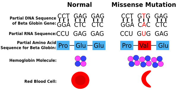
People with sickle-cell disease are born with a mutation in their beta-globin gene.

missense_mutation3

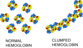
Beta-globin is one of the protein chains that compose the protein hemoglobin. Hemoglobin is the protein in red blood cells that ferries oxygen from the lungs to the tissues and then returns to the lungs to load up with oxygen again and then goes back to the tissues. Red blood cells, which are made in the bone marrow, are packed from stem to stern with hemoglobin molecules, and normally are round, and slightly dished and flexible enough to squeeze through small capillary beds in tissues. The mutation in the beta-globin gene that causes sickle-cell disease, however, causes hemoglobin to form long, stiff rods of protein rather than tight, compactly packed clusters of hemoglobin. These protein rods deform the red blood cells and make them stiff, sickle-shaped, and unable to pass through tissue capillary beds.

sickle-cell-hemoglobin

These abnormally shaped red blood cells not only move poorly through blood vessels, but they also do not sufficiently carry oxygen to vital organs.

Sickle_cell 2

The stem cell gene therapy method described by Kohn and his colleagues corrects the mutation in the beta-globin gene in the bone marrow-based stem cells so that they produce normal, circular-shaped blood cells. The technique uses specially engineered enzymes, called zinc-finger nucleases, to eliminate the mutation and replace it with a corrected version that repairs the beta-globin mutation. Kohn’s research showed that this method has the potential to treat sickle-cell the disease if the gene therapy achieves higher levels of correction.

“This is a very exciting result,” said Dr. Kohn, professor of pediatrics and microbiology, immunology and molecular genetics. “It suggests the future direction for treating genetic diseases will be by correcting the specific mutation in a patient’s genetic code. Since sickle-cell disease was the first human genetic disease where we understood the fundamental gene defect, and since everyone with sickle-cell has the exact same mutation in the beta-globin gene, it is a great target for this gene correction method.”

Forcing Sugars on the Surfaces of Cord Blood Cell Increases Their Engraftment


When a child or adult needs new bone marrow, a bone marrow transplant from a donor is usually the only way to save their life. Without properly functioning bone marrow, the patient’s blood cells will die off, and there will be too few red blood cells to ferry oxygen to tissues or white blood cells to fight off infections.

An alternative to bone marrow from a bone marrow donor if umbilical cord blood. Umbilical cord blood does not require the rigorous tissue matching that bone marrow requires because the blood making stem cells from cord blood are immature and not as likely to cause tissue rejection reactions.. However, umbilical cord blood cells suffer from two drawbacks: low numbers of stem cells in cord blood and poor engraftment efficiencies.

Fortunately, some progress has been made at expanding blood-making stem cells from umbilical cord blood, and it is likely that such technologies might be ready for common use in the future. As to the poor engraftment efficiencies, a new paper in the journal Blood from the laboratory of Elizabeth J. Shpall at the University of Texas MD Anderson Cancer Center, in Houston, Texas reports a new way to increase cord blood stem cells engraftment efficiencies.

As previously discusses, delayed engraftment is one of the major limitations of cord blood transplantation (CBT). Delayed engraftment seems to be due to the diminished ability of the cord blood stem cells to home to the bone marrow. How are cells channeled to the bone marrow? A protein receptor called P- and E-selectins is expressed on the surfaces of bone marrow blood vessels. Cells that can bind these selectin receptors will pass from the circulation to the bone marrow. Thus binding selectin receptors is kind of like having the “password” for the bone marrow.

What does it take to bind the selectin proteins? Selectins bind to specific sugars that have been attached to proteins. These sugars are called “fucose” sugars. As it turns out, cord blood stem cells do not express robust levels of these fucosylated proteins. Could increasing the levels of fucosylated proteins on the surfaces of cord blood stem cells increase their engraftment? Shpall and her colleagues tested this hypothesis in patients with blood-based cancers.

Patients with blood cancers had their cancer-producing bone marrow stem cells destroyed with drugs and radiation. Then these same patients had their bone marrows refurbished with two units of umbilical cord blood. However, these cells in these cord blood units were treated with the enzyme fucosyltransferase-VI and guanosine diphosphate fucose for 30 minutes before transplantation. This treatment should have increased the content of fucosylated proteins on the surfaces of cells in the hope of enhancing their interaction with Selectin receptors on the surfaces of bone marrow capillaries.

The results of 22 patients enrolled in the trial were then compared with those for 31 historical controls who had undergone double unmanipulated CBT. There was a clear decrease in the length of time it took for cells to engraft into the bone marrow.  For example, the median time to neutrophil (a type of white blood cell) engraftment was 17 days (range 12-34) compared to 26 days (range, 11-48) for controls (P=0.0023). Platelet (a cell used in blood clotting) engraftment was also improved: median 35 days (range, 18-100) compared to 45 days (range, 27-120) for controls (P=0.0520).  These are significant differences.

These findings support show that treating cord blood cells with a rather inexpensive cocktail of enzymes for a short period of time before transplantation is a clinically feasible means to improve engraftment efficiency of CBT.  This is a small study.  Therefore, these data, though very hopeful, must be confirmed with larger studies.

High-Quality Cartilage Production from Pluripotent Stem Cells


High-quality cartilage has been produced from pluripotent stem cells by workers in the laboratory of Sue Kimber and her team in the Faculty of Life Sciences at The University of Manchester. Such success might be used in the future to treat the painful joint condition osteoarthritis.

Kimber and her colleagues used strict laboratory conditions to grow and transform embryonic stem cells into cartilage cells known as chondrocytes.

Professor Kimber said: “This work represents an important step forward in treating cartilage damage by using embryonic stem cells to form new tissue, although it’s still in its early experimental stages.” Kimber’s research was published in Stem Cells Translational Medicine.

During the study, the team analyzed the ability of embryonic stems cells to become cartilage precursor cells. Kimber and her colleagues then implanted these pre-chrondrocytes into cartilage defects in the knee joints of rats. After four weeks, the damaged cartilage was partially repaired and following 12 weeks a smooth surface, which looked very similar to normal cartilage, was observed. More detailed studied of this newly regenerated cartilage demonstrated that cartilage cells from embryonic stem cells were still present and active within the tissue.

Developing and testing this protocol in rats is the first step in generating the information required to run such a study in people with arthritis. Before such a clinical trial can be run, more data will need to be collected in order to check that this protocol is effective and that there are no toxic side-effects.

However, Kimber and her coworkers say that this study is very promising as not only did this protocol generate new, healthy-looking cartilage but also importantly there were no signs of any side-effects such as growing abnormal or disorganized, joint tissue or tumors. Further work will build on this finding and demonstrate that this could be a safe and effective treatment for people with joint damage.

Chondrocytes created from adult stem cells are being used on an experimental basis, but, to date, they cannot be produced in large amounts, and the procedure is expensive.

With their huge capacity to proliferate, pluripotent stem cells such as embryonic stem cells and induced pluripotent stem cells can be manipulated to form almost any type of mature cell. Such cells offer the possibility of high-volume production of cartilage cells, and their use would also be cheaper and applicable to a greater number of arthritis patients, the researchers claim.

“We’ve shown that the protocol we’ve developed has strong potential for developing large numbers of chondrogenic cells appropriate for clinical use,” added Prof Kimber. “These results thus mark an important step forward in supporting further development toward clinical translation.”

Osteoarthritis affects more than eight million people in the UK alone, and is a major cause of disability. It and occurs when cartilage at the ends of bones wears away causing joint pain and stiffness.

Director of research at Arthritis Research UK Dr Stephen Simpson added: “Current treatments of osteoarthritis are restricted to relieving painful symptoms, with no effective therapies to delay or reverse cartilage degeneration. Joint replacements are successful in older patients but not young people, or athletes who’ve suffered a sports injury.

“Embryonic stem cells offer an alternative source of cartilage cells to adult stem cells, and we’re excited about the immense potential of Professor Kimber’s work and the impact it could have for people with osteoarthritis.”

New Gene Therapy for Hemophilia


According to a multi-year, ongoing study, a new kind of gene therapy for hemophilia B could be safe and effective for human patients.

“The result was stunning,” said Timothy Nichols, MD, director of the Francis Owen Blood Research Laboratory at the University of North Carolina School of Medicine and co-senior author of the paper. “Just a small amount of new factor IX necessary for proper clotting produced a major reduction in bleeding events. It was extraordinarily powerful.”

Nichols published his work in the journal Science Translational Medicine, in which he showed that a genetically engineered retrovirus could successfully transfer new factor IX (clotting) genes into animals with hemophilia B to dramatically decrease spontaneous bleeding. To date, the new therapy has proven safe.

A new FDA-approved hemophilia treatment lasts longer than a few days but patients still require injections indefinitely at least once or twice a month. This new gene therapy only requires hemophilia patients to receive a one-time dose of new clotting genes instead of a lifetime of multiple injections of recombinant factor IX. This new gene therapy approach would involve a single injection and could potentially save money and provide a long-term solution to a life-long condition. A major potential advantage of this new gene therapy approach is that it uses lentiviral vectors, to which most people do not have antibodies that would reject the vectors and make the therapy less effective.

In human clinical studies, approximately 40 percent of the potential participants with hemophilia have antibodies in their blood against adeno-associated virus (AAV), which precludes them from entering AAV trials for hemophilia gene therapy treatment. Thus more people could potentially benefit from the lentivirus gene therapy approach.

Hemophilia is a bleeding disorder in which people lack a clotting factor. Therefore they bleed much more easily than people without the disease. People with hemophilia often bleed spontaneously into joints, which can be extremely painful and crippling. Spontaneous bleeds into soft tissues are also common and can be fatal if not treated promptly. Hemophilia A affects about one in 5,000 male births. These patients do not produce enough factor VIII in the liver. This leads to an inability to clot. Hemophilia B affects about one in 35,000 births; these patients lack factor IX.

The new method detailed in the Science Translational Medicine paper was spearheaded by Luigi Naldini, PhD, director of the San Raffaele Telethon Institute for Gene Therapy. Naldini and Nichols developed a way to use a lentivirus, a large retrovirus, to deliver factor IX genes to the livers of three dogs that have a naturally occurring form of hemophilia. They removed the genes involved in viral replication. “Essentially, this molecular engineering rendered the virus inert,” Nichols said. “It had the ability to get into the body but not cause disease.” This process turned the virus into a vector – simply a vehicle to carry genetic cargo.

Unlike some other viral vectors that have been used for gene therapy experiments, the lentiviral vector is so large that it can carry a large payload – namely, the clotting factor IX genes that people with hemophilia B lack. (This approach could also be used for hemophilia A where the FVIII gene is considerably larger.)

These viral vectors were then injected directly into the liver or intravenously. After more than three years, the three dogs in the study experienced zero or one serious bleeding event each year. Before the therapy, the dogs experienced an average of five spontaneous bleeding events that required clinical treatment. Importantly, the researchers detected no harmful effects.

“This safety feature is of paramount importance,” Nichols said. “Prior work elsewhere during the early 2000s used retroviruses for gene therapy to treat people with Severe Combined Immunodeficiency, but some patients in clinical trials developed leukemia.” Newer retroviral vectors, though, have so far proved safe for SCID patients.

To further demonstrate the safety of this new hemophilia treatment, Nichols and Naldini used three different strains of mice that were highly susceptible to developing complications, such as malignancies, when injected with lentiviruses. Fortunately, Nichols, Naldini and their coworkers found no harmful effects in these mice. Thus manipulating lentiviruses and converting them into lentiviral vectors made them safe for gene therapy.

“Considering the mouse model data and the absence of detectible genotoxicity during long-term expression in the hemophilia B dogs, the lentiviral vectors have a very encouraging safety profile in this case,” Nichols said.

This gene therapy approach requires more work before it can be used in human trials. For instance, researchers hope to increase the potency of the therapy to decrease spontaneous bleeding even more while also keeping the therapy safe.

Before the treatment, the hemophilia dogs had no sign of factor IX production. After the treatment, they exhibited between 1 and 3 percent of the production found in normal dogs. This slight increase was enough to substantially decrease bleeding events.

Nichols wants to try to boost factor IX production to between 5 and 10 percent of normal while still remaining safe. This amount of factor IX expression could potentially eliminate spontaneous bleeding events for people with hemophilia B.

Umbilical Cord Blood Mesenchymal Stem Cells Relieve the Symptoms of Interstitial Cystitis by Activating the Wnt Pathway and EGF Receptor


Interstitial tissue refers to the tissue that lies between major structures in an organ. For example, the tissue between muscles is an example of interstitial tissue.

Interstitial cystitis, otherwise known as painful bladder syndrome is a chronic condition that causes bladder pressure, bladder pain and sometimes pelvic pain, ranging from mild discomfort to severe pain.

The bladder is a hollow, muscular organ that stores urine and expands until it is full, at which time it signals the brain that it is time to urinate, communicating through the pelvic nerves. This creates the urge to urinate for most people. In the case of interstitial cystitis, these signals get mixed up and you feel the need to urinate more often and with smaller volumes of urine than most people. Interstitial cystitis most often affects women and can have a long-lasting impact on quality of life. Unfortunately no treatment reliably eliminates interstitial cystitis, but medications and other therapies may offer relief. There is no sign of bacterial infection in the case of bacterial cystitis.

A new study evaluated the potential of umbilical cord blood-derived mesenchymal stem cells or (UCB-MSCs) to treat interstitial cystitis (IC). In this study, Dr. Miho Song and colleagues from the Asan Medical Center, Seoul, South Korea, established a rat model of IC in 10-weeks-old female Sprague-Dawley rats by instilling 0.1M HCl or PBS (sham). After 1-week, human UCB-MSCs (IC+MSCs) or PBS (IC) were directly injected into the submucosal layer of the bladder.

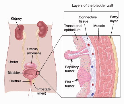
To clarify this part of the experiment, the urinary bladder is made of several distinct tissue layers: a) The innermost layer of the bladder is the mucosa layer that lines the hollow lumen. Unlike the mucosa of other hollow organs, the urinary bladder is lined with transitional epithelial tissue that is able to stretch significantly to accommodate large volumes of urine. The transitional epithelium also provides protection to the underlying tissues from acidic or alkaline urine; b) Surrounding the mucosal layer is the submucosa, a layer of connective tissue with blood vessels and nervous tissue that supports and controls the surrounding tissue layers; c) The visceral muscles of the muscularis layer surround the submucosa and provide the urinary bladder with its ability to expand and contract. The muscularis is commonly referred to as the detrusor muscle and contracts during urination to expel urine from the body. The muscularis also forms the internal urethral sphincter, a ring of muscle that surrounds the urethral opening and holds urine in the urinary bladder. During urination, the sphincter relaxes to allow urine to flow into the urethra.

Bladder histology

Now a single subcutaneous injection of human UCB-MSCs significantly attenuated the irregular and decreased voiding interval in the IC group. In addition, the denudation of the epithelium that is characteristic of IC and increased inflammatory responses, mast cell infiltration, neurofilament production, and angiogenesis observed in the IC bladders were prevented in the IC+MSC group. Therefore, the injected UBC-MSCs prevented the structural changes in the bladder associated with the pathology of IC.

How did these cells do this? Further examination showed that the injected UCB-MSCs successfully engrafted to the stromal and epithelial tissues of the bladder and activated the Wnt signaling cascade. In fact, if the Wnt activity of these infused cells was blocked, the positive effects of the UCB-MSCs were also partially blocked. Additionally, activation of the epidermal growth factor receptor (EGFR) also helped UCB-MSCs heal the bladder. If the activity of the EGF receptor was inhibited by small molecules, then the benefits of MSC therapy were also abrogated. Also if both the Wnt pathway and EGFR were inhibited, the therapeutic capacities of UCB-MSCs were completely wiped out.

These data show the therapeutic effects of MSC therapy against IC in an orthodox rat animal model. However, this study also elucidates the molecular mechanisms responsible for these therapeutic effects. Our findings not only provide the basis for clinical trials of MSC therapy to IC, but also advance our understanding of IC pathophysiology.

Stem Cells Lurk in Tumors and Can Resist Treatment


Regenerative medicine seeks to train stem cells to transform into nearly any kind of cell type. Unfortunately, this ability that makes stem cells so useful also is cause for concern in cancer treatments. Malignant tumors contain resident stem cells, which prompts worries among cancer experts that the cells’ transformative powers help cancers escape treatment.

Data from new research shows that the threat posed by cancer stem cells is more prevalent than previously thought. Until now, stem cells had been identified only in aggressive, fast-growing tumors. However, a mouse study at Washington University School of Medicine in St. Louis has revealed that slow-growing tumors also have treatment-resistant stem cells.

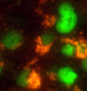
Brain tumor stem cells (orange) in mice express a stem cell marker (green). Researchers at Washington University School of Medicine in St. Louis are studying how cancer stem cells make tumors harder to kill and are looking for ways to eradicate these treatment-resistant cells. Credit: Yi-Hsien Chen

In mice, low-grade brain cancer stem cells were less sensitive to anticancer drugs. When compared to healthy stem cells, tumor-based stem cells from brain tumors, revealed the reasons behind their resistance to treatments, which points to new therapeutic strategies.

“At the very least, we’re going to have to use different drugs and different, likely higher dosages to make sure we kill these tumor stem cells,” said senior author David H. Gutmann, MD, PhD, the Donald O. Schnuck Family Professor of Neurology.  Their data were published in the March 12 edition of Cell Reports.

First author Yi-Hsien Chen, who is a senior postdoctoral research associate in Gutmann’s laboratory, used a mouse model of neurofibromatosis type 1 (NF1), which forms low-grade brain tumors, to identify cancer stem cells and demonstrate that they could form tumors when transplanted into normal, cancer-free mice.

Neurofibromatosis type I is caused by mutations in the NF1 genes, and such mutations affect about 1 in every 2,500 babies. Neurofibromatosis type I can cause an array of physical problems, including brain tumors, impaired vision, learning disabilities, behavioral problems, heart defects and bone deformities.

In children with NF1 mutations, the most common brain tumor is optic gliomas. Treatment for NF1-related optic gliomas usually includes drugs that inhibit a cell growth pathway originally identified by Gutmann. In laboratory tests conducted as part of the new research, it took 10 times the dosage of these drugs to kill the low-grade cancer stem cells.

Compared with healthy stem cells from the brain, cancer stem cells made multiple copies of a protein called Abcg1 that helps those cells survive stress.

“This protein blocks a signal from inside the cells that should make them more vulnerable to treatment,” Gutmann explained. “If we can identify a drug that disables this protein, it would make some cancer stem cells easier to kill.”

Even though these laboratory mice were bred to model NF1 optic gliomas, Gutmann and others said that their findings could be applied more broadly to other brain tumors.

“Because stem cells haven’t differentiated into specialized cells, they can easily activate genes to turn on new developmental programs that allow the cells to survive cancer treatments,” said Gutmann, who directs the Washington University Neurofibromatosis Center. “Based on these new findings, we will have to develop additional strategies to keep these tumors from evading our best treatments.”

Bone Marrow Stem Cell Treatment Plus Immunosuppression are Superior to Immunosuppression Alone in Multiple Sclerosis Patients


Multiple Sclerosis (MS) is a debilitating autoimmune disease in which the immune system attacks elements of the central nervous system. There are different types of MS, but more progressive cases can leave patients unable to walk and may require rather extreme immunosuppressive treatments that can predispose a patient to illness and cancer.

However, a new study that was published in the journal Neurology has shown that stem cell transplantation could be a more effective therapy in severe cases of multiple sclerosis (MS) than the drug mitoxantrone.

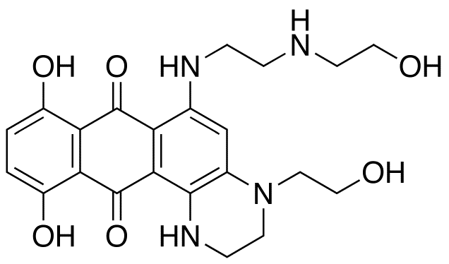
Mitoxanthone is a “type II topoisomerase inhibitor” that disrupts DNA synthesis and DNA repair by inserting between the bases in DNA. Mitoxanthone can cause nausea, vomiting, hair loss, heart damage, and suppression of the immune system. Some side effects may have delayed onset. Heart damage (cardiomyopathy) is a particularly concerning effect with this drug, since it is irreversible. Therefore, because of the risk of cardiomyopathy, mitoxantrone carries a limit on the cumulative lifetime dose, which is based on the body surface area of patients.

Mitoxantrone
Mitoxantrone

Because MS is an immune-mediated disorder, and because immune cells are made by stem cells in the bone marrow, bone marrow transplants (hematopoietic stem cell transplantation), which are routinely used in the treatment of leukemia and lymphoma, are being considered as a treatment for MS.

A clinical trial conducted by Giovanni Mancardi from the University of Genova, Italy designed a randomized phase II clinical trial study that included 21 MS patients, whose average age was 36 and whose disability due to the disease had worsened in the previous year despite the fact that the patients were under conventional medication treatment. The average disability level of the participants was represented by the need of a crutch or cane to walk. The goal of the study was to determine the efficacy of intense immunosuppression followed by either a bone marrow transplant with the patient’s own bone marrow, or mitoxantrone (MTX) in MS disease activity.

Giovanni Mancardi
Giovanni Mancardi

All participants in this clinical trial received immune-suppressive medication. MTX was given to 12 of the patients while the remaining 9 received hematopoietic stem cells harvested from their own bone marrow. After treatment with MTX, the stem cells were intravenously reintroduced into their donors and the stem cells migrated back to the bone marrow where they generated new immune cells. All participants were followed-up for a period of up to four years after their treatment.

“This process appears to reset the immune system,” said the lead study author Dr. Giovanni Mancardi. “With these results, we can speculate that stem cell treatment may profoundly affect the course of the disease.”

Mancardi and his team found that treatment of MS patients with robust immunosuppression followed by stem cell treatment resulted in a significantly higher decrease in disease progression in comparison with MTX treatment alone. MS patients under stem cell treatment reduced the number of new areas of brain damage (T2 lesions) by 79% compared to patients under MTX treatment. Another type of lesion seen in MS patients – gadolinium-enhancing lesions – were not detected in patients under stem cell treatment during the study, whereas 56% of patients receiving MTX exhibited at least one new gadolinium-enhancing lesion.

Mancardi and his team concluded that an intense immunosuppression followed by autologous hematopoietic stem cell transplantation is more efficient than MTX to reduce MS activity in severe cases.

“More research is needed with larger numbers of patients who are randomized to receive either the stem cell transplant or an approved therapy, but it’s very exciting to see that this treatment may be so superior to a current treatment for people with severe MS that is not responding well to standard treatments,” concluded study author Dr. Mancardi.

Gene Discoveered That Drives Fertility in Male Mice


Workers in the laboratory of Richard Freiman, associate professor of medical science at Brown University have discovered a specific gene in human males that seems to be essential to sperm production later in life.

A paper published in the journal Stem Cells details how the loss of a protein called TAF4b in male mice causes premature infertility. According to Freiman, mutations that prevent the continuous production of TAF4b leave mice incapable of sustaining spermatogenesis after only a few months of sexual maturity.

This study began when Freiman’s team discovered that TAF4b was expressed at high levels in the ovaries and the testes. Later, Freiman and his colleagues used homologous recombination to specifically modify the TAF4b gene. Freiman explained that homologous recombination can “knock specific genes out of the mouse genome,” after which you can “examine the mice that are born and see what function the gene serves in normal development.” Such experiments showed that male mice whose TAF4b gene was synthetically modified so that it expressed TAF4b initially, but did not sustain its expression were only fertile for a month or two, whereas mice with intact TAF4b remained fertile for several years.

“Cells that are involved in initial fertility are different than cells involved in subsequent rounds of sperm production. The first set undergoes meiosis and become sperm by a direct route, but the other set develops into precursor cells that become stem cells,” Freiman said. “What we hypothesized about our mice is that they’re able to go through this initial round of spermatogenesis, but they can’t make the stem cell population, so they can’t set themselves up for long-term fertility,” he added.

Since humans have a TAF4b gene that is very similar to the mouse gene, the results of Freiman’s laboratory might be applicable to human fertility, said Eric Gustafson, a postdoctoral research fellow in Freiman’s laboratory and first author of the paper.

These results interact with another study that was published last year that examined a population of four infertile brothers in eastern Turkey; each of whom each had a homozygous mutation in their TAF4b gene similar to the one created in the mice. According to Gustafson, these men had very low or no sperm counts. “So we think these genes have many similar, if not identical functions in humans. What we learn about in the mouse gene may be used to address or diagnose reproductive defects in humans as well,” he added.

With couples having children later and later in life, this study has important implications for family planning. “If we could learn how this process is regulated normally, clinicians might be able to devise better strategies to either monitor or even intervene with cases of infertility,” said Freiman. To give an example, Freiman suggested that if scientists could detect the mutation in teenage boys early on, then doctors could freeze their patients’ sperm for later in life.

The study also has important outcomes in terms of stem cell research, said Professor of Biology Gary Wessel, who was not involved in the study. “This research shows us that this particular transcription factor, TAF4b, is involved in the transcription process involved in maintaining the stem cell itself,” Wessel said. “As a consequence, it now gives the investigators a more careful view of what stem cell decisions are like,” he added.

The study’s results are relevant to all stem cell research, Wessel said. “Everything in biology is connected. If you make any kind of breakthrough, it’s going to have ripple effects throughout the entire discipline.”

How does TAF4b affect fertility? That’s the next goal of Freiman’s research. Freiman said. “We now know that TAF4b performs this function, but we don’t know how it does it,” he added. “Once we figure that out, it might reveal new areas of intervention for fertility preservation.”

NG2-Expressing Neural Lineage Cells Derived from Embryonic Stem Cells Penetrate Glial Scar and Promote Axonal Outgrowth After Spinal Cord Injury


After a spinal cord injury, resident stem cells in the spinal cord contribute to the production of a glial scar that is rich in chondroitin sulfate proteoglycan (CSPG). The glial scar is a formidable barrier to axonal regeneration in the injured spinal cord, since CSPG actively repels growing axonal growth cones. Even though the glial scar seals off the spinal cord from further damage from inflammation, the long-term effects of the glial scar are to prevent regeneration of spinal nerves, which have the ability to regenerate in culture.

The major components of the site of injury include myelin debris, the scar-forming astrocytes, activated resident microglia and infiltrating blood-borne immune cells, chondroitin sulfate proteoglycans (CSPGs) and other growth-inhibitory matrix components. All of them are potential targets for therapeutic intervention. Many of the interventions can be optimized by considering the beneficial aspects of the scar tissue and fine-tuning the optimal time window for their application. Each target and the strategies directed at its modulation are shown.

New work by Sudhakar Vadivelu, in the laboratory of John McDonald at the International Center for Spinal Cord Injury, Hugo W. Moser Research Institute at the Kennedy Krieger Institute, Baltimore, Maryland has discovered new ways to breach the glial scar. Vadivelu and colleagues used a cell culture system that tested the ability of particular cells to help growing axonal growth cones penetrate glial scar material. This culture system showed that embryonic stem cell-derived neural lineage cells (ESNLCs) with prominent expression of nerve glial antigen 2 (NG2) survived, and passed through an increasingly inhibitory gradient of CSPG. These cells also expressed matrix metalloproteinase 9 (MMP-9) at the appropriate stage of their development, which helped poke holes in the CSPG. The outgrowth of axons from ESNLCs followed the NG2-expressing cells because the migrating cells chiseled pathways through the CSPG for the outgrowth of new axons.

To confirm these results in a living animal, Vadivelu and others transplanted embryonic stem cell-derived ESNLCs directly into the cavities of a contused spinal cord of laboratory animals 9 days after injury. One week later, implanted ESNLCs survived and expressed NG2 and MMP-9. The axons of these neurons had grown through long distances (>10 mm), although they preferred to grow across white rather than gray matter.

These data are consistent with CSPG within the injury scar acting as an important impediment to neuronal regeneration, but that NG2+ progenitors derived from ESNLCs can alter the microenvironment within the injured spinal cord to allow axons to grow through such a barrier. This beneficial action seems to be due, in part, at least, to the developmentally-regulated expression of MMP-9. Vadivelu and others conclude from these data that it might be possible to induce axonal regeneration in the human spinal cord by transplanting ESNLCs or other cells that express NG2.

Regenerating Dead Cells In the Brain with Stem Cells


Neuroscientists at the Université Libre De Bruxelles (ULB) in Belgium have taken a very important step in cell therapy for diseases of the brain. This team generated cortical neurons from embryonic stem cells, which they then used to treat adult with brain problems. This research was recently published in the journal Neuron.

The ULB team was led by Pierre Vanderhaeghen, Kimmo Michelsen and Sandra Acosta (ULB Neuroscience Institute, in collaboration with the laboratory of Afsaneh Gaillard (INSERM/U. Poitiers, France). These results open new perspectives for the repair of damaged cells in the brain and replacing damage neurons.

The cerebral cortex is definitely the most complex and essential structure of our brain. The nerve cells or neurons that compose the cerebral cortex are the basic building blocks that help it do every job that it does. The loss of loss of cortical neurons is the cause of many neurological diseases as a result of stroke, Alzheimer disease, or physical trauma to the brain can seriously compromise brain function.

Previously, these same ULB researchers discovered how to generate cortical neurons in the laboratory cortical neurons from embryonic stem cells. Despite the triumph of these findings, it was completely unclear whether these findings could be translated into a living creature.

Now, the ULB team has successfully tested the use of their laboratory-generated cortical neurons in a living animal. In this study, Vanderhaeghen and others transplanted cortical pyramidal neurons made from embryonic stem cells into the brains of adult mice who had undergone chemically induced brain damage. This experiment cause rather massive neuronal losses in the visual cortex.

Remarkably, the implanted neurons integrated effectively into the brain after injury, but most importantly they could connect with the host brain, and some of them even responded to visual stimuli, like the visual cortex.

Integration only occurred, if the types of implanted neurons were matched to the lesioned area. In other words, since visual cortex neurons were lost, only the implantation of other cortical neurons allowed the cells to properly engraft into the visual cortex. However the grafted neurons displayed long-range patterns of connectivity with the host neurons.

This remains an experimental approach that has, to date, only been successfully performed with laboratory mice. A good deal more much research is required before any clinical application in humans will come to the clinic. Regardless, the success of these experiments combining cell engineering to generate nerve cells in a controlled and unlimited fashion, together with transplantation in to damaged brain, opens new avenues to repair the brain following damage or degeneration, such as following stroke or brain trauma.

Stem Cell Structure and Obesity


New research conducted at Queen Mary University of London (QMUL) has discovered that the regulation of the length of primary cilia, which are small hair-like projections on the surfaces of most cells, can prevent the production of fat cells taken from adult human bone marrow. Such a discovery might be used to develop a way of preventing obesity.

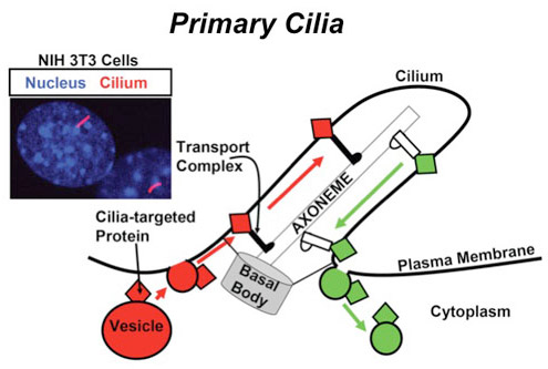
What are primary cilia?  For many years, almost all attention was focused on cilia that moved because their function was readily observable.  However, Alexander Kowalevsky first reported in 1867 the presence of single (nonmotile) cilia in a variety of vertebrate cells.  These solitary and nonmotile cilia are far more widespread than the motile type.  In humans, only a few cell types have motile cilia, namely epithelial cells in the bronchi and oviducts, and ependymal cells that line brain vesicles.  However, virtually all other cells have a primary cilium.

What makes primary cilia different from the motile form? First, they lack the central pair of microtubules, which would explain the lack of motility.  Primary cilia also seem to lack dynein, one of the molecular motors needed for motility.  In addition, some primary cilia do not project beyond the cell surface, and most, but not all, are very short.  What do these organelles do if they are not sticking out of the cell, or motile?

Further work has shown that primary cilia are important in intracellular transport and also in sensory function for cells.  Now it seems that primary cilia are also important in the process of adipogenesis.

Primary cilia

Adipogenesis refers to the differentiation of stem cells into fat cells. The QMUL research team showed that during adipogenesis, the length of primary cilia increases, which increases the movement of specific proteins associated with the cilia. When the QMUL team genetically restricted primary cilia elongation by genetic means, they were able to stop the formation of new fat cells.

One of the lead authors or this study, Melis Dalbay, said that it was the first time that subtle changes in primary cilia structure can influence the differentiation of stem cells into fat.

Since the length of primary cilia can be influenced by various factors including pharmaceuticals, inflammation and even mechanical forces, this study provides new insight into the regulation of fat cell formation and obesity.

This research points toward a new type of treatment known as “cilia-therapy” where manipulation of primary cilia may be used in the future to treat a growing range of conditions including obesity, cancer, inflammation and arthritis.

Testing Stem Cell Quality


A new paper published in the journal EMBO Molecular Medicine by a team from the Lausanne University Hospital describes a protocol that can ensure the safety of adult epidermal stem cells before they are used as treatments for patients. The approach devised by this team takes cultivated, genetically modified stem cells and isolates single cells that are then used to make clonal cell cultures. These cloned cells are then rigorously tested to ensure that they meet the highest possible safety criteria. This protocol was inspired by approaches designed in the biotechnology industry and honed by regulatory authorities for medicinal proteins produced from genetically engineered mammalian cells.

“Until now there has not been a systematic way to ensure that adult epidermal stem cells meet all the necessary requirements for safety before use as treatments for disease,” says EMBO Member Yann Barrandon, Professor at Lausanne University Hospital, the Swiss Federal Institute of Technology in Lausanne and the lead author of the study. “We have devised a single cell strategy that is sufficiently scalable to assess the viability and safety of adult epidermal stem cells using an array of cell and molecular assays before the cells are used directly for the treatment of patients. We have used this strategy in a proof-of-concept study that involves treatment of a patient suffering from recessive dystrophic epidermolysis bullosa, a hereditary condition defined by the absence of type VII collagen which leads to severe blistering of the skin.”

Barrandon and co-workers have cultivated epidermal cells from patients who suffer from epidermolysis bullosa. These cells were then genetically engineered in order to insert a normal copy of the type VIII collagen gene. Then the genetically fixed cells were grown in culture so that they can be used to regenerate skin. Barrandon and others subjected these cells to an array of tests in order to determine which of the genetically engineered cells meet the requirements for safety and “stemness,” which refers to the stem cell characteristics that distinguish it from regular cells; its developmental immaturity and its ability to grow and self-renew. Clonal analysis revealed that the cultured, genetically engineered stem cells varied in their ability to produce functional type VII collagen. When the most viable, modified stem cells were selected and transplanted into the skin of immunodeficient mice, the cells regenerated skin and produced skin that did not blister in the mouse model system for recessive dystrophic epidermolysis bullosa. Furthermore, the cells produced functional type VII collagen. The safety of the cells was assessed by mapping the sites of integration of the viral vector. Because such viruses and produce gene rearrangements other mutations, the chosen cell lines were subjected to whole genome sequencing. Only the cells with insertions in benign locations were considered for use in their mouse model.

Barrandon concluded: “Our work shows that at least for adult epidermal stem cells it is possible to use a clonal strategy to deliver a level of safety that cannot be obtained by other gene therapy approaches. A clonal strategy should make it possible to integrate some of the more recent technologies for targeted genome editing that offer more precise ways to change genes in ways that may further benefit the treatment of disease. Further work is in progress in this direction.”

This work is certainly fascinating, but I think that using integrating viral vectors is asking for trouble. Certainly it should be possible to fix or replace the abnormal type VII collagen gene. Viruses that randomly insert genes into the genome can cause genetic problems, and even sequencing the genome may not properly address the safety concerns of the use of such viral vectors.

Safer Culture Conditions for Stem Cells


Jeanne Loring from the Scripps Institute is the senior author of a very important study that examined the culture conditions for pluripotent stem cells.

Several scientists have discovered that induced pluripotent stem cells (iPSCs) and embryonic stem cells (ESCs) can accumulate cancer-causing mutations when grown in culture for extended periods of time (for example, see Uri Weissbein, Nissim Benvenisty, and Uri Ben-David, J Cell Biol. 2014 Jan 20; 204(2): 153–163). However, some laboratories have managed to keep ESCs in culture for extended periods without observing instabilities.

To try to tease apart why this might be the case, Loring and her group examined various culture methods and determined that some stem cell culture methods are associated with increased incidence of mutations in the DNA of stem cells.

“This is about quality control; we’re making sure these cells are safe and effective,” said Loring, who is a professor of developmental neurobiology at Scripps Research Institute (SRI) in San Diego, CA.

All cells run the risk of accumulating mutations when they divide, but previous research from Loring and her colleagues showed that particular culture conditions could potentially select for faster growth and mutations that accelerate growth. Such growth-enhancing mutations are sometimes associated with tumors.

“Most changes will not compromise the safety of the cells for therapy, but we need to monitor the cultures so that we know what sorts of changes take place,” said Ibon Garitaonandia, who is a postdoctoral research fellow in Loring’s laboratory at SRI.

New research from Loring’s group has shown how particular culture conditions can reduce the likelihood of mutations. Loring and her colleagues tested several different types of surfaces upon which the cells were grown. They also used different ways of propagating or “passaging” the cultures. When cells are grown in culture, the culture dishes must be scraped to get the cells off them and then the cells must be transferred to a fresh culture dish. How you do this matters: do you use enzymes to detach the cells, or do you mechanically scrape them off? Other culture techniques use layers of “feeder cells” that do not divide, but are still able to secrete growth factors that improve the health of the growing stem cells.

Loring and her crew tested various combinations of surfaces, passaging methods and feeder cell populations and grew the cells for three years with over 100 passages. Over the course of this experiment, the cells were sampled and analyzed for the presence of new mutations in their genomes.

It turns out that stem cells grown on feeder cells that are passaged by hand (manually) show the fewest growth-enhancing mutations after being cultured for three years.

Loring’s study also demonstrated the importance of monitoring cell lines over time. In particular, deletion of the TP53 gene, a tumor suppressor gene, in whose absence cancer develops, should be closely watched.

“If you want to preserve the integrity of the genome, then grow your cells under those conditions with feeder cells and manual passaging,” said Loring. “Also, analyze your cells. It’s really easy, she added.

When Thomson made the first human ESC lines, he used feeder cells derived from mouse skin cells.  However, the use of animal materials to make ESCs might pollute them with animal viruses and specific sugars from the surfaces of the animal cells might also contaminate the surfaces of the ESCs, making them unsuitable for regenerative medicine (see Stem Cells 2006; 24:221-229).  To address this problem, several laboratories have made “Xeno-free” ESC lines that were made without touching any animal products.  Some of these Xeno-free lines were made without feeder cells (see C. Ellerström, et al., Stem Cells. 2006 Oct;24(10):2170-6)., but others were made with human feeder cell lines (see K Rajala, et al., Hum Reprod. 2007 May;22(5):1231-8). Therefore, it appears, that the use of human feeder cell lines are preferable to feeder-free systems, given Loring’s findings.  However, it is also possible that such culture systems are also preferable for iPSCs, which do not have the problem of immunological rejection for patients, and do not require the killing of the youngest members of humanity.  Therefore, Loring’s work could very well benefit iPSC cultures as well.

Monkey’s Own Cells Are Used to Treat Parkinson’s Disease


Neurologist Ole Isacson and his Harvard Medical School team successfully implanted neurons made from a monkey’s own cells to treat Parkinson’s disease in those animals. The implanted neurons were watched for two years, and they proved to be both safe and effect in the treatment of Parkinson’s disease.

Induced pluripotent stem cells or iPSCs are made from mature, adult cells by means of a combination of genetic engineering and cell culture techniques. The cells resemble embryonic stem cells in many of their growth characteristics and gene expression patterns, but they are have several differences as well. One of the biggest differences between iPSCs and embryonic stem cells is that the reprogramming process that makes iPSCs places cells under stresses that increase the mutation rate and makes iPSCs, on average, more likely to cause tumors than embryonic stem cells. However, it is also clear that not all iPSC lines are the same and careful screen protocols that determine safe lines from less safe lines.

A distinct advantage of iPSCs over embryonic stem cells is that they have the same set of cell surface proteins as the patient from whom they were made, which makes them less likely to be rejected by the patient’s immune system. Even though some experiments had shown that cells derived from iPSCs can be rejected by the patient’s immune system, these experiments used poor-quality iPSC lines. High-quality iPSCs lines are much less likely to be rejected by the immune system. Therefore, using a patient’s own stem cells has distinct advantages as opposed to embryonic stem cells.

Isacson and his colleagues made patient-specific iPSCs from cynomolgus monkeys and used them to produce midbrain dopamine-making neurons – the kind that die off in patients with Parkinson’s disease – and used them to treat those same monkeys that suffered from Parkinson’s disease.

Such an experiment is potentially risky because even though differentiation of pluripotent stem cells into midbrain dopamine-making neurons is feasible, getting pure cultures of these cells that do not have any non-differentiated cells that can cause tumors is not all that easy to do. Fortunately, some advances in these techniques in the past few years have increased the ability of laboratories to not only produce large quantities of midbrain dopamine-making neurons, but screen them properly before transplantation.

In this experiment, Isacson and his team analyzed their implants for up to 2 years. The implanted animals were subjected to routine observations and tests, and in one animal, with the most successful protocol, they observed that lateral engraftment of CM-iPSCs on one side of the animal’s brain produced a gradual onset of functional motor improvement on the side opposite to the that of dopamine neuron transplantation, and increased motor activity. These implantation also did not require any immunosuppression and the implants caused to evidence of graft rejection. Postmortem analyses of these implanted animals revealed robust survival of midbrain-like dopaminergic neurons and extensive outgrowth into the tissue into which the cells were transplanted; the putamen, which is one of the “basal ganglia” that help control voluntary movements.

This remarkable proof-of-concept experiment supports further development of iPSC-derived cell transplantation for treatment of Parkinson’s Disease.

Preconditioning Your Way to Better Stem Cells


When stem cells are implanted into injured tissues, they often face a hostile environment that is inimical to their survival. A stroke, for example, can produce brain tissue without ample blood flow, low oxygen levels, and lots of cell debris and inflammation. The same can be said for the heart after a heart attack. If stem cells are going to help anyone we have to find a way for them to survive.

The first hints came in the form of genetically-engineered stem cells that expressed a host of genes that can help cells survive in low oxygen, high stress environments. However, the FDA is unlikely to approve genetically engineered cells for therapeutic purposes. Therefore, a more “user-friendly” way to precondition cells was sought, and found. Instead of loading cells up with extra genes, all you had to do was grow the cells under low oxygen, high stress conditions, and they would adapt and survive when implanted into damaged tissue. This, however, has a drawback: if you want to treat a patient, you do not always have the time it takes for extract and isolate their cells, grow them in culture over a week or two, and then implant them. Is there a better way?

The answer turns out to be yes. Treating cells with particular compounds or growth factors can induce resistance to low-oxygen, high-stress conditions, and two papers show us how it’s done.

The first paper is from the laboratory of Ling Wei at Emory University School of Medicine in Atlanta who has shown in the past that low-oxygen adaptation of mesenchymal stem cells from bone marrow made them better able to treat acute heart attacks in laboratory animals. In this paper, Wei and her colleagues exploited a biochemical pathway known to induce resistance of low-oxygen conditions known as the HIF-1 pathway. The HIF-1 pathway consists of two proteins that work as a pair; HIF1alpha and HIF1beta. HIF1beta is made all the time and HIF1alpha is oxygen sensitive. In the presence of oxygen, enzymes called prolyl hydroxylases modify HIF1alpha, marking it for destruction. In the absence of oxygen, the prolyl hydroxylases do not have enough oxygen to modify HIF1alpha and the HIF1alpha/beta complex activates the expression of a host of genes necessary for increased tolerance to low oxygen levels. Therefore, to make cells more tolerant to low oxygen levels, we need to turn on the HIF1 pathway and to do that we need to inhibit the prolyl hydroxylases.

This turns out to be pretty straight forward. A small molecule called dimethyloxalyglycine or DMOG can effectively inhibit prolyl hydroxylase and induce survival in low-oxygen, high-stress environments. Therefore Wei and her group used DMOG to treat cells and test them out.

In culture, the DMOG-treated cells made proteins known to be important for the establishment of new blood vessels and for survival. When they were compared to cultured stem cells that had not been treated with DMOG, the DMOG-treated cells expressed significantly more of VEGF, Glut-1 and HIF1alpha, all of which are important for surviving in low-oxygen environments. In a Matrigel assay, the DMOG-treated cells also made more blood vessels that were longer than their non-DMOG-treated counterparts.

When used in laboratory animals that had suffered heart attacks, the DMOG-treated cells distinguished themselves once again. They survived better than the control cells and hearts that had received the DMOG-treated cells had much smaller heart scars after heart attacks. Functional assays of heart function illustrated that the DMOG-treated cells helped their heart perform above and beyond what was shown observed in the animals implanted with stem cells that had not bee treated with DMOG.

Thus it is possible to precondition cells without long culture periods or genetic engineering. One compound can accomplish it and the cells only needed to be exposed to DMOG for 24 hours.

In a similar vein, Genshan Ma and others from Zhongda Hospital in Nanjing, China used a small peptide called bradykinin to precondition human umbilical cord endothelial progenitor cells (EPCs); the cells that form blood vessels. In this paper, Ma and colleagues used bradykinin-treated EPCs to treat heart attacks in mice. One nice aspect of this paper is the large number of controls they ran with their experimental runs.

The bradykinin-treated cells outperformed their untreated counterparts when it came to the size of the heart scar, the number of dead cells in the heart, and heart performance parameters. Cell culture experiments established that the bradykinin-treated cells expressed the Akt kinase at high levels, and expressed higher levels of VEGF, the blood vessel-inducing growth factor. Bradykinn-treated cells also were more resistant to being starved for oxygen, and survived better under unusual culture conditions. All of these benefits could be abrogated by inhibiting the activity of the Akt kinase by treating cells with LY294002, a compound that specifically inhibits the activator of Akt.

In this case, cells were treated with bradykinin for 10 minutes to 12 hours.

Two papers, two success stories. Stem cell preconditioning certainly works in laboratory animals. Since stem cell trials have been completed in human patients, it might be time to try preconditioned stem cells in human patients.