Better Ways to Make Dopamine-Producing Neurons From Stem Cells


Producing dopamine-making neurons from stem cells for transplantation into Parkinson’s disease patients remains challenging. Differentiating stem cells into dopaminergic neurons is not as efficient a process as we would like it to be. While several laboratories have managed to make pretty good batches of dopaminergic neurons, reliably producing large and pure batches of dopamine-making neurons from pluripotent stem cells is still somewhat problematic. Secondly, transplanting dopamine-making neurons into either the midbrain or the striatum of the brain represents another patch of problems because the production of too much dopamine can cause unwanted, uncontrollable movements. Preclinical assessments of stem cell-derived dopamine neurons in laboratory animals have produced positive, but highly varied results, even though the transplanted cells are very similar at the time of transplantation.

“This has been frustrating and puzzling, and has significantly delayed the establishment of clinical cell production protocols,” said Malin Parmar, who led the study at Lund University.

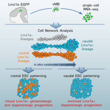
To address this issue, Parmar and his colleagues used modern global gene expression studies to gain a better understand the molecular changes that drive the differentiation of stem cells into dopamine-making neurons. Parmar conducted these experiments in collaboration with a team of scientists at Karolinska Institute. In their paper, which appeared in the journal Cell Stem Cell, Parmar and his colleagues used single-cell RNA seq to construct the neuronal development of dopaminergic neurons.

lmx1a-expressing-cells

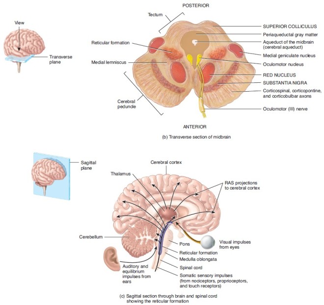
These neurons are characterized by the expression of a gene called LMX1a. However, it turns out that LMX1a-expressing neurons includes not only midbrain dopaminergic neurons (see below at the substantia nigra), but also subthalamic nuclear neurons.

midbrain

These findings reveal that markers used to identify midbrain dopaminergic neurons do not specifically isolate midbrain dopaminergic neurons, but isolate a mixture of cells. Is there a way to separate these two populations?

subthalamic-nucleus

Indeed, there is. Parmar and his colleagues in the laboratory of Thomas Perlmann showed that although dopaminergic neurons from the midbrain and subthalamic nuclear neurons are related, they do express a distinct profile of genes that are specific to the two cell types. The authors argue that the application of these distinct marker genes can help optimize those protocols that differentiate dopaminergic neurons from pluripotent stem cells.

See Nigel Kee and others, “Single-Cell Analysis Reveals a Close Relationship between Differentiating Dopamine and Subthalamic Nucleus Neuronal Lineages,” Cell Stem Cell, 2016; DOI: 10.1016/j.stem.2016.10.003.
Making Connections Between Neural Structures in Culture


Even though it is now possible to fabricate organs and tissues in the laboratory, the vast majority of these structures can be made in isolation without compromising their functionality. Brain cells are quite different, since they required connections known as synapses with other brain cells. Synapses are also responsible for the functional interactions between different regions of the brain. Even though nerve cells (neurons) can be made in the laboratory, engineering connections between neurons is not trivial. Furthermore, some laboratories have used pluripotent stem cells to make portions of the brain in the laboratory, but getting those portions to properly connect with other regions of the brain has proven stultifying.

A new study by William Freed at the National Institutes of Health and his colleagues has designed a way to successfully grow multiple brain structures in the laboratory that form proper connections with each other in culture. This report is the first of its kind.

In particular, Freed and his co-workers defined a culture system for human pluripotent stem cells that produced connected human midbrain and neocortex.

The midbrain houses dopaminergic neurons (mDAs). These neurons use the neurotransmitter dopamine to signal to other neurons that reside elsewhere in the brain. Abnormalities of mDAs or connections between mDAs and other neurons are thought to play intimate roles in disorders like schizophrenia, Parkinson disease, attention-deficit disorder, Roulette’s syndrome, Lesch-Nyhan syndrome, and maybe even eating disorders.

Unfortunately, studying neocortical neurons and mDAs in isolation reveal little about the connections between them or their interactions. However, this new data from Freed that shows that it is possible to grow and interconnect these two types of neurons in culture provides neuroscientists with a powerful model system for examining this system and the abnormalities that afflict it.

The encourage connections between the two neuronal populations, mDAs and neocortical neurons were grown in special containers called “ibidi wound healing” dishes. Ibidi wound healing dishes contain two chambers separated by a removable barrier. Neocortical neurons were grown on one side and mDAs were grown on the other side. Both neuron populations were derived from human pluripotent stem cells. Once the cell cultures had properly formed, the barrier was removed and the two cell populations formed synapses across the barrier.

Freed is eager to examine human pluripotent stem cells derived from patients with neurological disorders that have been traced to abnormalities with connections between mDAs and other neuronal populations to study if neurons made from these patient’s cells properly synapse.

Clearly, this model system has great potential. This work was published in Restorative Neurology and Neuroscience, 2015 DOI: 10.3233/RNN-1140488.