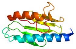
Friedrich’s ataxia (FA) results from insufficient concentrations of a protein called Frataxin. Frataxin serves as an iron metabolism protein that puts iron into proteins that need it. Because several proteins that play crucial roles in energy metabolism in cells use iron, Frataxin is a very busy molecule and without sufficient quantities of Frataxin, energy metabolism decreases and metabolically active cells, such as nerves and muscles, weaken and die.

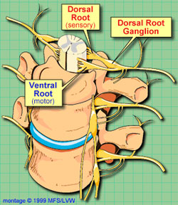
In patients with FA, the dorsal root ganglia, which lie just in front of the spinal cord, are the first to die off and degenerate. Can stem cell treatments provide relief from the ravages of FA?
To test this possibility, and his colleagues from the University Miguel Hernández in Alicante, Spain examined two mouse populations, both of which harbored loss-of-function mutations in the Frataxin (FXN) gene. Mice from both groups were injected with bone marrow-derived mesenchymal stem cells isolated from either wild-type or YG8 mice. YG8 mice a genetically manipulated so that they suffer from a mouse form of FA that shows several similarities to human FA. The mesenchymal stem cells injections were “intrathecal” injections, which means that they were directly injected into the nervous system.
As a result of the stem cell injections, both groups of mice showed improved motor skills compared to nontreated mice. The dorsal root ganglia also showed increased frataxin expression in the treated groups, and less cell death.
Why did the stem cell-injected mice fare better? Further investigations revealed that the injected mesenchymal stem cells expressed the following growth factors: NT3, NT4, and BDNF. All of these growth factors can bind to specific receptors embedded in the membranes of those sensory neurons located within the dorsal root ganglia and buck up their survival, thus preventing them from dying. The stem cell-treated mice also had increased levels of “antioxidant enzymes.”. These are enzymes found in our own cells that dispose of dangerous molecules. Enzymes such as catalase, superoxide dismutase and so on are examples of antioxidant enzymes. The stem cell-treated mice had higher levels of catalase and GPX-1 in their dorsal root ganglia, which is significant because YG8 mice show decreased levels of these antioxidant enzymes.
Interestingly, the results were not significantly different if the injected stem cells were isolated from wild-type or YG8 mice. In both cases injected mesenchymal stem cells ameliorated the condition of the FA mice.
In conclusion, transplantation of bone marrow mesenchymal stem cells, either the patient’s own stem cells or donated stem cells, is a feasible therapeutic procedure that might delay the onset of cell death in the dorsal root ganglia of patients with Friedreich’s ataxia.
