Minneapolis Heart Institute Foundation Tests Stem Cell Combination in Heart Attack Patients


The Minneapolis Heart Institute Foundation has announced a new clinical trial that will examine the ability of a stem cell combination to treat patients with ischemic heart failure.

In patients who have suffered from former heart attacks, clogged coronary blood vessels and heart muscle that hibernates can result in a heart that no longer works well enough to support the life of the patient. The lack of blood flow to vital parts of the heart and an increasing work load can result is so-called “Ischemic heart failure.” Such heart failure after a previous heart attack is one of the leading cause of death and morbidity in the world. According to the World Health Organization, ischemic heart disease affects more than 12% of the world’s population.

Stem cell therapy has been tested as a potential treatment for ischemic heart disease. Despite flashes of remarkable success, the overall efficacy of these treatments has been relatively modest. Most clinical trials have used the patient’s own bone marrow cells. In this case, the cell population is very mixed and it might not even be stem cell populations in the bone marrow that are eliciting recovery. Also, the quality of each patient’s bone marrow is probably quite varied, which makes standardizing such experiments remarkably difficult. Other clinical trials have used bone marrow derived mesenchymal cells [MSCs]. Several clinical trials with MSCs have seen some improvement in patients. MSCs seem to induce the formation of new blood vessels and also seem to induce endogenous stem cell populations in the heart to come to life and fix the heart. Other trials have used cardiac stem cells (CSCs) that were derived from biopsies of the heart. Even though fewer clinical trials have tested the efficacy of CSCs in human patients, the trials that have been conducted suggest that these cells can truly regenerate damaged heart tissue.

The Minneapolis Heart Institute Foundation® (MHIF) has announced a new clinical trial which will examine the combination of MSCs with CSCs to treatment patients with ischemic heart failure. This clinical trial, the CONCERT study, will be led by Principal Investigator Jay Traverse, MD. The CONCERT study will implant MSC’s and CSC’s in order to determine if the combination would be more successful than using either alone based on pre-clinical studies in swine demonstrating an enhanced synergistic effect of the combination.

CONCERT is sponsored by the National Institutes of Health and the Cardiovascular Cell Therapy Research Network (CCTRN), of which MHIF is a charter member. This will be a phase II clinical trial, which means that the focus of this leg of the study is to assess the relative safety of CSCs and MSCs, delivered either alone, or in combination, in comparison to placebo, and to measure the efficacy of the stem cell cocktail as well. To that end, researchers will measure and note any change or improvement in left ventricular (LV) function by cardiac MRI as well as changes in various clinical outcomes (survival, 6-minute walking, blood pressure, etc.), and quality of life.

This phase II study is a randomized, blinded, placebo-controlled study that will enroll 160 subjects at seven different CCTRN sites throughout the U.S. All recruited subjects will have ischemic cardiomyopathy and an ejection fraction 5%). This is significant, because some work in animals suggests that CSCs can make new heart muscle tissue that can shrink the heart scar. The first 16 patients were recently enrolled in a FDA-required safety run-in phase, but the remaining patients will be enrolled in the fall after a three-month safety analysis is performed. Incidentally, this is the first cardiac stem cell trial to perform MRIs on patients with defibrillators and pacemakers

“This combination of cells represents the most potent cell therapy product ever delivered to patients,” said Dr. Traverse. “Confirming that both types of stem cells together work better than either individual cell type could lead to improved patient outcomes and better quality of life for ischemic heart failure patients.”
Stem Cell-Based Skin Graft for Severe Burns


Severe wounds are typically treated with full thickness skin grafts. Some new work by researchers from Michigan Tech and the First Affiliated Hospital of Sun Yat Sen University in Guangzhou, China might provide a way to use a patient’s own stem cells to make split thickness skin grafts (STSG). If this technique pans out, it would eliminate the needs for donors and could work well for large or complicated injury sites.

This work made new engineered tissues were able to capitalize on the body’s natural healing power. Dr. Feng Zhao at Michigan Tech and her Chinese colleagues used specially engineered skin that was “prevascularized, which is to say that Zhao and other designed it so that it could grow its own veins, capillaries and lymphatic channels.

This innovation is a very important one because on of the main reasons grafted tissues or implanted fabricated tissues fail to integrate into the recipient’s body is that the grafted tissue lacks proper vascular support. This leads to a condition called graft ischemia. Therefore, getting the skin to form its own vasculature is vital for the success of STSG.

STSG is a rather versatile procedure that can be used under unfavorable conditions, as in the case of patients who have a wound that has been infected, or in cases where the graft site possess less vasculature, where the chances of a full thickness skin graft successfully integrating would be rather low. Unfortunately, STSGs are more fragile than full thickness skin grafts and can contract significantly during the healing process.

In order to solve the problem of graft contraction and poor vascularization, Zhao and others grew sheets of human mesenchymal stem cells (MSCs) and mixed in with those MSCs, human umbilical cord vascular endothelial cells or HUVECs. HUVECs readily form blood vessels when induced, and growing mesenchymal stem cells tend to synthesize the right cocktail of factors to induce HUVECs to form blood vessels. Therefore this type of skin is truly poised to form its own vasculature and is rightly designated as “prevascularized” tissue.

Zhao and others tested their MSC/HUVEC sheets on the tails of mice that had lost some of their skin because of burns. The prevascularized MSC/HUVEC sheets significantly outperformed MSC-only sheets when it came to repairing the skin of these laboratory mice.

When implanted, the MSC/HUVEC sheets produced less contracted and puckered skin, lower amounts of inflammation, a thinner outer skin (epidermal) thickness along with more robust blood microcirculation in the skin tissue. And if that wasn’t enough, the MSC/HUVEC sheets also preserved skin-specific features like hair follicles and oil glands.

The success of the mixed MSC/HUVEC cell sheets was almost certainly due to the elevated levels of growth factors and small, signaling proteins called cytokines in the prevascularized stem cell sheets that stimulated significant healing in surrounding tissue. The greatest challenge regarding this method is that both STSG and the stem cell sheets are fragile and difficult to harvest.

An important next step in this research is to improve the mechanical properties of the cell sheets and devise new techniques to harvest these cells more easily.

According to Dr. Zhao: “The engineered stem cell sheet will overcome the limitation of current treatments for extensive and severe wounds, such as for acute burn injuries, and significantly improve the quality of life for patients suffering from burns.”

This paper can be found here: Lei Chen et al., “Pre-vascularization Enhances Therapeutic Effects of Human Mesenchymal Stem Cell Sheets in Full Thickness Skin Wound Re-pair,” Theranostics, October 2016 DOI: 10.7150/ thno.17031.

Activation of the Proteasome Enhances Stem Cell Function and Lifespan


As we age, the capacity of our stem cells to heal and replace damaged cells and tissues decline. This age-associated decrease in adult stem cell function seems to be a major contributor to the physiological decline during aging. A new paper, by Efstathios Gonos and his colleagues at the National Hellenic Research Foundation in Athens, Greece gives one possible technique that might improve the function of stem cells in an aging body.

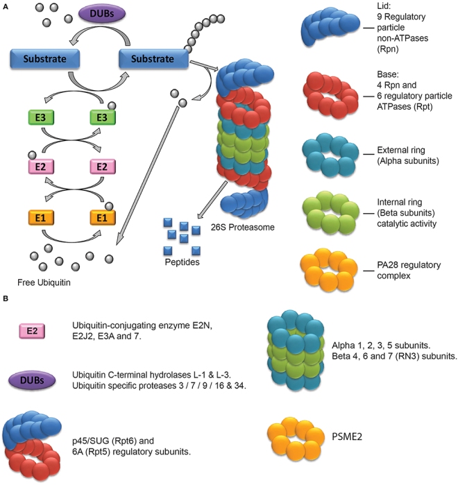
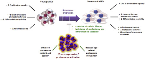
Cells contain a multiprotein complex called the “proteasome” that degrades unneeded or defective proteins. The proteasome controls protein half-lives, function, and the protein composition of the cell. Functional failure of the proteasome has been linked to various biological phenomena including senescence and aging. The role of the proteasome in stem cells aging, however has received little attention to date.

Proteasome figure

Gonos and his coworkers used mesenchymal stem cells from umbilical cord Wharton’s Jelly and human fat. Because they were able to compare the proteasome activity in very young and aged stem cells, Gonos and others discovered a significant age-related decline in proteasome content and activity between these two types of stem cells. The proteasome from Warton’s Jelly mesenchymal stem cells were consistently more active and displayed more normal function and activity than those from human fat.  In fact, not only were the protease activities of the proteasomes from the aging stem cells decreased, but they also displayed structural alterations.

These differences in proteasomal activity were not only reproducible, but when the proteasome of young stem cells were compromised, the “stemness,” or capacity of the stem cells to act as undifferentiated cells, was negatively affected.

Even more surprisingly, once after mesenchymal stem cells from human donors lost their ability to proliferate and act as stem cells (their stemness, that is) their decline could be counteracted by artificially activating their proteasomes. Activating the proteasome seems to help the cell “clean house,” get rid of junk proteins, and rejuvenate themselves.

proteasomes-and-stem-cells

Gonos and his team found that the stem cell-specific protein, Oct4, binds to the promoter region of the genes that encode the β2 and β5 proteasome subunits. Oct4 might very well regulate the expression of these proteasome-specific genes.

From this paper, it seems that a better understanding the mechanisms regulating protein turnover in stem cells might bring forth a way to stem cell-based interventions that can improve health during old age and lifespan.

This paper was published in Free Radical Biology and Medicine, Volume 103, February 2017, Pages 226–235.

Positive Results from Mesoblast’s Phase 2 Trial of Cell Therapy in Diabetic Kidney Disease


Mesoblast Limited has announced results from its Phase 2 clinical Trial that evaluated their Mesenchymal Precursor Cell (MPC) product, known as MPC-300-IV, in patients who suffer from diabetic kidney disease. In short, their cell product was shown to be both safe and effective. The results of their trial were published in the peer-reviewed journal EBioMedicine.  Researchers from the University of Melbourne, Epworth Medical Centre and Monash Medical Centre in Australia participated in this study.

The paper describes a randomized, placebo-controlled, and dose-escalation study that administered to patients with type 2 diabetic nephropathy either a single intravenous infusion of MPC-300-IV or a placebo.

All patients suffered from moderate to severe renal impairment (stage 3b-4 chronic kidney disease for those who are interested).  All patients were taking standard pharmacological agents that are typically prescribed to patients with diabetic nephropathy.  Such drugs include angiotensin-converting enzyme inhibitors (e.g., lisinopril, captopril, ramipril, enalapril, fosinopril, ect.) or angiotensin II receptor blockers (e.g., irbesartan, telmisartan, losartan, valsartan, candesartan, etc.).  A total of 30 patients were randomized to receive either a single infusion of 150 million MPCs, or 300 million MPCs, or saline control in addition to maximal therapy.

Since this was a phase 2 clinical trial, the objectives of the study were to evaluate the safety of this treatment and to examine the efficacy of MPC-300-IV treatment on renal function.  For kidney function, a physiological parameter called the “glomerular filtration rate” or GFR is a crucial indicator of kidney health.  The GFR essentially indicates how well the individual functional units within the kidney, known as “nephrons,” are working.  The GFR indicates how well the blood is filtered by the kidneys, which is one way to measure remaining kidney function.  The decline or change in glomerular filtration rate (GFR) is thought to be an adequate indicator of kidney function, according to the 2012 joint workshop held by the United States Food and Drug Administration and the National Kidney Foundation.

nephronanatomy

Diabetic nephropathy is an important disease for global health, since it is the single leading cause of end-stage kidney disease.  Diabetic nephropathy accounts for almost half of all end-stage kidney disease cases in the United States and over 40% of new patients entering dialysis treatment.  For example, there are almost 2 million cases of moderate to severe diabetic nephropathy in 2013.

Diabetic nephropathy can even occur in patients whose diabetes is well controlled – those patients who manage to keep their blood glucose levels at a reasonable level.  In the case of diabetic nephropathy, chronic infiltration of the kidneys by inflammatory monocytes that secrete pro-inflammatory cytokines causes endothelial dysfunction and fibrosis in the kidney.

Staging of chronic kidney disease (CKD) is based on GFR levels.  GFR decline typically defines the progression to kidney failure (for example, stage 5, GFR<15ml/min/1.73m2).  The current standard of care (renin-angiotensin system inhibition with angiotensin converting enzyme inhibitors or angiotensin II receptor blockers) only delays the progression to kidney failure by 16-25%, which leaves a large residual risk for end-stage kidney disease.  For patients with end-stage kidney disease, the only treatment option is renal replacement (dialysis or kidney transplantation), which incurs high medical costs and substantial disruptions to a normal lifestyle.  Due to a severe shortage of kidneys, in 2012 approximately 92,000 persons in the United States died while on the transplant list.  For those on dialysis, the mortality rate is high with an approximately 40% fatality rate within two years.

The main results of this clinical trial were that the safety profile for MPC-300-IV treatment was similar to placebo.  There were no treatment-related adverse events.  Secondly, patients who received a single MPC infusion at either dose had improved renal function compared to placebo, as defined by preservation or improvement in GFR 12 weeks after treatment.  Third, the rate of decline in estimated GFR at 12 weeks was significantly reduced in those patients who received a single dose of 150 million MPCs relative to the placebo group (p=0.05).  Finally, there was a trend toward more pronounced treatment effects relative to placebo in a pre-specified subgroup of patients whose GFRs were lower than 30 ml/min/1.73m2 at baseline (p=0.07).  In other words, the worse the patients were at the start of the trial, the better they responded to the treatment.

The lead author of this publication, Dr David Packham, Associate Professor in the Department of Medicine at the University of Melbourne and Director of the Melbourne Renal Research Group, said: “The efficacy signal observed with respect to preservation or improvement in GFR is exciting, especially given that this trial was not powered to show statistical significance. Patients receiving a single infusion of MPC-300-IV showed no evidence of developing an immune response to the administered cells, suggesting that repeat administration is feasible and may in the longer term be able to halt or even reverse progressive chronic kidney disease. I hope that this very promising investigational therapy will be advanced to rigorous Phase 3 clinical trials to test this hypothesis as soon as possible.”

Patients who received s single IV infusion of MPC-300-IV cells showed no evidence of developing an immune response to the administered cells.  This suggests that repeated administration of MPCs is feasible and might even have the ability to halt, or even reverse progressive chronic kidney disease.

Packham and his colleagues hope that this cell-based therapy can be advanced to a rigorous Phase 3 clinical trial to further test this treatment.

Bone Marrow Mesenchymal Stem Cells Spontaneously Make Cartilage After Blockage of VEGF Signaling


Bone marrow-derived mesenchymal stem cells (MSCs) can be induced to make cartilage by incubating the cells with particular growth factors.  Unfortunately, batches of MSCs show respectable variability from patient-to-patient.  Therefore the growth factor-dependent method suffers from poor efficacy, limited reproducibility from batch-to-batch, and the cell types that are induced are not always terribly stable.  Finding a better way to make cartilage would certainly be a welcome addition to regenerative treatments,

Cartilage that coats the ends of bones is known as articulate cartilage, and articular cartilage lacks blood vessels.  Therefore, is it possible that inhibiting blood vessel formation could conveniently push MSCs to differentiate into cartilage-making chondrocytes?

A new paper by Ivan Martin and Andrea Basil from the University Hospital Basel and their colleagues have used this very strategy to induce cartilage formation in MSCs from bone marrow.

Martin and others isolated MSCs from bone marrow aspirates from human donors.  These cultured human MSCs were then genetically engineered with modified viruses to express a receptor for soluble vascular endothelial growth factor (VEGF) that binds this growth factor, but fails to induce any intracellular signals.  Such a receptor that binds the growth factor but does not induce any biological effects is called a “decoy receptor,” and decoy receptors efficiently sequester or vacuum up all the endogenous VEGF.  VEGF is the major blood vessel-inducing growth factor and it is heavily expressed during development, by cancer cells, and during healing.

After expressing the decoy VEGF receptor in these human MSCs, these genetically engineered cells were grown on collagen sponges and then implanted in immunodeficient mice.  If the implanted MSCs were not genetically engineered to express decoy VEGF receptors, they induced for formation of vascularized fibrous tissue.  However, the implantation of genetically engineered MSCs that expressed the decoy VEGF receptor efficiently and reproducibly differentiated into chondrocytes and formed hyaline cartilage. This is significant because headline cartilage is the very type of cartilage found at articular surfaces where the ends of bones come together to form joints.

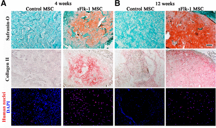
In vivo chondrogenesis. Histological staining with Safranin-O for glycosaminoglycans and immunohistochemistry for type II collagen of engineered tissue generated by naïve (control) or sFlk-1 MSCs after 4 (A) or 12 (B) weeks in vivo. Fluorescence staining with DAPI (in blue) and a specific anti-human nuclei antibody (in red) of constructs generated by control or sFlk-1 MSCs after 4 (A) or 12 (B) weeks in vivo. Scale bar = 100 µm. Abbreviations: DAPI, 4′,6-diamidino-2-phenylindole; MSC, bone marrow-derived mesenchymal stromal/stem cell.

This articular cartilage was quite stable and showed no signs of undergoing the chondrocytes enlargement found in terminally differentiated cartilage that is ready to form bone.  This stability was maintained for up to 12 weeks.

In vivo cartilage stability. Immunohistochemistry for type X collagen, BSP, and MMP-13 on sections of hypertrophic cartilage generated in vitro by MSCs (as a positive control) and on sections of the cartilaginous constructs generated in vivo by sFlk1 MSCs 12 weeks after implantation. Scale bar = 50 µm. Abbreviations: BSP, bone sialoprotein; MMP-13, metalloproteinase-13; MSC, bone marrow-derived mesenchymal stromal/stem cell.

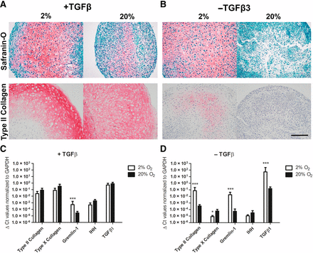
Why did inhibition of VEGF signaling induce cartilage?  Inhibition of angiogenesis induced low oxygen tensions, which activated a growth factor called transforming growth factor-β.  Activation of the TGF-beta pathway robustly enhanced the formation of articular cartilage.

In vitro chondrogenesis at different oxygen tensions. Histological staining with Safranin-O and immunohistochemistry for type II collagen on constructs generated in vitro by naïve MSC cultured with (A) or without (B) TGFβ3 supplementation at 2% or 20% of oxygen tension. Scale bar = 50 µm. Expression levels of the mRNA for type II and X collagen, Gremlin-1, IHH TGFβ1 were quantified in pellets generated by naïve bone marrow-derived mesenchymal stromal/stem cells (C, D) cultured in the two different oxygen tensions. ∆Ct values were normalized to expression of the GAPDH housekeeping gene, and results are shown as mean ± SD (n = 6 samples/group from 3 independent experiments). ∗, p < .05, ∗∗∗, p < .001. Abbreviations: GAPDH, glyceraldehyde-3-phosphate dehydrogenase; IHH, Indian hedgehog; TGFβ, transforming grown factor-β.

Cartilage formation from MSCs was induced by blocking VEGF-mediated angiogenesis.  These results represent a remarkable advance in cartilage formation that can be used for regenerative treatments.  This cartilage formation was spontaneous and efficient and if it can be carried out with VEGF-inhibiting drugs rather than genetic engineering techniques, then we might have a transferable technique for making cartilage in the laboratory to treat osteoarthritis and other joint-based maladies.  Clinical trials will be required, but this is certainly an auspicious start.

Fat-derived cells Enhance the Bone-Forming Capacity of Hypertrophic Cartilage Matrices


Treating particular bone defects or injuries present a substantial challenges for clinicians. The method of choice usually involves the use of an “autologous” bone graft (“autologous” simply means that the graft comes from the patient’s own bone). However, autologous bone grafts have plenty of limitations. For example, if a patient has a large enough bone defect, there is no way the orthopedist and take bone from a donor site without causing a good deal of risk to the donor site. Even with small bone grafts, so-called “donor site morbidity” remains a risk. Having said that, plenty of patients have had autologous bone grafts that have worked well, but larger bone injuries or defects are not treatable with autologous bone grafts.

The answer: bone substitute materials. Bone substitute materials include tricalcium phosphate, hydroxyapatite, cement, ceramics, bioglass, hydrogels, polylactides, PMMA or poly(methy methacrylate) and other acrylates,, and a cadre of commercially available granules, blocks, pastes, cements, and membranes. Some of these materials are experimental, but others do work, even if do not work every time. The main problem with bone substitute materials is that, well, they are not bone, and, therefore lack the intrinsic ability to induce the growth of new bone (so-called osteoinductive potential) and their ability to integrate into new bone is also a problem at times.

We must admit that a good deal of progress has been made in this area and it’s a good thing too. Many of our fabulous men and women-at-arms have returned home with severe injuries from explosives and wounds from large-caliber weapons that have shattered their bones. These courageous men and women have been the recipient of these technologies. However, the clinician is sometimes left asking herself, “can we do better?”

A new paper from the laboratories of Ivan Martin and Claude Jaquiery from the University Hospital of Basel suggests that we can. This paper appeared in Stem Cells Translational Medicine and describes the use of a hypertrophic cartilage matrix that was seeded with cells derived from the stromal vascular faction of fat to not only make bone in the laboratory, but to also heal skull defects in laboratory animals. While this work benefitted laboratory animals, it was performed with human cells and materials, which suggests that this technique, if it can be efficiently and cheaply scaled up, might be usable in human patients.
The two lead authors of this paper, Atanas Todorov and Matthias Kreutz and their colleagues made hypertrophic cartilage matrices from human bone marrow mesenchymal stem cells (from human donors) that were induced to make cartilage. Fortunately, protocols have been very well worked out and making cartilage plugs with chondrocytes that are enlarged (hypertrophic) is something that has been successfully done in many laboratories. After growing the mesenchymal stem cells in culture, half a million cells were induced to form cartilage with dexamethasone, ascorbic acid 2-phosphate, and the growth factor TGF-beta1. After three weeks, the cartilage plugs were subjected to hypertrophic medium, which causes the cartilage cells to enlarge.

Chondrocyte enlargement is a prolegomena to the formation of bone and during development, many of our long bones (femur, humerus, fibula, radius, etc.), initially form as cartilage exemplars that are replaced by bone as the chondrocytes enlarge. Ossification begins when a hollow cylinder forms in the center of the bone (known as the periosteal collar). The underlying chondrocytes degenerate and die, thus releasing the matrix upon which calcium phosphate crystals accrete. The primary ossification center commences with the calcification of the central shaft of the bone and erosion of the matrix by the invasion of a blood vessel. The blood vessels bring osteoprogenitor cells that differentiate into osteoblasts and begin to deposit the bone matrix.

Next, Todorov and his crew isolated the stromal vascular fraction from fat that was donated by 12 volunteers who had fat taken from them by means of liposuction. The fat is then minced, digested with enzymes, centrifuged, filtered and then counted. This remaining fraction is called the stromal vascular fraction or SVF, and it consists of a pastiche of blood vessel-forming cells, mesenchymal stem cells, and bone-forming cells (and probably a few other cells types too). These SVF cells were seeded onto the hypertrophic cartilage plugs and used for the experiments in this paper.

First, the SVF-seeded plugs were used to grow bone in laboratory rodents. The cartilage plugs were implanted into the backs for nude mice. Different cartilage plugs were used that had been seeded with gradually increasing number of SVF cells. The implanted plugs definitely made ectopic bone, but the amount of bone they made was directly proportional to the number of SVF cells with which they had been seeded. Staining experimental also showed that some of the newly-grown bone came from the implanted SVF cells.

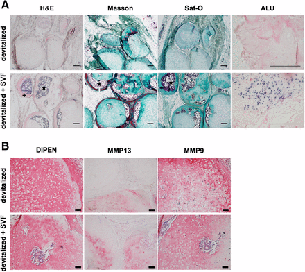
Ectopic bone formation. Grafts based on devitalized hypertrophic cartilage pellets were embedded in fibrin gel without or with stromal vascular fraction cells from adipose tissue and implanted subcutaneously in nude mice. (A): Representative hematoxylin and eosin, Masson-Tri-Chrome, and Safranin-O (Saf-O) staining and in situ hybridization for human ALU sequences (dark blue = positive) after 12 weeks in vivo. Saf-O stainings are blue-green because of lack of glycosaminoglycans and counterstaining with fast green. Osteoid matrix and bone marrow are visible. Scale bars = 200 µm. (B): Stainings for metalloproteinase (MMP)13 and MMP9, as well as for the N-terminal neoepitope at the major MMP cleavage site (DIPEN) after 12 weeks in vivo (red/pink = positive). Scale bars = 50 µm. +, osteoid matrix; ⋆, bone marrow. Abbreviations: ALU, Arthrobacter luteus; H&E, hematoxylin and eosin; Masson, Masson’s trichrome; MMP, metalloproteinase; Saf-O, Safranin-O; SVF, stromal vascular fraction.

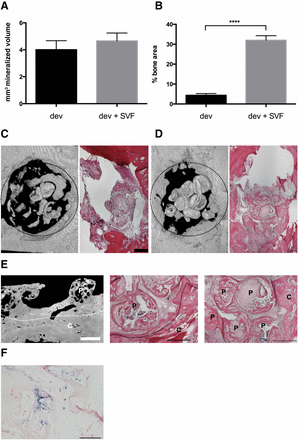
In the second experiment, Todorov and Kreutz used these SVF-seeded cartilage plugs to repair skull lesions in rats. Once again, the quantity of bone produced was directly proportional to the number of SVFs seeded into the cartilage matrices prior to implantation. In both experiments, the density of SVF cells positively correlates with the bone-forming cells in the grafts and the percentage of SVF-derived blood vessel-forming cells correlates with the amount of deposited mineralized matrix.

Bone repair capacity. Devitalized hypertrophic cartilage pellets were embedded in fibrin gel without or with stromal vascular fraction (SVF) cells from adipose tissue and implanted in rat calvarial defects. (A): Mineralized volume quantified by microtomography (n = 9 grafts assessed per group). (B): Bone area assessed in histological sections, expressed as percentage of total defect area (n = at least 24 sections assessed per group). ∗∗∗∗, p < .0001. (C, D): Representative three-dimensional microtomography reconstructions (left) and hematoxylin/eosin (H&E) staining (right) of the calvarial defects filled with devitalized grafts, implanted without (C) or with (D) activation by SVF cells after 4 weeks. Dotted circles indicate the defect borders (4 mm diameter). Scale bars = 500 µm. (E): Microtomography (left) and H&E staining (middle and right) displaying the bridging between hypertrophic matrix and bone of the calvarium, or the fusion of single pellets (right) in activated grafts. White bar = 850 µm; black bars = 500 µm. Dotted lines indicate the edge of the calvarium. (F): In situ hybridization for Arthrobacter luteus sequences showing the presence of human cells (dark blue, positive) in the explants. Scale bar = 200 µm. Abbreviations: C, calvarium; dev, fibrin gel without stromal vascular fraction; dev + SVF, fibrin gel with stromal vascular fraction; P, hypertrophic matrix; SVF, stromal vascular fraction.

This is not the first time scientists have proposed the use of cartilage plugs to induce the formation of new bone. Van der Stok and others and Bahney and colleagues were able to repair segmental bone defects in laboratory rodents. Is this technique transferable to human patients? Possibly. Hypertrophic cartilage is relatively easy to make and it is completely conceivable that hypertrophic cartilage wedges can be sold as “off-the-shelf” products for bone treatments. SVF can also be derived from the patient or can be derived from donors.

Furthermore, the protocols in this paper all used human cells and grew the products in immunodeficient rats and mice. Therefore, in addition to scaling this process up, we have a potentially useful protocol that might very well be adaptable to the clinic.

The efficacy of this technique must be confirmed in larger animal model system before human trials can be considered. Hopefully human trials are in the future for this fascinating technique.

Fat-Based Stem Cell Treatment Suggests a New Way to Slow Scarring in Scleroderma Patients


Scleroderma is an autoimmune disease that causes chronic scarring of the skin and internal organs. The deposition of massive quantities of collagen decrease the pliability and elasticity of the skin, lungs, and blood vessels. As you might guess, the prognosis of scleroderma patients is quite poor and this disease causes a good deal of suffering and morbidity.

Treatments options usually include steroids, and other drugs that suppress the immune system, all of which have severe side effects.

New research from scientists at the Hospital for Special Surgery in New York City and other collaborating institutions, led by Dr. Teresa T. Lu, may have identified a new mechanism in operation during the onset and maintenance of scleroderma. This work was published in the Journal of Clinical Investigation.

In this study, scleroderma patients were shown to possess diminished numbers of “adipose-derived stromal cells” (ADSCs) in the layer of fat that underlies the upper layers of the skin. These fatty tissues are referred to as “dermal white adipose tissue.” The loss of these dermal white adipose tissue ADSCs tightly correlates with the onset of scarring in two different mouse model systems that recapitulate scleroderma in laboratory mice. These observations may show that ADSC loss contributes to scarring of the skin.

Why do these ADSCs die? Lu and her coworkers discovered that ADSC survival depends on the presence of particular molecules secreted by immune cells called “dendritic cells.” Skin-based dendritic cells secrete a molecule called lymphotoxin B. Although this molecule is called a toxin, it is required for ADSC survival. In laboratory mice that suffered from a scleroderma-like disease, artificial stimulation of the lymphotoxin B receptor in ADSCs amplified and eventually restored the numbers of ADSCs in the skin. Could stimulating ADSCs in this manner help treat scleroderma patients?

According the Dr. Lu, the administrating author of this publication, injecting “ADSCs is being tried in scleroderma; the possibility of stimulating the lymphotoxin B pathway to increase the survival of these stem cells is very exciting.” Dr. Lu continued, “By uncovering these mechanisms and targeting them with treatments, perhaps one day we can better treat the disease.”

Lu also thinks that a similar strategy that targets stem cells from other tissues might provide a treatment for other rheumatological conditions – such as systemic lupus erythematosis and rheumatoid arthritis. Additionally, bone and cartilage repair might also benefit from such a treatment strategy.

In the coming years, Dr. Lu and her colleagues hope to test the applicability of this work in human cells. If such a strategy works in human cells, then the next stop would be trial in human scleroderma patients. The success of such a treatment strategy would be a welcome addition to the treatment options for scleroderma patients, but only if this treatment is shown to be proven safe and effective.

“Improving ADSC therapy would be a major benefit to the field of rheumatology and to patients suffering from scleroderma,” said Lu.

Intravenous Preconditioned Mesenchymal Stem Cells from Donors Improve the Heart Function of Heart Failure Patients


CardioCell is a global biotechnology company that was founded in 2013 in San Diego, California. CardioCell specializes in ischemia-tolerant mesenchymal stem cells (itMSCs). These stem cells are derived from bone marrow-derived mesenchymal stems extracted from healthy donors. However, after isolation, these cells are grown in low-oxygen conditions, which induces the expression of genes that allow cells to adapt to stressful, oxygen-poor conditions.

Non-ischemic dilated cardiomyopathy (NIDCM) is a progressive disorder with no current cure, often culminating in heart transplantation. Because the heart has enlarged, there are areas where the blood supply of the heart fails to properly provide oxygen to the tissues. Without proper muscular support, the walls of the heart begin to thin and the blood supply becomes less and less adequate to the task of feeding the heart muscle. Also, the heart of a patient experiencing chronic heart failure also seems to have some low-level of inflammation that slowly damage the heart (Circ Res. 2016;119(1):159-76). Stem cell treatments might help ameliorate the physiological quandary in which the heart finds itself, but these oxygen-poor areas of the heart are inimical to stem cell survival and flourishing. Therefore, itMSCs stand a better chance of surviving when implanted into a damaged heart than non-conditioned stem cells. Experiments in laboratory animals have confirmed that itMSCs show a greater ability to seek out and find the damaged heart and engraft into the heart at higher rates than MSCs grown under normal culture conditions (see PLoS One. 2015 Sep 18;10(9):e0138477; Stem Cells. 2015 Jun;33(6):1818-28). These itMSCs also secrete higher levels of growth factors and angiogenic factors than normal MSCs. On the strength of these laboratory and animal-based studies itMSCs are now in the process of being tested as a treatment for heart attack patients.

CardioCell has sponsored a single-blind, placebo-controlled, crossover, randomized phase II-a trial of patients with NIDCM who have an ejection below 40% (the ejection fraction refers to the average percentage of blood pumped from the left ventricle at each contraction. The average ejection from for a healthy individual is about 65% or so).  The results of this study were published in the journal Circulation Research (;

Patients who volunteered for this study were randomly assigned to group I or group II. Group I patients received intravenous infusions of one and a half-million itMSCs per kilogram body weight. Group II received the placebo. There were 22 patients in all, and 10 received the itMSCs and 10 received the placebo. Since this was a crossover trial, after 90 days, patients in group I received he placebo and group II received the intravenous itMSCs. After crossover, safety and efficacy data were available for all 22 itMSC patients.

With respect to safety issues, there were no major differences in the number of deaths, hospitalizations, or serious adverse events between the two treatments. With respect the efficacy, the data is but more difficult to analyze. In the first place, when it comes to changes in the ejection fraction of the left ventricle from the originally measured baseline, there were no statistically significant changes between the two treatments. The same could be said for the volume of the left ventricle. This is an unfortunately finding, since heart failure includes a decrease in the ejection fraction of the heart and stretching and dilation of the ventricles. Stem cell treatments, if they are to properly treat heart failure, should increase the ejection fraction of the heart and reduce the dilation of the left ventricle. However, there might be more to these data than originally meets the eye. When it came to patient performance, the data was much more hopeful. Compared to patients who received the placebo, patients who received the itMSCs significantly increased the distance they were able to walk during 6-minutes. Patients who had received the itMSCs walked an average of 36.47 longer meters than patients who had received the placebo. Additionally, patients were also given a commonly-used survey, called the Kansas City Cardiomyopathy clinical summary. This survey is a 23-item, self-administered instrument that quantifies physical function, symptoms (frequency, severity and recent change), social function, self-efficacy and knowledge, and quality of life. Administration of this survey to both sets of patients revealed that patients who had received the itMSCs consistently and statistically significantly scored higher on this survey than those patients who received the placebo. The same was also demonstrated for particular functional status tests. Therefore, when it came to how well patients felt and well they functioned, itMSC treatments seemed to excel significantly better than placebo.

Given the ability of MSCs to suppress inflammation, and given the tendency for patients with heart failure to suffer from chronic inflammation of the heart, individual patients were measured for their degree of inflammation. There was an inverse relationship between the degree of inflammation in a patient and their ejection fraction; the lower their level of inflammation, the higher their ejection fraction.

Thus this study seems to suggest that treatment of heart failure with itMSCs is indeed safe. These treatments also did reduce inflammation in heart failure patients and these reductions in inflammation were also associated with improvements in health status and functional capacity.

Donor Fat-Based Stem Cells May Provide Augmented Healing of Rectovaginal Fistulas of Crohn’s Disease Patients


Fistulas are openings in organ systems that connect with another system. They usually result from wounds or erosions in the lining of a tube or duct that gets deeper and deeper and eventually opens into another tube or duct. Physical injuries can cause fistulas, but so can diseases such as Crohn’s disease. Anal fistulas result from erosions of the rectum that open to the outside and are typically very painful and do not readily heal.

Damián García-Olmo and his colleagues at the Universidad Autónoma de Madrid have conducted several clinical trials that have examined the ability of adipose-derived stem cells (ASCs) to facilitate the healing of fistulas in Crohn’s disease patients. A phase I study, which primarily examines safety, was published in 2005 (see García-Olmo D., et al., Dis Colon Rectum 2005; 48:1416-1423). According to this study, “No adverse effects were observed in any patient at the end of the follow-up period (minimum follow-up, 12 months; maximum follow-up, 30 months; follow-up average, 22 months).” The Phase II study was published in 2009 (García-Olmo, D., et al., Dis Colon Rectum 2009; 52:79-86). According to the results of this study, fistula healing was observed in 71 percent of patients who were treated with ASCs in combination with fibrin glue compared with 16 percent of patients who received fibrin glue alone. Quality of life scores were also higher in patients who received ASCs than in those who received fibrin glue alone. Once again, the stem cell treatments were well tolerated. The third study was a multicenter, randomized, single-blind clinical trial that enrolled 200 adult patients from 19 centers that were randomly assigned to three groups. The first group received 20 million stem cells (group A, 64 patients). The second group received 20 million adipose-derived stem cells plus fibrin glue (group B, 60 patients). The third group received only fibrin glue (group C, 59 patients). In treatment of anal fistulas in Crohn’s disease patients, a dose of 20 or 60 million adipose-derived stem cells alone or in combination with fibrin glue were demonstrably safe and did promote healing. However, there were no statistically significant differences between the three groups once the 3 groups were compared.

These studies suggest that stem cells from fat might have a place in the treatment of fistulas in Crohn’s disease patients. The application of the stem cells is feasible and safe, and requires no new equipment or skill. The stem cells also might augment the healing of these fistulas.

Unfortunately, anal fistulas are not the only type of fistulas that Crohn’s disease patients can experience. Female Crohn’s disease patients can have fistulas that open from their rectum into their birth canal. These rectovaginal fistula can deposit the contents of the gastrointestinal tract into the lower reproductive tract. While Crohn’s disease is not the only cause rectovaginal fistulas, Crohn’s disease patients are at higher risks for complications, which include: loss of control over stool deposition (fecal incontinence), hygiene problems combined with recurrent vaginal or urinary tract infections, inflammation of the birth canal and skin around the anus (perineum), abscess formation, which can become life-threatening if not treated, and recurrence of the fistula. Surgical treatment of rectovaginal fistulas requires that the tissue be free of inflammation before surgery, which can take time and cause extensive amounts of patient suffering.

Garcia-Olmo and his colleagues have conducted a small phase I-IIa clinical trial to evaluate the possibility of banked fat-based stem cells to treat recto-vaginal fistulas in female Crohn’s patients. This study has several limitations because it is so small and they have to exclude at least half of the participants because of complications beyond their control. Therefore, this study is not statistically significant. However, it does show what might be the beginnings of a stem-based treatment for this horrid condition.

The design of the study included 11 subjects who were initially enrolled in the study, but one of those recruited patients did not meet the criteria for the study. Therefore, ten subjects, all of whom suffered from Crohn’s disease and had rectovaginal fistulas were treated with 20 million fat-based stem cells that had been donated by a healthy volunteer in addition to surgical repair of their fistulas. These donated fat-based stem cells were provided by a Spanish biotechnology company called Cellerix S.L. Three months after this stem cell treatment, two patients were healed and the other either were given an additional treatment of 40 million fat-based stem cells. Of this group, four were healed. However, five of these patients experienced severe flare-ups of their Crohn’s disease that required treatment with biological agents, which disqualified these patients from further consideration from this study. The biological agents used to treat the Crohn’s disease flare-ups are very powerful medicines and can significantly influence the outcome of this study. Thus half of the subjects in this study had to be excluded. Of the five subjects that remained, 3 showed healing of their fistulas, and 2 did not.

The authors present the data as a “final efficacy rate of 60%.” However, given the high rate of exclusion and the very low numbers of subjects in this study, all we can say with any confidence is that based on the previous successes of this treatment in other studies, there is precedent for such a technique to be safe and somewhat effective, and that the data in this study are in a favorable direction. However, that’s about it.

One feature of this study that differs from the other clinical trials done by this same group is that the previous studies utilized the patient’s own fat-based stem cells, whereas this study used stem cells from a healthy donor. The authors stress that this modification greatly simplifies the procedure and decreases its expense. Because of the ease of the treatments, it reduces postoperative hospitalization and is minimally invasive. This new trial suggests that further work is warranted and the results or even minimally hopeful.

This work was published in the journal Stem Cells Translational Medicine 2016; 5(11): 1441-1446.

Cynata’s MSC Technology Produces Significant Relief of Asthma in Preclinical Study


An Australian stem cell company called Cynata Therapeutics Limited is in the process of developing a therapeutic stem cell platform technology that they called “Cymerus.” The idea for Cymerus originated at the University of Wisconsin-Madison, but Cymerus would generate a protocol by which clinical laboratories could produce very immature mesenchymal stem cells from induced pluripotent stem cells. Such cells would be personalized for patients and their needs, and Cynata’s goal is to produce a platform that is economically feasible and relatively fast so that patients can receive infusions of the cells they so badly need in a timely fashion. These are very ambitious goals to say the least, but Cynata has been hacking away at this problem for some time, and we certainly wish them the best.

Cynata has recently released some very encouraging data in which their personalized mesenchymal stem cells were used to treat laboratory animals with a laboratory-induces form of asthma. Briefly, female mice (BALB/c mice for those who are interested) were injected with a yolk-protein called “ovalbumin.” Ovalbumin is a protein found in egg whites, and because it is an egg-specific protein, mice do not have it and their immune systems have never seen it before. Such an injection causes the mice to mount an immune response to the ovalbumin, and these mice are then administered aerosolized ovalbumin by means of a nebulizer. This causes the animals to develop a rather severe asthmatic attack against ovalbumin.

In this study, Cynata scientists and their collaborators used 48 mice that were divided into six different groups. The first group was untreated animals that did not suffer from ovalbumin asthma. The second group contained eight animals that had no asthma but were treated intravenously with one million mesenchymal stem cells. The third group also had no asthma, but were treated with an intranasal infusions of one million mesenchymal stem cells. The fourth group contain eight asthmatic animals that were untreated during the course of the experiment. The fifth group contain eight asthmatic animals that were treated intravenously with one million mesenchymal stem cells. The final group contained eight asthmatic animals that were treated with intranasal infusions of one million mesenchymal stem cells. As a note, all animals that were treated mesenchymal stem cells were treated three times. So-called airway hyperresponsiveness (AHR) is a measure of the sensitivity and irritability of the bronchial tissues. AHR is an important measure of the tendency of the lungs to undergo constriction during an asthma attack and AHR is usually measured by administering a drug that can cause bronchoconstriction. The greater the degree of bronchoconstriction in such an experiment is indicative of great AHR. The successful treatment of asthma results in reduction in AHR.

The results of this experiment were wonderfully successful. Exposing mice to the ovalbumin caused them to exhibit significantly increased AHR. However, intravenous administration of Cynata’s MSCs in asthmatic animals caused a statistically significant (60-70%) decrease in AHR compared to untreated, sensitized animals. Additionally, intranasal administration of Cynata’s MSCs completely normalized AHR. The AHR in these asthmatic mice was brought down to a level that was largely the same as the non-asthmatic mice. Also, importantly, no adverse side effects were observed during the study.

This study was conducted under the supervision of Associate Professor Chrishan Samuel and Dr. Simon Royce from the Department of Pharmacology at Monash University, Melbourne, Australia. Because the features of this model asthma system closely resemble the clinical manifestations of asthma in humans, these results provide excellent evidence that such a treatment stands a chance of working in human patients.

“We are very excited by these results, which indicate that Cymerus™ MSCs could have a profound effect in the treatment of asthma. This is a debilitating condition, which affects about 10% of the population, resulting in close to 40,000 hospitalizations and several hundred deaths each year, in Australia alone,” said Cynata Vice President of Product Development, Dr. Kilian Kelly. “Although a number of drugs are approved for the treatment of asthma, studies have shown that conventional treatments result in as few as 5% of asthma patients achieving full control of their condition. Consequently, there is a widely recognized need for novel treatments that address – and potentially eliminate – the underlying disease”, added Dr. Kelly.

“This study has clearly demonstrated that Cynata’s MSCs have a dramatic effect on AHR in our model, particularly when directly administered into the allergic lung. We look forward to continuing our analysis of the effects of these unique cells on markers of inflammation and airway remodeling, and we are optimistic of building on the very positive data we have generated so far,” said Associate Professor Samuel.

Asthma is a condition characterized by the inflammation, narrowing, and swelling of the airways, accompanied by excessive mucous production that makes it difficult to breathe. According to the Global Asthma Network, asthma affects over 330 million people globally. Cynata had partnered with Monash University to examine the potential of its Cymerus technology as an alternate treatment for asthma sufferers.

Cymerus™ makes us of induced pluripotent stem cells (iPSCs) that are then differentiated into a specific type of mesenchymal stem cell precursor known as a “mesenchymoangioblast” or MCA. Cymerus potentially provides a source of MSCs that can be made for so-called “off-the-shelf” therapeutic uses.

Fat-Based Mesenchymal Stem Cell-Seeded Matrix Heals Bronchopleural Fistula in Female Cancer Patient


Bronchopleural fistulae, mercifully abbreviated as BPF, refers to an opening or hole in the respiratory tree that causes continuity between the pleural space that surrounds the lungs and the bronchial tree. BPH is a highly feared complication of surgery on the respiratory system.

BPH can complicate surgical resection of the pulmonary system. Patients with lung cancers may require lung resection in order to remove tumorous lung tissue. The rate of BPH incidence after lung surgery varies widely, with reported incidences ranging from 1.5 to 28%. Necrosis or death of lung tissue as a result of infection can also cause BPH, as can tuberculosis. Chemotherapy or radiation therapy for lung cancers can also result in BPF. Finally, BPF may caused by persistent spontaneous pneumothorax, which refers to an abnormal build up of air or other gases in the pleural space, which causes an uncoupling of the lung from the chest wall.

To date, treatment for BPF is only partially effectively. The main treatment includes surgery, but the rate of recurrence of the fistulae remains rather high as do the rate of mortality. Can stem cells show us a better way?

Perhaps they can. Dennis A. Wigle, a surgeon at Mayo Clinic, and his collaborators used a synthetic bioabsorbable matrix seeded with the patients one fat-based mesenchymal stem cells to heal a BPF in a 63-yr old woman. Mind you, this is a case study (the lowest quality clinical evidence) and not a controlled study,. However, the success of this case study is at least suggestive that such an approach might prove useful for patients who suffer from BPFs.

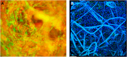
Microscopic assessment of matrix cell seeding. (A): Ethidium bromide (red) and Syto-13 (green) costain demonstrating live and dead cells on mesenchymal stem cell seeding on matrix. (B): Confocal microscopy with CD90 (Thy-1) fluorescein isothiocyanate (green) and Hoechst 33342 (trihydrochloride trihydrate) (blue) fluorescent nuclear staining. These images were captured using a ×20 objective and a ×10 eyepiece, for a combined magnification of ×200. Scale bar = 150 µm.

A 63-yr old woman who had surgical resection of the lung in order to treat her lung cancer had, as a consequence of her surgery, a BPF. Some 30 different surgical attempts were made to repair the BPF, but all of them failed. The woman’s health declined and her medical team started to think of alternative treatments.

Fortunately, Mayo Clinic has been participating in an ongoing clinical trial to use fat-based mesenchymal stem cells to treat anal fistulae in Crohn’s disease patients. Therefore Dr. Wigle and his team considered using the protocol utilized with Crohn’s patients to repair this woman’s BPF.

Fat biopsies were taken from the patient and the fat was washed, minced, digested with enzymes, and then grown in special culture media. The adipose tissue-derived mesenchymal stem cells (AD-MSCs) grew and were isolated, characterized and shown to be MSCs.

These cells were then seeded on a matrix of synthetic bioabsorbable poly(glycolide-trimethylene carbonate) copolymer and then placed in a bioreactor to grow. After about 4 days, the matrix was flush with AD-MSCs, and this cell-seeded patch was then used in a subsequent surgery to seal the opening in the respiratory tree. This time the surgery worked. The patient was discharged 25 days after the surgery and sent home.

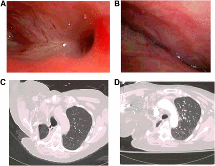
MRIs of the respiratory system showed that the BPF had indeed closed and properly resolved.

Preoperative imaging showing size and location of fistula, and postoperative imaging demonstrating disease resolution. (A): Preoperative bronchoscopy demonstrating large bronchopleural fistula (BPF) cavity and lateral extension of fistula tracts. (B): Postoperative bronchoscopy (3 months) demonstrating progressive healing of BPF site. (C): Preoperative computed tomography scan demonstrating large BPF with connection to atmosphere (additional axial slices inferiorly). (D): Postoperative computed tomography scan (16 months) demonstrating resolution of BPF.

This case study might confirm what was previously observed in large animal studies by Petrella and others, namely that AD-MSCs can be used to heal BPF. Petrella and others theorized that implanted MSCs induce the proliferation of fibroblasts that then deposit collagen, which seals the BPF (see Ann Thorac Surg 97:480483.  Alternatively, AD-MSCs might differentiate into cell types  required for regeneration of the airways (Dominici M, and others, Cytotherapy 8:315317).  Either way, this paper seems to suggest that AD-MSCs can be isolated from a patient’s fat (even a very sick patient like this one) without incident and used for tissue engineering applications that can repair very serious wound like BPF. 

This paper was published in: Johnathon M., Aho, et Al., “Closure of a Recurrent Bronchopleural Fistula Using a Matrix Seeded With Patient-Derived Mesenchymal Stem Cells.” Stem Cells Trans Med October 2016 vol. 5 no. 10 1375-1379. 

Computer Simulations of MSC-Heart Muscle Interactions Identify A Family of MSCs that Produce Few Side Effects


A research team at the Icahn School of Medicine at Mount Sinai has utilized a mathematical modeling to simulate the delivery of human mesenchymal stem cells to a damaged heart. In doing so, they found that a particular subset of harvested MSCs minimizes the risks associated with this therapy. This study represents a development that could lead to novel strategies to repair and regenerate heart muscle and might even improve stem cell treatments for heart attack patients.

In the United States alone, one person suffers a myocardial infarction or heart attack every 43 seconds (on the average). The urgency of this situation has motivated stem cells scientists and cardiologists to develop novel therapies to repair and regenerate heart muscle. One of these therapies includes the implantation of human mesenchymal stem cells (hMSCs). However, in clinical trials the benefits of hMSC implantation have often been modest and even transient. This might reflect our understanding of the mechanism by which hMSCs influence cardiac function.

Kevin D. Costa and his colleagues at the Icahn School of Medicine have used mathematical modeling to simulate the electrical interactions between implanted hMSCs and endogenous heart cells. They hoped to eventually understand the possible adverse effects of hMSC transplantation and new methods for reducing some potential risks of this therapy.

Implanted hMSCs can disrupt the electrical connections between heart muscle cells and can even cause the heart to beat irregularly; a condition called “arrhythmias.” One particular type of hMSCs, however, did not express an ion channel called EAG1 (which stands for “ether-a-go-go”). The EAG1-less hMSCs did not cause arrhythmias at nearly the rate as the EAG1-containing hMSC, in computer simulations run by Costa’s group.

These EAG1-less hMSCs, also known as “Type C” MSCs, minimized electrochemical disturbances in cardiac single-cell and tissue-level electrical activity. The benefits of these EAG1-less hMSCs may enhance the safety of hMSC treatments in heart attack patients who receive stem cell therapy. This advance could therefore lead to new clinical trials and future improvements in treatment of patients with heart failure.

Costa’s study might provide a template for future computational studies on mesenchymal stem cells. It also provides novel insights into hMSC-heart cell interactions that can guide future experimental studies to understand the mechanisms that underlie hMSC therapy for the heart.

This work was published in Joshua Mayourian et al., “Modeling Electrophysiological Coupling and Fusion between Human Mesenchymal Stem Cells and Cardiomyocytes, PLOS Computational Biology, 2016; 12 (7): e1005014. DOI: 10.1371/journal.pcbi.1005014.

Adjustable Gels Used to Determine Those Molecules That Drive Stem Cell Differentiation


Scientists have been very interested in the details of stem cell differentiation. To that end, several laboratories have designed hydrogels that mimic the stiffness of biological tissue in order to grow stem cells and study their differentiation.

In one enterprising laboratory, led by Rein Ulijn of the City University of New York and the University of Strathclyde, scientists have used a novel culture-based gel system to study mesenchymal stem cell differentiation and identify those metabolites used by stem cells when they select bone and cartilage cell fates. When these molecules are provided to standard stem cell cultures, these molecules can guide stem cells to generate desired cell types. This new study illustrates how new biomaterials can provide an exacting model system that can help scientists precisely determine those identifying factors that drive stem cell differentiation.

Stem-cell scientists have known that the rigidity of a hydrogel surface can instruct stem cells to differentiate. A rigid surface, as it turns out, can result in bone cell formation, whereas soft surfaces induce the differentiation of cells into neuron-like cells. With this information, Ulijn and others developed a protocol that generates gels by combining small building-block molecules that spontaneously form a network of nanosized fibers. Furthermore, by varying the concentration of these building blocks, the stiffness of these gels can be adjusted. By mimicking the stiffness of bone (40 kilopascal) or cartilage (15 kilopascal), the gel stimulates stem cells applied to its surface to differentiate accordingly.

“This paper is a great example of how chemistry can help make step changes in biology,” said Matthew Dalby of the University of Glasgow and co-senior author. “As a biologist, I needed simple yet tunable cell-culture gels that would give me a defined system to study metabolites in the laboratory. Rein had developed the chemistry to allow this to happen.”

The available gels for growing stem cells are typically derived from animal products. Unfortunately, this can affect the reproducibility of results, since different preparations of particular animal products can have rather different properties. Synthetic components usually require coatings or coupling of cell-adhesive ligands. However, the gel developed by Ulijn’s group is composed of two simple synthetic peptide derivatives. One component binds to copies of itself with high directional preference, which results in the spontaneous formation of nanoscale fibers when the molecules are dissolved in water. The second components consists of a surfactant-like molecule that binds to the fiber surface and presents simple, cell-compatible chemical groups to any cells.

Print

The components are held together by relatively weak and reversible interactions, e.g., hydrogen bonding and aromatic stacking. Interestingly, variants of these gels are commercially available through a spinoff company called Biogelx, Ltd., where Ulijn serves as chief scientific officer.

“We wanted a platform that provides nanofiber morphology and as-simple-as-possible chemistry and tunable stiffness to serve as a blank-slate background so that we could focus on changes in stem cell metabolism,” said Ulijn. “Matt and his team performed metabolomics analysis to find out how the key metabolites within a stem cell are used up during the differentiation process.”

Particular transcription factors are often the ingredients scientists use to induce stem cell fate in the case of induced pluripotent stem cells. However, Dalby and Ulijn think that certain metabolites might drive those pathways that cause the different intracellular concentrations of transcription factors that drive the various differentiation pathways.

One metabolite featured in the study is cholesterol sulfate. Cholesterol sulfate is used up during osteogenesis on a rigid matrix and can also be used to convert stem cells into bone-like cells in cell culture.

In their paper, Ulijn and his coworkers showed how small molecules, like cholesterol sulfate, can put into motion those cell-signaling pathways that culminate in the activation of the transcription factors that drive the transcription of major bone-related genes. The expression of these bone-specific genes drives bone formation, and this demonstrates a connection between the metabolites and the activation of transcription factors.

It must be noted that this gel does not precisely recapitulate the microenvironment inside the body. Therefore, it is unclear if the stem cells grown on it behave differently on the designed gel surfaces than they would in the body.

Although the full list of metabolites derived from the analysis is preliminary, “it could certainly point researchers in the right direction,” Ulijn said. “Our ambition is to simplify drug discovery by using the cell’s own metabolites as drug candidates,” Dalby said.

This paper was published here: Alakpa et al., “Tunable Supramolecular Hydrogels for Selection of Lineage Guiding Metabolites in Stem Cell Cultures,” Chem, 2016 DOI:10.1016/j.chempr.2016.07.001.

STEMTRA Trial Tests The Efficacy of Genetically-Modified SB623 Mesenchymal Stem Cells in Stroke Patients


SanBio, Inc., has announced the randomization of the first patient in their STEMTRA Phase 2 clinical trial study for traumatic brain injury. The STEMTRA trial is presently enrolling patients in both the United States and Japan, and the first patient was randomized at Emory University Hospital in Atlanta, Ga.

STEMTRA stands for “Stem cell therapy for traumatic brain injury,” and this trial will examine the effects of SB623 stem cells to treat patients with chronic motor deficits that result from traumatic brain injury (TBI).

SB623, a proprietary product of SanBio, are bone marrow-derived mesenchymal stem cells that have been genetically engineered to express the intracellular domain of Notch-1. When injected into neural tissue, SB623 cells seem to reverse neural damage. Since SB623 cells come from donors, a single donor’s cells can be used to treat thousands of patients. In cell culture and animal models, SB623 cells restore function to neurons damaged by strokes, spinal cord injury and Parkinson’s disease. There have been no serious adverse events attributable to the cell therapy product and patients benefit on all three stroke scales.

Traumatic brain injuries (TBIs) can be caused by a wide range of events, including falls, fights, car accidents, gunshot wounds to the head, blows to the head from falling objects, and battlefield injuries. These events often result in permanent damage, including significant motor deficits; leaving more than 5.3 million people living with disabilities in the United States alone.

Damien Bates of SanBio, said, “This modified stem cell treatment has improved outcomes in patients with persistent limb weakness secondary to ischemic stroke. Our preclinical data suggest it may also help TBI patients. For people suffering from the often debilitating effects of TBI, this milestone brings us one step closer to proving whether it’s an effective treatment option.”

The STEMTRA trial follows a Phase 1/2a clinical trial in patients afflicted with chronic motor deficit secondary as a result of an ischemic stroke were treated with SB623 cells. In this trial, SB623 cells statistically significantly improved motor function following implantation. The STEMTRA study will evaluate the tolerability, efficacy, and safety of the SB623 cell treatment and the administration process in those patients who have suffered a TBI.  As a Phase 2 trial, STEMTRA will evaluate the clinical efficacy and safety of intracranial administration of SB623 cells in patients with chronic motor deficit from TBI.

STEMTRA will be conducted across approximately 25 clinical trial sites throughout the United States and five sites in Japan. Total enrollment is expected to reach 52 patients in total, and all enrolled patients must have suffered their TBI at least 12 months ago.

Key Molecules Tha Control Stem Cell Fate Identified


Adult stem cells, such as mesenchymal stem cells and blood-vessel-associated pericytes represent patient-specific stem cells that are excellent candidates for regenerative medicine. To that end being able to control the differentiation of these stem cells with drugs or small molecules is extremely desirable for eliciting targeted tissue and organ regeneration.

However, identifying these stem-cell-inducing molecules is time-consuming, expensive, and fraught with dead ends. Is there an easier way to control the behavior of stem cells in culture or in your own body?

Research from the City University of New York (CUNY) suggests that the answer to this question might be “yes.” According to Rein Ulijn from CUNY, “Simple small metabolites present in the body already can dictate cell behavior.”

In collaboration with Matthew Dalby from the University of Glasgow, Ulijn and his colleagues discovered that when they grew stem cells on a gel-like medium, the stiffness of which could be easily adjusted, they found molecules that could direct the differentiation of cultured stem cells. As an added bonus, they could direct the differentiation of cultured stem cells much more cheaply.

Ulijn and Dalby began their collaboration in 2011 after other laboratories had demonstrated that the stiffness of the medium could affect the differentiation of stem cells. “On a stiff gel you might get bone-like differentiation,” Ulijn explained. “On a softer gel differentiation into neurons is more likely.” They wanted to use such a system to identify small molecules that can control stem cell differentiation in culture. Such a finding could also “aid the discovery of natural metabolite-based drugs,” added Ulijn added. Such natural-based drugs could be used to, for example, reinforce bones in osteoporosis.

Dalby was interested in the role metabolites played in this stem cell differentiation. Unfortunately, these metabolites are present in fleetingly low concentrations. To complicate the picture, the different formulations of stiffer and floppier materials can mask subtle changes in metabolite concentration. Ulijn found a way around this problem by turning to the two-component peptide gels made by Biogelx (full disclosure: Ulijn serves as the chief scientific officer for Biogelx). Fine-tuning the concentration of the two different gel components changes the rigidity of the gel without changing any other components of the gel that might mask metabolite variation.

The researchers therefore studied concentration changes of hundreds of metabolites during stiffness-controlled stem cell differentiation of stem cells into bone or cartilage. Several metabolites that seemed to make a significant difference for stem cell differentiation were lysophosphatidic acid, which drove stem cells to form cartilage and cholesterol sulfate, which helped stem cells form bone. When Ulijn and his coworkers fed these metabolites to standard stem cell cultures, they differentiated into the desired cell type.

Helena Azevedo of Queen Mary University of London, said, “We will see, for sure, studies exploiting these metabolites for inducing controlled differentiation of stem cells.” She went on to called this study “highly innovative” and said that it might directly influence future stem cell differentiation experiments; particularly those that involve the formation of cartilage or bone.

NurOwn, Modified Mesenchymal Stem Cells, Show Clinical Benefit in Phase 2 Trial in ALS Patients


BrainStorm Cell Therapeutics Inc. (BCLI) has developed a cell-based product they call “NurOwn.” NurOwn consists of mesenchymal stem cells that have been cultured to secrete a variety of neurotrophic factors (NTFs). These NTFs are a collection of different growth factors that promote the survival of neurons. NurOwn cells were originally developed in the laboratories of Professor Dani Offen and the late Professor Eldad Melamed, at Tel Aviv University. NurOwn cells have been studied extensively and they clearly have the capacity to migrate to damaged areas in the central nervous system (Sadan O, et al., Stem Cells. 2008 Oct;26(10):2542-51), decrease dopamine depletion in a Parkinson’s disease model system (Barhum Y, et al., J Mol Neurosci. 2010 May;41(1):129-37), can promote the survival of photoreceptors in the retina of animals who optic nerves were damaged (Levkovitch-Verbin H, et al., Invest Ophthalmol Vis Sci. 2010 Dec;51(12):6394-400), decrease quinolinic acid toxicity in an animal model of Huntington’s disease (Sadan O, et al., Exp Neurol. 2012 Apr;234(2):417-27), and improve motor function and survival in a genetic model of Huntington’s disease.

On the strength of these experiments, NurOwn cells have also been tested in clinical trials. Because NTF-secreting MSCs (or, MSC-NTF cells) are designed specifically to treat neurodegenerative diseases, most of the clinical trials, to date, have examined of safety and efficacy of MSC-NTFs in patients with neurological disorders. The safety of NurOwn cells was established in a small phase I/II trial with amyotrophic lateral sclerosis (ALS) patients. This was a small study (12 patients), but showed that, at least in this patients population, intrathecal (injected into the central nervous system) and intramuscular administration of MSC-NTF cells in ALS patients with is safe and patients even showed some indications of clinical benefits, but the study was too small to be definitive about the efficacy of these cells.

Now a recently completed randomized, double-blind, placebo-controlled phase 2 study of NurOwn in ALS patients has found that NurOwn is safe and well tolerated and may also confer clinical benefits upon ALS patients.

According to BrainStorm, this phase 2 study achieved its primary objective (safety and tolerability). No deaths were reported in the study and no patients discontinued participation because of an adverse event. All patients in both active treatment and placebo groups experienced at least one treatment-emergent adverse event that tended to be mild-to-moderate in intensity in both groups. Treatment-related adverse events, as determined by a blinded investigator, occurred slightly more frequently in active-treated patients than in placebo-treated patients (97.2 percent vs. 75.0 percent). The largest differences in frequencies were for the localized reactions of injection site pain and back pain, and fever, headache, and joint pain.

However, NurOwn also achieved multiple secondary efficacy endpoints in this trial. NurOwn showed clear evidence of a clinically significant benefit. Most significantly, the response rates were higher for NurOwn-treated subjects compared to placebo at all time points in the 24 weeks during which when the study was conducted.

This clinical trial conducted at three sites in the U.S: Massachusetts General Hospital, UMass Medical School and the Mayo Clinic. 48 patients were randomized to receive NurOwn cells administered via combined intramuscular and intrathecal injection (n= 36), or placebo (n=12). They were followed monthly for approximately three months before treatment and six months following treatment, and were assessed at 2, 4, 8, 12, 16 and 24 weeks.

The primary investigator in this trial, Robert H. Brown of the University of Massachusetts Medical Center and Medical School said, “These exciting findings clearly indicate that it is appropriate to conduct a longer study with repetitive dosing.”

Subjects treated with NurOwn in this trial showed slowing of progression of ALS and no safety concerns. NurOwn-treated patients also displayed increased levels of growth factors in the cerebrospinal fluid and decreased signs of inflammation after two weeks. These are good indicators that the MSC-NTF cells are orchestrating some kind of beneficial biological effect.

Based on these results, new trials are warranted that will examine repeat dosing at 8 to 12 weeks and employ a larger number of subjects.

Umbilical Cord Blood Mesenchymal Stem Cells do Not Cause Tumors in Rigorous Tests


Human umbilical cord blood mesenchymal stem cells (hUCB-MSCs) have the ability to self-renew and also can differentiate into a wide range of cell types. However, many clinicians and scientists fear that even these very useful cells might cause tumors.

To that end, Moon and colleagues from the Korean Institute of Toxicology have rigorously tested the tendency for hUBC-MSCs to cause tumors. They used a large battery of tests in living organisms and in culture. hUCB-MSCs were compared to MRC-5 and HeLa cells. MRC-5 cells are known to have no ability to cause tumors and HeLa cells have a robust ability to form tumors, and therefore, constitute negative and positive controls,

To evaluate the ability of cells to cause tumors, Moon and others examined the tendency of these cells to grow without being attached to a substratum. This is a hallmark of tumor cells and is called “anchorage-independent growth” (AIG). To assess AIG, the cells were grown in soft agar, which is a standard assay for AIG. hUCB-MSCs and MRC-5 cells formed few colonies in soft agar, but HeLa cells formed a greater number of larger colonies. This indicated that hUCB-MSCs and MRC-5 cells do not show AIG, a common trait of tumorigenic cells.

The next assay implanted these cells into live laboratory animals. hUCB-MSCs were implanted as a underneath the skin of BALB/c-nu mice (nasty creatures – they bite). All the mice implanted with hUCB-MSCs and NRC-5 cells showed any sign of tumors. Both gross and microscopic examination failed reveal any tumors. However, all mice transplanted with HeLa cells developed tumors that were clearly derived from the implanted cells.

These experiments, though somewhat mundane, rigorously demonstrate that hUCB-MSCs do not exhibit tumorigenic potential. This provides further evidence of these cells clinical applications.

The paper appeared in Toxicol Res. 2016 Jul;32(3):251-8. doi: 10.5487/TR.2016.32.3.251.

New Stem Cell Treatment for Bronchopleural Fistulas


Mayo Clinic researchers have made history by using a patient’s own stem cells to heal an open wound on the upper chest of a patient that had been caused by postoperative complications of lung removal.

A hole in the chest that opens to the outside is called a bronchopleural fistulae. Such wounds are holes that lead from large airways in the lungs to the membrane that lines the lungs.

Unfortunately, present treatments for bronchopulmonary fistulae tend to be terribly successful and death from such injuries are all too common.

According to Dr. Dennis Wigle, a Mayo Clinch Researcher, “Current management is not reliably successful. After exhausting therapeutic options, and with declining health of the patient, we moved toward a new approach. The protocol and approach were based on an ongoing trial investigating this method to treat anal fistulas in Cohn’s disease”.

So Dr. Wigle and his colleagues harvested stem cells from the belly fat of their patient and seeded onto a bioabsorbable mesh that was surgically implanted at the site of the fistula.

Follow-up imaging of the patient showed that the fistula had closed and remained healed. More than a year-and-a-half later, the patient remains asymptomatic and has been able to resume activities of daily living.

In their paper, Wigle and others describe their patient, a 63-year-old female patient, who was referred to Mayo Clinic for treatment of a large bronchopleural fistula.

Because present therapies offer little relief, Wigle and his team turned to regenerative therapies in order to try a more innovative treatment.

“To our knowledge, this case represents the first in human report of surgically placed stem cells to repair a large, multiple recurrent bronchopleural fistula. The approach was well tolerated suggesting the potential for expanded use,” said Dr. Wigle.

While this procedure was successful in this case, it is unclear if this treatment was the main contributor to the healing of the wound. Since this is a single-patient case study and not a double blinded, placebo-controlled study, it is lower-quality evidence.

However, Wigle and others hope to further examine this technique, and in particular, the use a patient’s own stem cells, to treat fistulae in the respiratory system.

This case study was published in Stem Cells Translational Medicine, June 2016 DOI:10.5966/sctm.2016-0078.

C-Cure Shows Positive Trends in Phase 3 Trial but Fails to Meet Primary Endpoints


Celyad has pioneered a stem cell treatment for the heart called C-Cure. C-Cure consists of bone marrow stem cells that are isolated from a bone marrow aspiration that are then treated with a proprietary concoction that drives the cells to become cardiac progenitor cells, After this treatment, the cells are administered to the patient by means of a catheter where they will hopefully regenerate dead heart muscle tissue, make new blood vessels to replace clogged and dead blood vessels, and also smooth muscle cells to regulate the diameter of the newly-formed blood vessels.

The first clinical trial for C-Cure was announced in the Journal of the American College of Cardiology in June 2013. At this time, Celyad reported in their published data that all the mesenchymal stem cells (MSCs) had been successfully primed with their cocktails and successfully delivered to each patient. The desired cell dose was achieved in 75% of patients in cell delivery without complications occurred in 100% of cases. Fortunately, there were incidents of increased cardiac or systemic toxicity induced by the therapy.

Patients also showed some improvements. For example, left ventricular ejection fraction was improved by cell therapy (from 27.5 ± 1.0% to 34.5 ± 1.1%) versus standard of care alone (from 27.8 ± 2.0% to 28.0 ± 1.8%, p = 0.0001) and was associated with a reduction in left ventricular end-systolic volume (−24.8 ± 3.0 ml vs. −8.8 ± 3.9 ml, p = 0.001). Patients was received MSC therapy also improved their 6-min walk distance (+62 ± 18 m vs. −15 ± 20 m, p = 0.01) and had a superior composite clinical score encompassing cardiac parameters in tandem with New York Heart Association functional class, quality of life, physical performance, hospitalization, and event-free survival. The initial trial examined 13 control patients who received standard care and 20 patients who received their own MSCs and followed them for 2 years.

The strategy surrounding C-Cure is based on preclinical experiments in laboratory mice in which animals that had suffered heart attacks were treated with human MSCs that had been isolated from volunteers and pretreated with a cocktail that consisted of transforming growth factor-beta1, bone morphogenetic protein-4, activin A, retinoic acid, insulin-like growth factor-1, fibroblast growth factor-2, alpha-thrombin, and interleukin-6. This cocktail apparently drove the cells to form a heart-like fate. Then the cocktail-treated MSCs were implanted into the hearts of the mice and in the words of the paper’s abstract, the cells “achieved superior functional and structural benefit without adverse side effects. Engraftment into murine hearts was associated with increased human-specific nuclear, sarcomeric, and gap junction content along with induction of myocardial cell cycle activity.”. must say that I did not see definitive proof in this paper that the implanted cells actually formed new myocardium as opposed to inducing native cardiac stem cell population to form new myocardial cells.

This present trial is a Phase 3 clinical trial and it examined changes in patient mortality, morbidity, quality of life, six-minute walk test, and left ventricular structure and function at nine months after the treatment was given, The trial recruited 271 evaluable patients with chronic advanced symptomatic heart failure in 12 different countries in Europe and Israel. Like the trial before it, it was double blinded, placebo controlled.

First the good news: the procedure was well tolerated with no safety concerns.

The bad news was that a statistically-significant difference between the control group and treatment group was not observed 39 weeks after treatment. There is a silver lining to all this though: a positive trend was seen across all treatment groups. More interestingly, the primary endpoint was met (p=0.015) for a subset of the patients treated with their own MSCs. This subset represents 60% of the population of the CHART-1 study (baseline End Diastolic Volume (EDV) segmentation), which is pretty significant subset of the subject group. These patients showed less mortality and worsening of heart failure, better quality of life, an improved 6-minute walk test, end systolic volume and an improved ejection fraction.

On the strength of these data, Celyad thinks that this 60^ might represent the patient population for whom C-Cure is a viable treatment. What remains is to determine exactly who those patients are, the nature of their disease, and how much patients might be identified.

Dr. Christian Homsy, CEO of Celyad, commented: “For the first time in a randomized, double-blind, controlled, Phase III cell therapy study, a positive effect, consistent across all parameters tested, was observed for a substantial, clearly definable, group of heart failure patients.

CHART-1 has allowed us to better define the patient population that would benefit from C-Cure®. We are excited by the prospects for C-Cure® as a new potential treatment option for a highly relevant heart failure population. We are confident that the results will generate interest from potential partners that could accelerate the development and commercialization of C-Cure®.”

Prof. Jozef Bartunek, CHART-1 principal co-investigator, said: “This pioneering study has contributed greatly to our understanding of heart failure disease and the place of regenerative medicine in its management. The results seen for a large clinically relevant number of the patients are ground breaking. We look forward to completing the full analysis and making the data available to the medical community at ESC.

On behalf of the CHART 1 steering committee we wish to thank the patients and families who were enrolled in the study as well as all the physicians and medical teams that made this study possible.”

Prof. Gerasimos Filippatos, Immediate Past-President of the Heart Failure Association of the European Society of Cardiology, member of the CHART-1 dissemination committee, said, “The CHART-1 results have identified a well-defined group of patients with symptomatic heart failure despite optimal therapy. Those patients are a large subset of the heart failure population and present specific therapeutic challenges. The outcome of CHART-1 indicate those patients could benefit from this therapy”.

The Company will use their CHART-1 results as the foundation of their CHART-2 US trial, which will test the target patient group with C-CURE. Celyad is also in the process of seeking partnerships to accelerate further development and commercialization of C-Cure®.

Do C-CURE cells make new heart muscle cells?  Count me skeptical.,  Just because cells form something that looks like cardiac cells in culture is no indication that they form tried and true heart muscle cells.  This is especially true, since bone marrow-based cells lack the calcium handling machinery of heart muscle cells and until someone definitely shows that bone marrow cells can be transdiferentiated into cells that possess the calcium handling proteins of heart muscle cells, I will remain skeptical,

Having said that, this is a very interesting clinical trial despite the fact that it failed to meet its primary endpoints.  Further work might even make more of it.  Here’s to hoping.

Phase I Clinical Trial of Fat-Based Mesenchymal Stem Cells for Severe Osteoarthritis


In the July 2016 edition of the journal Stem Cells Translational Medicine, a report has been published that lays out the results of a phase I clinical trial that used mesenchymal stem cells from a patient’s own fat tissues to treat osteoarthritis of the knee.  This study was not placebo controlled, but did examine the effects of escalated doses on the patient.  The main  investigator for this trial was Dr. Christian Jorgensen from Lapeyronie University Hospital in Montpellier, France.

Osteoarthritis (OA) is the most common musculoskeletal condition in adults and it can cause a good deal of pain and disability.

Joints like the knee consist of a junction between two or more bones.  The ends of these bones are capped by layer of cartilage called “hyaline cartilage” that serves as a shock absorber.  Larger joints like the knee, shoulder, and hip are encased in a sac called the “bursa” that is filled with lubricating synovial fluid.

Knee

OA involves damage and/or destruction of the cartilage caps at the ends of long bones, and erosion and ultimately permanent changes in the structure of bone that underlies the cartilage at the end of the bone. The knee loses its shock absorbers and lubricators and becomes a grinding, inflamed, painful caricature of its former self.

To treat OA, most orthopedic surgeons will replace the damaged knee with an artificial knee that is attached the upper (femur) and lower (tibia and fibula) bones of the leg.  This procedure, arthroplasty, reconstructs the knee with artificial materials that form synthetic joints.  Alternatively, some enterprising physicians have tried to use stem cells from bone marrow to repair eroded cartilage in the knees of OA patients.  Christopher Centeno and his colleagues at his clinic near Denver, CO and affiliated sites have pioneered procedures for OA patients.  However, Dr. Centeno remains skeptical of the ability of stem cells from fat to treat patients with OA.

In animal studies, OA of the knee can be induced by injected tissue-destroying enzymes.  If laboratory mice that received injectionof these enzymes into their knees are then treated with fat-based mesenchymal stem cells, the effects and symptoms of OA do not appear (ter Huurne M, et al. Arthritis Rheum 2012; 64:3604-3613).  In another study in rabbits, injections of 2-6 million fat-derived mesenchymal stem cells into the knee-joint of rabbits suffering from OA improved cartilage health and inhibited cartilage degradation.  These administered cells also reduced inflammation in the knee (Desando G., et al., Arthritis Res Ther 2013; 15:R22).  Therefore, fat-based mesenchymal stem seem to have some ability to ameliorate the effects and consequences of OA, at least in preclinical studies.  This trial is the beginnings of what will hopefully be a series of experiments that will assess the ability (or inability) to treat OA patients.

18 patients were enrolled from an initial pool of 48 candidates who all suffered from severe, symptomatic OA of the knee.  Six patients received 2 million mesenchymal stem cells isolated from their own fat, 6 others received ten million mesenchymal stem cells isolated from their own fat, and the final group of 6 OA patients received 50 million mesenchymal stem cells isolated from their own fat tissues.  These mesenchymal stem cells were isolated from the patient’s fat that was collected by means of liposuction.  The fat was then processed by means of a standard protocol that is used to isolated mesenchymal stem cells from human fat (see Bura A, et al., Cytotherapy 2014; 16:245-257).  All patients received their stem cells by means of injection into the knee-joint (inter-articular injections).

Because this is a Phase I clinical trial, assessing the safety of the procedure is one of the main goals of this study.  No adverse effects were associated with either the liposuction or the interarticular injections.  The article even states: “Laboratory tests, vital signs and electrocardiograms indicated no local or systemic safety concerns.”. Four patients experienced slight knee pain and joint effusion that either resolved by itself or with treatment with a nonsteroidal antinflammatory drug (think ibuprofen).  Therefore it seems fair to conclude that this procedure seems safe, but a larger, placebo-controlled study is still required to confirm this.

As to the patient’s clinical outcomes, 17 of the 18 patients elected to forego total knee replacement.  All patients showed improvement in pain and knee functionality at 1 week, 3 months and 6 months after the procedure.  However, only the low-dose group showed improvements that were statistically significant.

F2.medium
WOMAC pain and function improvement during the study (WOMAC = Western Ontario and McMaster Universities Arthritis Index)

WOMAC pain and function improvement during the study. Abbreviation: WOMAC, Western Ontario and McMaster Universities Arthritis Index.

Seven of the patients treated in Germany (11 patients were treated in France and 7 were treated in Germany) were also examined with Magnetic Resonance Imaging (MRI) before and 4 months after the procedure.  Six of the seven patients showed what could be interpreted as improvements in cartilage.

F3.medium (2)
Enter a caption

dGEMRIC and T1rho magnetic resonance imaging (MRI) of selected patients. The graphs on the left show the dGEMRIC (n = 6) and T1rho (n = 5) values before and 4 months after cell therapy. Increasing dGEmRIC and decreasing T1rho values are each known to correspond to increasing glycosaminoglycan/proteoglycan content and thus improved cartilage condition. On the right, the corresponding dGEMRIC and T1rho maps are shown as a color-coded overlay on an anatomical MRI for a patient receiving a low cell dose. The observed values in the cartilage change in the time course can be easily seen and correspond to an increase in cartilage condition. Abbreviation: dGEMRIC, delayed gadolinium-enhanced magnetic resonance imaging of cartilage.

Tissue biopsies of 11 of the 18 patients revealed an absence of significant inflammation, but some patients (4-5) showed signs of weak or moderate inflammation.  One patient showed what seemed to be a sheet of MSC cells on the surface of the cartilage.

F4.medium (2)
Enter a caption

Histologic findings. (A): Vascular congestion and weak lymphocytic infiltrate of the synovial (case 8) (magnification, ×50). (B): Osteoarthritic cartilage OARSI grade >3 (case 4) (×25). (C): Toluidine blue staining (case 2) (magnification, ×100). (D): Stem cell stroma shows an Alcian blue depleted matrix compared with the strong staining of osteoarthritic cartilage (case 2) (magnification, ×100). (E): Weak PS100 staining of possible stem cells on the cartilage surface and strong PS100 staining of chondrocytes (case 2) (magnification, ×100). Abbreviations: OARSI, Osteoarthritis Research Society International.

The primary outcome of this study – the safety of interarticular injections of fat0-based mesenchymal stem cells – seems to have been satisfied.  This is similar to the safety profiles of such cells in clinical trials that have used fat-based mesenchymal stem cells to treat fistulae in inflammatory bowel disease (Bura A, et al., Cytotherapy 2014; 16: 245-257) or critical limb ischemia (Lee WY and others, Stem Cells 2013; 31:2575-2581).  Also, patients showed improvements in pain and functionality.  Even though there was no placebo in this study, a double-blinded, placebo-controlled study that examined the use of efficacy of interarticular hyaluronic acid injections showed a smaller decreased in pain score that what was observed in this case (22.9 ± 1.4 vs 30.7 ± 10.7).  It is doubtful that the injected mesenchymal stem cells made much cartilage but instead quelled inflammation and stimulated resident stem cell populations to repair damage in the knee.

This study is small and is not placebo controlled, however, the hopeful results do warrant a larger, phase 1/2 placebo-controlled study that is apparently already underway.

An even more intriguing project might be to prime the isolated mesenchymal stem cells to make cartilage and then use live fluoroscopy to overlay the cells on the actual cartilage lesions.  While this is a more exacting procedure, it is the way Centeno and his group are using stem cells to treat their patients, and a true head-to-head study of the efficacy of fat-based mesenchymal stem cells versus bone marrow-based mesenchymal stem cells would be immensely useful.