Controlling Mesenchymal Stem Cell Activity With Microparticles Loaded With Small Molecules


Mesenchymal stem cells are the subject of many clinical trials and show a potent ability to down-regulate unwanted immune responses and quell inflammation. A genuine challenge with mesenchymal stem cells (MSCs) is controlling the genes they express and the proteins they secrete.

A new publication details the strategy of one enterprising laboratory to control MSC function. Work by Jeffery Karp from the Harvard Stem Cell Institute and Maneesha Inamdar from the Institute for Stem Cell Biology and Regenerative Medicine in Bangalore, India and their colleagues had use microparticles that are loaded with small molecules and are readily taken up by cultures MSCs.

In this paper, which appeared in Stem Cell Reports (DOI: http://dx.doi.org/10.1016/j.stemcr.2016.05.003), human MSCs were stimulated with a small signaling protein called Tumor Necrosis Factor-alpha (TNF-alpha). TNF-alpha makes MSCs “angry” and they pour out pro-inflammatory molecules upon stimulation with TNF-alpha. However, to these TNF-alpha-stimulated, MSC, Karp and others added tiny microparticles loaded with a small molecule called TPCA-1. TPCA-1 inhibits the NF-κB signaling pathway, which is one of the major signal transduction pathways involved in inflammation.

fx1 (7)

Delivery of these TPCA-1-containing microparticles thinned-out the production of pro-inflammatory molecules by these TNF-alpha-treated MSCs for at least 6 days. When the culture medium from TPCA-1-loaded MSCs was given to different cell types, the molecules secreted by these cells reduced the recruitment of white blood cells called monocytes. This is indicative of the anti-inflammatory nature of TPCA-1-treated MSCs. The culture medium from these cells also prevented the differentiation of human cardiac fibroblasts into collagen-making cells called “myofibroblasts.” Myofibroblasts lay down the collagen that produces the heart scar after a heart attack. This is a further indication of the anti-inflammatory nature of the molecules made by these TPCA-1-treated MSCs.

These results are important because it shows that MSC activities can be manipulated without gene therapy. It is possible that such non-gene therapy-based approached can be used to fine-tune MSC activity and the types of molecules secreted by implanted MSCs. Furthermore, given the effect of these cells on monocytes and cardiac fibroblasts, perhaps microparticle-treated MSCs can prevent the adverse remodeling that occurs in the heart after a heart attack.

Individual Cells of Four Cell-Stage Embryos Show Distinct Genetic Signatures


University of Cambridge and EMBL-EBI researchers have revealed that differences in gene expression begin emerge earlier in human development than originally thought.  According to the Cambridge and EMBL teams, genetic differences arrive as early as the second day after the completion of fertilization.  These four cell-stage embryos consist of four “blastomeres” that appear identical in size and shape.  However, even at these early stages, these four blastomeres are already beginning to display subtle differences in gene expression.

Fertilization of an egg (oocyte) by a sperm is a multistep process that begins with the contact of the sperm with the jelly layer that surrounds the egg (zona pellucida), and the acrosomal reaction of the sperm, contact of the egg and sperm membranes, followed by fusion of the egg and sperm membranes, egg activation, disassembly of the sperm and remodeling of the sperm and egg pronuclei, contact of the sperm and egg pronuclei, and culminating in the initiation of the first mitotic division.  The first cell division or “cleavage” occurs approximately 24 hours after the initiation of fertilization, and forms the two-cell embryo.  The next cleavage occurs about 12 hours later, and the blastomeres initially divide synchromously (at the same time), but eventually divide asynchronously (at different times).  During these early cleavages of the zygote, special embryonic cell cycles and include S phases and M phases that alternate without any intervening G1 or G2 phases.  Therefore individual cell volume decreases.  About day 4, the embryo is a solid ball of 16-20 cells with peripheral cells flattened against the zona pellucida, and compaction occurs forming a cavity that leads to the next blastocyst stage, which is a large free-floating ball of stem cells.

At first, the blastomeres of the early embryo are “totipotent,” which means that each blastomere can potentially divide and grow and produce every single cell of the whole body and the placenta.  After compaction, two cell populations emerge that include, round, slow-dividing cells in the center and fast-growing flatter cells on the outside.  The central cells of the inner cell mass have a “pluripotent” status, which means that they can generate the cells of the whole body, but not the placenta.  However, the point during development at which cells begin to show a preference for becoming a specific cell type is unclear.

At this point, the new study, which was published in the journal Cell, presents rather convincing data that even as early as the four-cell embryo stage, the cells are indeed different.

The EMBL/Cambridge teams utilized the latest sequencing technologies to model embryo development in mice and examined the activity of individual genes at a single cell level.  This analysis showed that some genes in each of the four blastomeres showed distinct genetic signatures.  The expression of one gene in particular, Sox21, differed the most between cells.  Sox21 is part of the so-called “pluripotency network.”  The pluripotency network consists of a cascade of genes that are essential both in culture (in vivo) and in vitro (in the organism) for early development and maintenance of pluripotency.  The EMBL/Cambridge teams discovered that when the activity of Sox21 was reduced, the activity of a master regulator that directs cells to develop into the placenta increased.

“We know that life starts when a sperm fertilizes an egg, but we’re interested in when the important decisions that determine our future development occur,” says Professor Magdalena Zernicka-Goetz from the Department of Physiology, Development and Neuroscience at the University of Cambridge. “We now know that even as early as the four-stage embryo – just two days after fertilization – the embryo is being guided in a particular direction and its cells are no longer identical.”

Dr John Marioni of EMBL-EBI, the Wellcome Trust Sanger Institute and the Cancer Research UK Cambridge Institute, adds: “We can make use of powerful sequencing tools to deepen our understanding of the molecular mechanisms that drive development in individual cells. Because of these high-resolution techniques, we are now able to see the genetic and epigenetic signatures that indicate the direction in which early embryonic cells will tend to travel.”

This research tends to diffuse one of the arguments embryonic stem cell proponents use to justify the destruction of human embryos.  Namely, the early human embryos consist of cells that are all the same and have no interactions with each other.  The embryo is, then, not an individual organism, but a collection of many potential organisms that eventually becomes as unified organism.  This turns out to be incorrect, since the cells of the early blastomere are not all equivalent.  Instead, the blastomeres are interacting with each other and using these interactions to figure what kind of cells their progeny will form.  This is the hallmark of an entity with a unified purpose that has a distinct goal.  Folks, that sounds like a unified organism.  It is simply young.

Gladstone Institute Scientists Devise New Way to Make Heart Cells from Skin Cells Opening the Door to the Possibility of Personalized Medicine for Heart Attack Patients


Gladstone Institute research scientists have devised a new way to make heart replacement cells. This novel protocol generates cells that lie in between embryonic stem cells and adult heart cells. These induced expandable cardiovascular progenitor cells (ieCPCs) might very well hold the key to treating heart disease. Even though ieCPCs can develop into heart cells, they still have the ability to grow and expand in culture to produce the large numbers of cells required for clinical purposes. When these ieCPCs are injected directly into the hearts of laboratory mice that have recently suffered a heart attack, they formed heart muscle cells and other heart-specific cell types and significantly improved heart function.

Yu Zhang, MD, PhD, lead author on the study and a postdoctoral scholar at the Gladstone Institutes said, “Scientists have tried for decades to treat heart failure by transplanting adult heart cells, but these cells cannot reproduce themselves, and so they do not survive in the damaged heart.” Zhang continued, “Our generated ieCPCs can prolifically replicate and reliably mature into the three types of cells in the heart, which makes them a very promising potential treatment for heart failure.”

CPCs or cardiovascular progenitor cells are the result of embryonic development and help form the embryonic heart. In the embryo, CPCs can differentiate into a wide variety of different heart-specific cells. This Gladstone Institute study, which was published in the journal Cell Stem Cell, Zhang and his colleagues reprogrammed mouse embryonic fibroblasts into CPCs in the laboratory. Once the mouse embryonic fibroblasts had been reprogrammed into CPCs, Zhang and others used a special medium to keep the cells from differentiating into fully-mature, functional heart cells that no longer were able to divide.

CPCs constitute so-called “organ-specific stem cells.” Organ-specific stem cells are special because they can differentiate into adult cells and, under the right conditions, grow, expand and proliferate in culture indefinitely. Zhang and his colleagues were able to expand their ieCPC cultures for over a dozen generations. This generated more than enough cells to treat several patients.

The importance of the ability of these cells to expand in the laboratory cannot be undersold. When a patient suffers a heart attack, over one billion heart cells can die off. Robust cell renewal means ieCPCs can play the role of a sustainable source of cells that can replace the cells that died as a result of the heat attack. Furthermore, ieCPCs can also develop into each of the three different types of heart cells: cardiomyocytes (heart muscle cells), endothelial cells (blood vessel cells), and smooth muscle cells (that surround the blood vessels and regulate their diameter).. When ieCPCs were injected into a mouse hearts, they spontaneously differentiated into each of these heart-specific cell types without requiring any further coaxing or signals.

Previous attempts to treat heart failure by transplanting adult heart cells have produced, for the most part, modest results. Implanted cells tend to survive poorly and do not self-renew, which seriously compromises their ability to repopulate and heal a damaged heart. An additional caveat is that regenerating the heart after a heart attack requires that the heart be supplied with more than just heart muscle cells (cardiomyocytes). Instead the heart needs all three cell types;

Clinical trials that have tested the ability of non-cardiac stem cells to heal the heart after a heart attack have also shown modest, though limited success. In this case, the implanted cells only differentiate into heart-specific cells types rather poorly. Such transdifferentiation events require complex signals that are absent in an adult heart. ieCPCs circumvent these issues since they are already heart-specific progenitor cells that are committed to forming heart-specific cell types.

In this study, 90% of the injected ieCPCs were retained in a mouse heart after a heart attack and successfully differentiated into functioning heart cells. The ieCPCs formed cardiomyocytes that integrated into the myocardium and formed functional connections with existing, surviving cardiomyocytes. The ability to connect with existing heart muscle cells is also crucial to minimize the risk of arrhythmias after a heart attack. The implanted ieCPCs also created new blood vessels that pumped blood and oxygen to newly-forming heart tissues. The ieCPCs significantly improved heart function. The mouse hearts pumped more efficiently, and the benefits lasted for at least three months. Because these cells are generated from skin cells, this procedure also opens the door for personalized medicine in which a heart patient’s own cells are used to treat their heart disease.

iceC-Figure6

ieCPCs Give Rise to CMs, ECs, and SMCs In Vivo and Improve Cardiac Function after MI

(A–E) Immunofluorescence analyses of RFP and CM (A), EC (B and C), and SMC (D and E) markers in tissue sections collected 2 weeks after transplanting RFP-labeled ieCPCs at passage 10 into infarcted hearts of immunodeficient mice. Scale bars represent 100 μm.

(F and G) Ejection fraction and fractional shortening of the left ventricle (LV) quantified by echocardiography. Results from two independent experiments were shown. D, days; W, weeks.

(H–J) Cardiac fibrosis was evaluated at eight levels (L1–L8) by Masson’s trichrome staining 12 weeks after coronary ligation. The ligation site is marked as X. Sections of representative hearts are shown in (I) with quantification in (J). Scar tissue (%) = (the sum of fibrotic area or length at L1–L8/the sum of LV area or circumference at L1–L8) × 100. Scale bars represent 500 μm.

(K) Quantification of LV circumference of mouse hearts 12 weeks after transplantation of 2nd MEFs or ieCPCs. Data were summarized from 48 sections for each group. Data are mean ± SE. p < 0.05.

“Cardiac progenitor cells could be ideal for heart regeneration,” said senior author Sheng Ding, PhD, a senior investigator at Gladstone. “They are the closest precursor to functional heart cells, and, in a single step, they can rapidly and efficiently become heart cells, both in a dish and in a live heart. With our new technology, we can quickly create billions of these cells in a dish and then transplant them into damaged hearts to treat heart failure.”


Did the city of Nazareth exist at the time of the birth of Jesus?


I was discussing a recent debate that a friend attended between an atheist musician named Dan Barker and a Christian with a doctorate in New Testament Studies named Justin Bass. According to my friend’s report, the atheist questioned the existence of Nazareth, and then went on from there to assert that everything we know about Jesus is legendary. This […]

http://winteryknight.com/2015/12/24/did-the-city-of-nazareth-exist-at-the-time-of-the-birth-of-jesus/

Scientists Create Injectable Foam To Repair Degenerating Bones


Researchers in France have developed a self-setting foam that can repair defects in bones and assist growth. Eventually, this advanced biomaterial could be used to quickly regenerate bone growth and treat degenerative diseases such as osteoporosis. Sourced through Scoop.it from: gizmodo.com See on Scoop.it – Cardiovascular Disease: PHARMACO-THERAPY

http://pharmaceuticalintelligence.com/2015/12/24/scientists-create-injectable-foam-to-repair-degenerating-bones/

Merry Christmas to all my readers!!!


From Luke 2:1-20

1 In those days Caesar Augustus issued a decree that a census should be taken of the entire Roman world. 2 (This was the first census that took place while Quirinius was governor of Syria.) 3 And everyone went to their own town to register.

4 So Joseph also went up from the town of Nazareth in Galilee to Judea, to Bethlehem the town of David, because he belonged to the house and line of David. 5 He went there to register with Mary, who was pledged to be married to him and was expecting a child. 6 While they were there, the time came for the baby to be born, 7 and she gave birth to her firstborn, a son. She wrapped him in cloths and placed him in a manger, because there was no guest room available for them.

8 And there were shepherds living out in the fields nearby, keeping watch over their flocks at night. 9 An angel of the Lord appeared to them, and the glory of the Lord shone around them, and they were terrified. 10 But the angel said to them, “Do not be afraid. I bring you good news that will cause great joy for all the people. 11 Today in the town of David a Savior has been born to you; he is the Messiah, the Lord. 12 This will be a sign to you: You will find a baby wrapped in cloths and lying in a manger.”

13 Suddenly a great company of the heavenly host appeared with the angel, praising God and saying,

14
“Glory to God in the highest heaven,
and on earth peace to those on whom his favor rests.”

15 When the angels had left them and gone into heaven, the shepherds said to one another, “Let’s go to Bethlehem and see this thing that has happened, which the Lord has told us about.”

16 So they hurried off and found Mary and Joseph, and the baby, who was lying in the manger. 17 When they had seen him, they spread the word concerning what had been told them about this child, 18 and all who heard it were amazed at what the shepherds said to them. 19 But Mary treasured up all these things and pondered them in her heart. 20 The shepherds returned, glorifying and praising God for all the things they had heard and seen, which were just as they had been told.

3D Printed Sugar Network to feed Engineered Organs


A 3-D sugar network to feed bioengineered organs: moldable, biodegradable, and useable. In a word – Sweet!!

Leaders in Pharmaceutical Business Intelligence (LPBI) Group

3D Printed Sugar Network to feed Engineered Organs

Reporter: Irina Robu, PhD

“Tissue engineers have long dreamed of building an organ in a dish. But without vessels running through the tissue, cells in the centre starve and suffocate.

Now, US researchers can build vessels into a cell-containing gel – the beginnings of a thick tissue. Scientists form the gel around a lattice of printed sugar fibres. The fibres dissolve after the gel sets, leaving a network of channels that carry nutrients like blood vessels.

For the past decade, tissue engineers have looked for ways to build a 3D tissue in such a way that vessels are immediately available to feed growing cells. One way to create these vessels uses a tiny silicon template to pattern grooves in a sheet of cell-containing gel. Covering these cut outs with another sheet of engineered tissue creates enclosed channels. While these sheets can be…

View original post 58 more words

Planned Parenthood and Fetal Tissue Procurement


Unless you have been without any internet access for the past month or so, you have probably heard about the undercover videos made by David Daleiden of the Center for Medical Progress that feature the chief medical director of Planned Parenthood, Dr. Deborah Nucatola,  discussing the sale of fetal tissue that results from an abortion, and Dr. Mary Gatter, the Medical Directors’ Council President for Planted Parenthood doing essentially the same thing.

The emotional impact of these videos are immense, but I would like to try to step back from that and discuss the legal side of these videos.  Fetal tissue procurement is heavily regulated by the Federal government.  The specific laws that regulate human fetal tissue procurement are shown below:

42 U.S. Code § 289g–2 – Prohibitions regarding human fetal tissue
a) Purchase of tissue
It shall be unlawful for any person to knowingly acquire, receive, or otherwise transfer any human fetal tissue for valuable consideration if the transfer affects interstate commerce.
(b) Solicitation or acceptance of tissue as directed donation for use in transplantation
It shall be unlawful for any person to solicit or knowingly acquire, receive, or accept a donation of human fetal tissue for the purpose of transplantation of such tissue into another person if the donation affects interstate commerce, the tissue will be or is obtained pursuant to an induced abortion, and—
(1) the donation will be or is made pursuant to a promise to the donating individual that the donated tissue will be transplanted into a recipient specified by such individual;
(2) the donated tissue will be transplanted into a relative of the donating individual; or
(3) the person who solicits or knowingly acquires, receives, or accepts the donation has provided valuable consideration for the costs associated with such abortion.
(c) Solicitation or acceptance of tissue from fetuses gestated for research purposes
It shall be unlawful for any person or entity involved or engaged in interstate commerce to—
(1) solicit or knowingly acquire, receive, or accept a donation of human fetal tissue knowing that a human pregnancy was deliberately initiated to provide such tissue; or
(2) knowingly acquire, receive, or accept tissue or cells obtained from a human embryo or fetus that was gestated in the uterus of a nonhuman animal.
(d) Criminal penalties for violations
(1) In general
Any person who violates subsection (a), (b), or (c) shall be fined in accordance with title 18, subject to paragraph (2), or imprisoned for not more than 10 years, or both.
(2) Penalties applicable to persons receiving consideration
With respect to the imposition of a fine under paragraph (1), if the person involved violates subsection (a) or (b)(3), a fine shall be imposed in an amount not less than twice the amount of the valuable consideration received.
(e) Definitions
For purposes of this section:
(1) The term “human fetal tissue” has the meaning given such term in section 289g–1 (g) of this title.
(2) The term “interstate commerce” has the meaning given such term in section 321 (b) of title 21.
(3) The term “valuable consideration” does not include reasonable payments associated with the transportation, implantation, processing, preservation, quality control, or storage of human fetal tissue.

If we wade through the legalese, we can see that you cannot sell fetal tissue.  It has to be donated and it cannot come from a pregnancy whose sole purpose was to provide a source of fetal tissue.  You may not sell it for a profit.  You may also not transplant it.  All of this is meant to prevent women from having babies so they can sell their parts for money.  For this reason, abortion clinics may not use the possibility of fetal tissue donation as an inducement to persuade women to have an abortion.

In both of these videos, Planned Parenthood executives, not people who run individual centers, medical directors, which makes this official Planned Parenthood policy, actively discuss the prices of fetal organs.  That reflects an intent to sell fetal organs and that means that these videos reflect an intent to break a Federal law.  If this reflects routine Planned Parenthood policy and/or practice, then they are routinely breaking the law.

As you can see, at the very least, this deserves an investigation.  If Planned Parenthood clinics routinely charge biotechnology companies beyond their normal administrative and medical costs for fetal tissue, then they are breaking the law.  Maybe that is not the case (I highly doubt it frankly, but that’s my take), but we do not know without an investigation.  The Justice Department should become involved quickly and all federal funding of Planned Parenthood should be suspended pending full cooperation with a Federal investigation.  This should be the minimal results of these troubling videos.

Allogeneic Stem Cell Transplantation [9.2]


Leaders in Pharmaceutical Business Intelligence (LPBI) Group

Allogeneic Stem Cell Transplantation

Larry H. Bernstein, MD, FCAP, Writer and Curator

http://pharmaceuticalintelligence.com/2015/lhbern/Allogeneic_Stem_Cell_Transplantation

9.2 Allogeneic Stem Cell Transplantation

9.2.1 Allogeneic Stem Cell Treatment

http://www.lls.org/treatment/types-of-treatment/stem-cell-transplantation/allogeneic-stem-cell-transplantation

Allogeneic stem cell transplantation involves transferring the stem cells from a healthy person (the donor) to your body after high-intensity chemotherapy or radiation.

Allogeneic stem cell transplantation is used to cure some patients who:

  • Are at high risk of relapse
  • Don’t respond fully to treatment
  • Relapse after prior successful treatment

Allogeneic stem cell transplantation can be a high-risk procedure. The high-conditioning regimens are meant to severely or completely impair your ability to make stem cells and you will likely experience side effects during the days you receive high-dose conditioning radiation or chemotherapy. The goals of high-conditioning therapy are to:

treat the remaining cancer cells intensively, thereby making a cancer recurrence less likely
inactivate the immune system to reduce the chance of stem cell graft rejection
enable…

View original post 7,485 more words

FDA approves Raplixa to help control bleeding during surgery


Spray-on fibrin for surgical use. Very cool.

New Drug Approvals

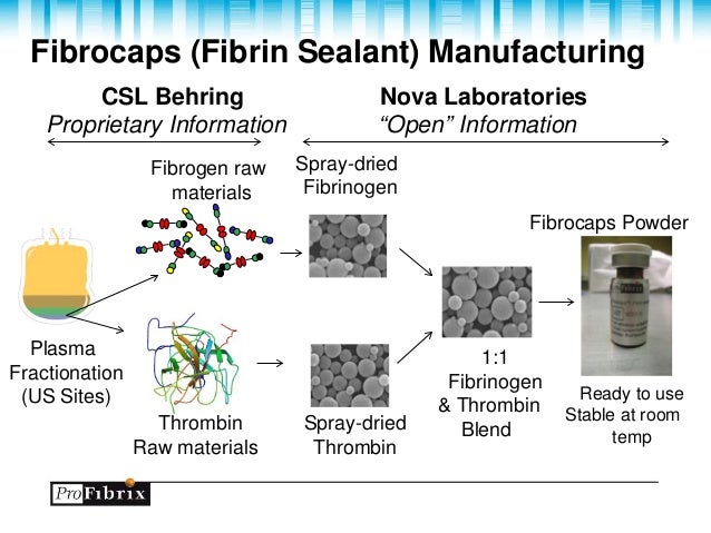
The U.S. Food and Drug Administration today approved Raplixa (fibrin sealant [human]), the first spray-dried fibrin sealant approved by the agency. It is used to help control bleeding during surgery.

April 30, 2015

Release

The U.S. Food and Drug Administration today approved Raplixa (fibrin sealant [human]), the first spray-dried fibrin sealant approved by the agency. It is used to help control bleeding during surgery.

Raplixa is a biological product approved for use in adults to help control bleeding from small blood vessels when standard surgical techniques, such as suture, ligature or cautery, are ineffective or impractical. When applied to a bleeding site, Raplixa is dissolved in the blood and a reaction starts between the fibrinogen and thrombin proteins. This results in the formation of blood clots to help stop the bleeding.

Raplixa contains fibrinogen and thrombin, two proteins found in…

View original post 246 more words

Genetically Engineered Bone Marrow Stem Cells Reverse Ischemic Cardiomyopathy


Muhammad Ashraf is a professor of Pharmacology at the University of Illinois in Chicago.  Dr. Ashraf and his colleagues have published some very fine papers that have examined the ability of stem cells to heal the heart after a heart attack.  Recently, Ashraf’s team examined the ability of a particular population of bone marrow cells, characterized by the presence of the Sca-1 protein on their cell surfaces, to heal the heart after a heart attack.  In particular, Ashraf’s group genetically engineered Sca-1-positive cells to secrete a cocktail of growth factors, and then they tested the ability of such cells to heal a damaged heart.

Sca-1 is a protein that is found on the surfaces of hematopoietic stem cells and other cell types as well.  Therefore, selecting Sca-1 cells will not necessarily give you a pure cell population.  However, such cells can be readily isolated and genetically engineered to make these growth factors.

Ashraf and his coworkers isolated Sca-1 cells from the bone marrow of mice that expressed a glowing green protein.  These cells were then genetically engineered with plasmids (small circles of DNA) to express the growth factors Insulin-like growth factor-1, vascular endothelial growth factor, hepatocyte growth factor, and stromal cell-derived factor-1alpha.  All four of these growth factors have been shown to play supportive roles in the healing of the heart after a heart attack.

When these genetically engineered Sca-1-positive cells were co-cultured with other cells and then grown in low oxygen conditions, the genetically engineered cells prevented the other cells from dying.  The genetically engineered cells are grew faster in culture than their non-genetically engineered counterparts.  When the culture medium in which these genetically engineered cells were grown was used to grow umbilical vein endothelial cells (UVECs), those UVECs showed increased rates of growth.  This suggests that the genetically-engineered Sca-1 cells secreted growth factors into their culture medium and these growth factors are the reason these cell grow faster than non-genetically engineered Sca-1 cells.

When these same cells were injected into the heart tissue of mice that had suffered from heart attacks, they were able to survival over twice as well as non-genetically engineered Sca-1 cells.  When the laboratory animals that received the injections of genetically engineered Sca-1 cells were put down 4 weeks after their stem cell treatments, their hearts were removed, sectioned and examined in detail.  These examinations clearly showed that hearts from those mice that had received injections of genetically-engineered Sca-1 cells had increased blood vessel densities.  There was an additional surprise, since the injected Sca-1 cells were taken from green-glowing mice, they also glowed green.  Green-glowing cells were shown to express Cx43 (the protein that links heart muscle cells together and lets them beat in harmony) and be connected to host heart muscle cells.  This suggests that a bone marrow cell population can actually electrically connect with a heart muscle cell, which is something bone marrow cells are not supposed to be able to do.  Mind you, the number of connected cells was small, and they could be doing this simply as a result of cell fusion.

Additionally, injection of genetically-engineered Sca-1 cells also decreased the infarct size in the hearts of these mice.  The size of the infarct in the treated mice was less than half that of the untreated mice.

This shows that Sca-1 cells from bone marrow have the capacity to augment recovery of the heart after a heart attack, and that this ability is further increased through the co-administration of growth factors.  Using such a system in human patients will require the determination of the precise dosage of these growth factors, since using genetically engineered cells in human patients will probably not be approved in the near future.

2014 in review


The WordPress.com stats helper monkeys prepared a 2014 annual report for this blog.

Here’s an excerpt:

Madison Square Garden can seat 20,000 people for a concert. This blog was viewed about 67,000 times in 2014. If it were a concert at Madison Square Garden, it would take about 3 sold-out performances for that many people to see it.

Click here to see the complete report.

Merry Christmas to All My Readers


Matthew 1:18-25:

18 This is how the birth of Jesus the Messiah came about: His mother Mary was pledged to be married to Joseph, but before they came together, she was found to be pregnant through the Holy Spirit. 19 Because Joseph her husband was faithful to the law, and yet did not want to expose her to public disgrace, he had in mind to divorce her quietly.

20 But after he had considered this, an angel of the Lord appeared to him in a dream and said, “Joseph son of David, do not be afraid to take Mary home as your wife, because what is conceived in her is from the Holy Spirit. 21 She will give birth to a son, and you are to give him the name Jesus, because he will save his people from their sins.”

22 All this took place to fulfill what the Lord had said through the prophet: 23 “The virgin will conceive and give birth to a son, and they will call him Immanuel” (which means “God with us”).

24 When Joseph woke up, he did what the angel of the Lord had commanded him and took Mary home as his wife. 25 But he did not consummate their marriage until she gave birth to a son. And he gave him the name Jesus.

Three-Dimensional Vaccines Sensitize the Immune System to Cancer


Cancer has the capacity to fool the immune system and evade attack from immune cells. This act of immune system evasion allows tumors to grow and spread throughout the body without any resistance. A relatively new strategy called immunotherapy attempts to stimulate the patient’s immune system to mount an immune response against the tumor and sensitize it to the tumor. However, a new protocol developed by a research teams at the Wyss Institute for Biologically Inspired Engineering at Harvard University and Harvard’s School of Engineering and Applied Sciences (SEAS) uses a three-dimensional structure to program the immune system to attack and destroy tumors.

The senior author of this study, David Mooney, who is a Wyss Institute Core Faculty member and the Robert P. Pinkas Professor of Bioengineering at Harvard SEAS, described this new technique: “We can create 3D structures using minimally–invasive delivery to enrich and activate a host’s immune cells to target and attack harmful cells in vivo.”

This 3-D structure consists of tiny, biodegradable rod–like structures made from silica, known as mesoporous silica rods (MSRs). These MSRs can be loaded with biological and chemical drug components, and then injected by needle just underneath the skin. These rods spontaneously assemble at the injection site to form a three–dimensional scaffold (think of pouring a box of match sticks into a pile on a table). The porous spaces between the MSRs are large enough to recruit and fill with dendritic cells. Dendritic cells are immune cells that play the part of surveillance cells that identify foreign cells and substances and trigger an immune response to those things identified as foreign.

Mesoporous silica rods (MSRs) spontaneously assemble to form a porous 3D scaffold, as seen in this SEM image. The 3D scaffold has many nooks and crannies and is large enough to house tens of millions of recruited immune cells. Credit: Wyss Institute at Harvard University
Mesoporous silica rods (MSRs) spontaneously assemble to form a porous 3D scaffold, as seen in this SEM image. The 3D scaffold has many nooks and crannies and is large enough to house tens of millions of recruited immune cells.
Credit: Wyss Institute at Harvard University

“Nano–sized mesoporous silica particles have already been established as useful for manipulating individual cells from the inside, but this is the first time that larger particles, in the micron–sized range, are used to create a 3D in vivo scaffold that can recruit and attract tens of millions of immune cells,” said co-lead author Jaeyun Kim, Ph.D., an Assistant Professor of Chemical Engineering at Sungkyunkwan University and a former Wyss Institute Postdoctoral Fellow.

MSRs are made in the lab with nanopores, which are small holes that can be filled with specific cytokines, oligonucleotides, large protein antigens, or any variety of drugs. Thus, these structures are excellent repositories that can present a vast range of possible combinations to treat a range of infections or stimulate the immune system to attack several different invading elements.

“Although right now we are focusing on developing a cancer vaccine, in the future we could be able to manipulate which type of dendritic cells or other types of immune cells are recruited to the 3D scaffold by using different kinds of cytokines released from the MSRs,” said co-lead author Aileen Li, a graduate student pursuing her Ph.D. in bioengineering at Harvard SEAS. “By tuning the surface properties and pore size of the MSRs, and therefore controlling the introduction and release of various proteins and drugs, we can manipulate the immune system to treat multiple diseases.”

Once the 3D scaffold has recruited dendritic cells from the body, the drugs contained in the MSRs are released. These drugs activate the dendritic cells and initiate an immune response. The activated dendritic cells then leave the MSR-based scaffolds and travel to the lymph nodes, where they raise alarm and direct the body’s immune system to attack specific cells, such as cancerous cells. Within a few months, the body naturally degrades the MSRs, and they dissolve and leave no trace of their presence.

To date, this team has only tested their 3D vaccines in mice, but these 3D vaccines have proven to be remarkably effective. Injectable 3D scaffolds recruited and attracted millions of dendritic cells in a host mouse, before dispersing the cells to the lymph nodes and triggering a powerful immune response.

These vaccines are easily and rapidly manufactured so that they could potentially be widely available very quickly in the face of an emerging infectious disease. “We anticipate 3D vaccines could be broadly useful for many settings, and their injectable nature would also make them easy to administer both inside and outside a clinic,” said Mooney.

Since the vaccine works by triggering an immune response, the method could even be used preventively by building the body’s immune resistance prior to infection.

“Injectable immunotherapies that use programmable biomaterials as a powerful vehicle to deliver targeted treatment and preventative care could help fight a whole range of deadly infections, including common worldwide killers like HIV and Ebola, as well as cancer,” said Wyss Institute Founding Director Donald Ingber, M.D., Ph.D. who is also Judah Folkman Professor of Vascular Biology at Harvard Medical School and Boston Children’s Hospital, and Professor of Bioengineering at Harvard SEAS. “These injectable 3D vaccines offer a minimally invasive and scalable way to deliver therapies that work by mimicking the body’s own powerful immune–response in diseases that have previously been able to skirt immune detection.”

The Patents for CRISPR, the DNA editing technology as the Biggest Biotech Discovery of the Century


A very nice article on the CRISPR nucleases that were discussed in the previous blog article.

Leaders in Pharmaceutical Business Intelligence (LPBI) Group

The Patents for CRISPR, the DNA editing technology as the Biggest Biotech Discovery of the Century

Reporter: Aviva Lev-Ari, PhD, RN

Who Owns the Biggest Biotech Discovery of the Century?

There’s a bitter fight over the patents for CRISPR, a breakthrough new form of DNA editing.

SOURCE

http://www.technologyreview.com/featuredstory/532796/who-owns-the-biggest-biotech-discovery-of-the-century/

Who Owns the Biggest Biotech Discovery of the Century?

There’s a bitter fight over the patents for CRISPR, a breakthrough new form of DNA editing.

By Antonio Regalado on December 4, 2014

WHY IT MATTERS

Control over genome editing could be worth billions.

These Streptococcus pyogenes bacteria use a DNA-cutting defense to battle viruses. The system, called CRISPR, is being harnessed to treat human genetic disease.

Last month in Silicon Valley, biologists Jennifer Doudna and Emmanuelle Charpentier showed up in black gowns to receive the $3 million Breakthrough Prize, a glitzy award put on by Internet billionaires including Mark Zuckerberg. They’d won…

View original post 1,496 more words

Human Amniotic Epithelial Cells Modulate Tooth Socket Restoration in Rats


Human amniotic epithelial cells have the capacity to differentiate into several different cell types. To that list, we can now add bone.

A study from Steve G.F. Shen at his colleagues at the Shanghai Jiao Tong University School of Medicine, Shanghai, China has used human amniotic epithelial cells to regenerate the tooth sockets in laboratory animals.

The first set of experiments examined the ability of human amniotic epithelial cells (hAECs) to form bone under controlled laboratory conditions. Then hAECs were loaded into artificial scaffolds that were then placed into the mouths of rats with tooth socket defects.

In culture, hAECs expressed bone-specific genes 10-14 days after induction. The cells also changed shape and made bone-specific proteins. When implanted into rat tooth sockets, the hAECs were embedded in a scaffold imbued with growth factors known to induce bone differentiation. These implants improved bone regeneration by directly participating in bone repair of the tooth socket defect. They also had an additional benefit in that they modulated the localized immune response against the implanted scaffolds. This immune response modulation augmented regeneration of the tooth sockets and allowed the implanted cells to get on with the job of fixing the surrounding bone without dealing with insults from the immune system.

This study has provided the first evidence that hAECs exhibit direct involvement in new bone regeneration and a localized modulatory influence on the early tissue remodeling process. These cells indirectly contributed to the bone-making process in the alveolar defect. Altogether, these results imply the potential clinical use of hAECs as an alternative stem cell-based for restoring tooth socket deformities.

Human Umbilical Mesenchymal Stem Cells Decreases Dextran Sulfate Sodium-Induced Colitis in Mice


Ulcerative colitis is one of the Inflammatory Bowel Diseases (IBDs) that features chronic inflammation of the large intestine. This is an autoimmune disease that features constant attacks by the immune system on the intestinal mucosae, and the inner layer of the large intestine undergoes constant damage and healing, which increases the risk of the patient to developing colorectal carcinoma.

Mesenchymal stem cells have the capability to suppress inflammation, which makes them promising tools for treating diseases like ulcerative colitis. Unfortunately, the lack of reproducible techniques for harvesting and expanding MSCs has prevented bone marrow- and umbilical cord blood-derived MSCs from being routinely used in clinical situations.

However, a study that was published in the journal Clinical and Experimental Pharmacology and Physiology has used Wharton’s jelly derived umbilical MSCs (UMSCs) to treat mice in which an experimental form of ulcerative colitis was induced. Dextran sulfate sodium (DSS) induced colitis in mice has many of the pathological features of ulcerative colitis in humans.

When mice treated with DSS were also given Wharton’s jelly derived UMSCs showed significant diminution of the severity of colitis. The structure of the tissue in the colon looked far more normal and the types of molecules produced by inflammation were significantly reduced. In addition, transplantation of UMSCs reduced the permeability of the intestine and also increased the expression of tight junction proteins, which help knit the colonic cells together and maintain the structural integrity of the colon. These results show that the anti-inflammatory properties of UMSCs and their capacity to regulate tight junction proteins ameliorates ulcerative colitis.

MSCs for Tissue Engineered Tracheas and Enhanced Fracture Healing


For all my readers who have ever broken a bone, this one’s for you.

Setting a broken bone properly can lead to the healing of a broken bone, but large fractures that generate gaps in bones are very hard to heal. Stem cell therapy in combination with small protein molecules called cytokines has the potential to improve bone repair, since cytokines summon resident stem cells to migrate and home to the injured site. Having said that, the engraftment, participation and recruitment of other cells within the regenerating tissue are equally important.

To stimulate stem cell-mediated healing, University College London scientists over-expressed the SDF-1 protein in mesenchymal stem cells. Since SDF-1 is a stem cell-recruitment protein, it seems reasonable to suspect that these engineered cells would effectively increase the migration of native cells to the site of fracture and enhance bone repair.

Once they made SDF-1-expressing mesenchymal stem cells, Chih-Yuan Ho and colleagues showed that these cells increased the migration of non-transfected cells in a cell culture system.

Once these SDF-1-expressing mesenchymal stem cells were implanted into rats with large bone defects, bone marrow mesenchymal stem cells that over-expressed SDF-1 showed significantly more new bone formation within the gap and less bone mineral loss at the areas next to the defect site during the early bone healing stage.

Thus, SDF-1 plays an important role in accelerating fracture repair and contributing to bone repair, at least in this rat model. SDF-1 does this by recruiting more host stem cells to the defect site and encouraging their differentiation into bone cells, which go on to produce good-quality bone.  This paper appeared the the journal Tissue Engineering, Part A.

In a second paper that appeared in the Annals of Biomedical Engineering, mesenchymal stem cells were used to tissue engineer tracheae. In this case a biocompatible scaffold was seeded various with various cells and this strategy could be a solution for tracheal reconstruction.

Yoo Seob Shin and colleagues seeded mesenchymal stem cells (MSCs) on a scaffold made from pig cartilage powder (PCP). The PCP was made with minced and decellularized pig joint cartilage and was molded into a 5 × 12 mm (height × diameter) scaffold. Mesenchymal stem cells from the bone marrow of young rabbits were grown in culture and then cultured with the PCP scaffold. After 7 weeks in culture, these tracheal implants were transplanted on a 5 × 10 mm tracheal defect in six rabbits, which were evaluated 6 and 10 weeks after the operation.

None of the six rabbits showed any sign of respiratory distress, and endoscopic examination of these tissue engineered tracheae showed that the a normal-looking respiratory epithelium completely covered the regenerated trachea. These trachea also displayed no signs of collapse or blockage.

The tissue engineered tracheae were also scanned and modeled on a computer model (luminal contour). The reconstructed areas of the trachea were the right width and dimensions compared to normal adjacent trachea and were not narrow.

Detailed microscopic tissue examinations of the tissue engineered tracheae showed that the new cartilage was successfully produced by the seeded mesenchymal stem cells and there was only a minimal degree of inflammation or granulation tissue that forms on the surfaces of wounds during the healing process. This shows that the implants did not trigger a massive inflammatory response that damaged resident or implanted tissue.

The outer surfaces of tracheal cells are decorated with tiny beating hairs called cilia that constantly beat to clear particles from the respiratory system. There are also cells that secrete mucus, which acts like fly paper for invading pollutants, particles or microorganisms. in the tissue engineered tracheae, ciliary beating frequency of the regenerated epithelium was not significantly different from the normal adjacent mucosa.

Thus, mesenchymal stem cells from bone marrow seeded on a PCP scaffold successfully restored not only the shape but also the function of the trachea without any signs of graft rejection.

Bones and trachea – mesenchymal stem cells pack a powerful healing punch!!

Mesenchymal Stem Cells Improve Movement and Decrease Neurodegeneration in Ataxic Mice


Friedrich’s ataxia (FA) results from insufficient concentrations of a protein called Frataxin.  Frataxin serves as an iron metabolism protein that puts iron into proteins that need it.  Because several proteins that play crucial roles in energy metabolism in cells use iron, Frataxin is a very busy molecule and without sufficient quantities of Frataxin, energy metabolism decreases and metabolically active cells, such as nerves and muscles, weaken and die.

Frataxin crystal structure.

In patients with FA, the dorsal root ganglia, which lie just in front of the spinal cord, are the first to die off and degenerate.  Can stem cell treatments provide relief from the ravages of FA?

DRG

To test this possibility, Salvador Martinez and his colleagues from the University Miguel Hernández in Alicante, Spain examined two mouse populations, both of which harbored loss-of-function mutations in the Frataxin (FXN) gene.  Mice from both groups were injected with bone marrow-derived mesenchymal stem cells isolated from either wild-type or YG8 mice.  YG8 mice a genetically manipulated so that they suffer from a mouse form of FA that shows several similarities to human FA.  The mesenchymal stem cells injections were “intrathecal” injections, which means that they were directly injected into the nervous system.

As a result of the stem cell injections,  both groups of mice showed improved motor skills compared to nontreated mice.  The dorsal root ganglia also showed increased frataxin expression in the treated groups, and less cell death.

Why did the stem cell-injected mice fare better?  Further investigations revealed that the injected mesenchymal stem cells expressed the following growth factors:  NT3, NT4, and BDNF.  All of these growth factors can bind to specific receptors embedded in the membranes of those sensory neurons located within the dorsal root ganglia and buck up their survival, thus preventing them from dying.  The stem cell-treated mice also had increased levels of “antioxidant enzymes.”. These are enzymes found in our own cells that dispose of dangerous molecules.  Enzymes such as catalase, superoxide dismutase and so on are examples of antioxidant enzymes.  The stem cell-treated mice had higher levels of catalase and GPX-1 in their dorsal root ganglia, which is significant because YG8 mice show decreased levels of these antioxidant enzymes.

Interestingly, the results were not significantly different if the injected stem cells were isolated from wild-type or YG8 mice. In both cases injected mesenchymal stem cells ameliorated the condition of the FA mice.

In conclusion, transplantation of bone marrow mesenchymal stem cells, either the patient’s own stem cells or donated stem cells, is a feasible therapeutic procedure that might delay the onset of cell death in the dorsal root ganglia of patients with Friedreich’s ataxia.