Using Cells from the Mouth to Cure Blindness


If we take tissue samples from the mouth and grow them in the laboratory and manipulate them, we might be able to cure the blind. Blind people who suffer from stem cell deficiency in the cornea might be able to see again by using stem cells isolated from the mouth. Furthermore, this treatment might not only restore vision, but it might also ameliorate pain in the cornea.

Ophthalmologist Tor Paaske Utheim has conducted research for over ten years on how to cure certain types of blindness by using stem cells harvested from tissue obtained from different parts of the body. He then transplants this cultured tissue into the damaged eye, and patients who suffer from blindness as a result of corneal stem cell deficiencies can regain their sight. Recently, Utheim’s research has utilized stem cells from the mouth to grow new corneal tissue, and has also tried to design optimal methods to store and transport this tissue to treat patients.

Utheim is the head of a research group at the Faculty of Dentistry at the University of Oslo (UiO) and the Department of Medical Biochemistry at Oslo University Hospital.

Using cells extracted from the mucous membrane lining the inside of the mouth (the oral mucosa) can restore vision is new to most people. Only ten years ago, this was considered impossible, but results confirm the potential of this method. Twenty clinical studies from various countries have, to date, shown good results, according to Utheim. These clinical trials, however, have only applied these cells to a group of diseases caused by stem cell deficiency in the cornea.

Utheim and his colleagues hope to treat patients with eye injuries caused by so-called limbal stem cell deficiencies. This disorder can be caused by such things as UV radiation, chemical burns, serious infections like trachoma, and various other diseases, some of which are heritable. The number of people worldwide affected by limbal stem cell deficiency is unknown, but in India alone there is an estimated 1.5 million. This disorder most often affects people living in developing countries.

Stem cells that are found at the outer edge of the cornea help to keep the surface of the cornea even and clear. In limbal stem cell deficiencies, the stem cells have been damaged, and they cannot renew the cornea’s outermost layer. Instead, other cells grow over the cornea, which clouds the cornea. The cornea can become fully or partially covered, explains Utheim, which leads to impaired vision or blindness.
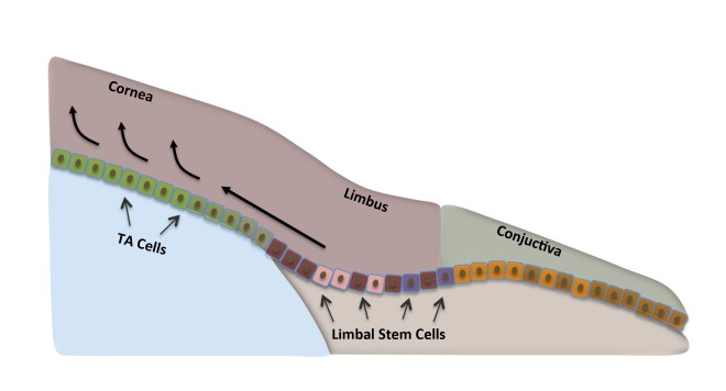
The stem cells are localized in the periphery of the cornea; an area known as the limbus.  They=se limbal stem cells renew the outermost layer of the cornea. Illustration: Amer Sehic, OD/UiO.
The stem cells are localized in the periphery of the cornea; an area known as the limbus. These limbal stem cells renew the outermost layer of the cornea. Illustration: Amer Sehic, OD/UiO.

Others suffer from severe pain as well. When one patient was interviewed by Norwegian national broadcaster NRK about his limbal stem cell deficiency, he responded: “I don’t know what’s worse: the pain, or losing my sight.”

Utheim explained that when stem cells do not work properly, ulcers can develop in the cornea, which exposed nerve fibers. Since the number of nerve fibers is far higher in the cornea than for example in the skin, it is not surprising that some patients experience severe pain.
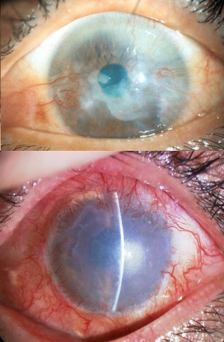
Eyes that suffer mildly from limbal stem cell deficiency. The stem cells stall and other cells grow over the cornea. The window of the eye, normally clear and transparent, is thus blurred, leading to reduced vision.Photo: Dr. Takahiro Nakamura, Department of Ophthalmology/Kyoto Prefectural University of Medicine.
Eyes that suffer mildly from limbal stem cell deficiency. The stem cells stall and other cells grow over the cornea. The window of the eye, normally clear and transparent, is thus blurred, leading to reduced vision. Photo: Dr. Takahiro Nakamura, Department of Ophthalmology/Kyoto Prefectural University of Medicine.

A breakthrough within the field occurred about ten years ago when Japanese researchers showed that cells from the oral mucosa could be used to replace limbal stem cells in patients with limbal stem cell deficiency. Although it had been possible since the late 1990s to cure the disorder using cultured stem cells. The available treatment relied on the patient having a healthy eye from which to collect cells.

Further developments made it possible to harvest cells from a relative or deceased individual, but using limbal stem cells from other patients required the use of strong immunosuppressive drugs for the patients, which could cause serious side effects.

A milestone seemed to be reached when it became possible to use a patient’s own cells to treat blindness in both eyes without the need for immunosuppressive drugs. Strangely, this makes some sense because there are similarities between the oral mucosae and the surface of the eye (see Utheim TP. Stem Cells. 2015;33:1685-1695). Originally, using mouth mucosal cells to treat the eye required that the laboratory where the cells are cultured and the clinic where the patients are treated be quite close together. Because there were no protocols for storing extracted oral mucosal cells so that they can be easily kept and transported. This has made the treatment virtually inaccessible to many of the patients who need it the most, namely those in developing countries. However, this may be about to change.

Utheim’s research group is now on the brink of a development that will make it possible to cure both severe pain and blindness in patients who are spread over a larger geographical area than before (see Islam R, et al. PLoS One. 2015;10:e0128306.). “Today, cells from the mouth are cultured for use in the treatment of blindness in only a few specialized centers in the world. By identifying the optimal conditions for storing and transporting the cultured tissue, we would allow for the treatment to be made available worldwide, and not just close to the cell culture centers,” said Rakibul Islam, who is a PhD candidate in the Department of Oral Biology at the Faculty of Dentistry.

Islam is collaborating with Harvard Medical School to introduce this method of treating blindness to clinics around the world. Islam’s findings could also help improve treatment outcomes. “Being able to store the cultured tissue in a small sealed container for a week increases the technique’s flexibility significantly. It makes it easier to plan the operation and allows for quality assurance through microbiological testing of the tissue before transplantation,” Islam explained.

One of the things that Islam and his colleagues have discovered is the specific temperature range at which cells from the mouth should ideally be stored at after culture. Islam has shown that cultured mouth stem cells retain their quintessential properties best between 12 and 16 degrees Celsius (See Dolgin, Elie. Nature Biotechnolgy, 2015;33:224-225.).

During a brief stint at Harvard University, Islam also examined which areas of the mouth are best suited to use in regenerative medicine. In other words, Islam and his colleagues wanted to know which parts of the mouth contain cell layers that regenerate the fastest. Islam explained this using this example: “If you burn any part of your mouth on hot coffee, it heals so quickly that by the next morning you have forgotten about it. This is because the oral mucosa contains cells that multiply quickly. We wanted to investigate whether there were regional differences in the mouth that we could exploit for the treatment of limbal stem cell deficiency.”

Islam continued, “Our results show that the location from which the mucosal tissue is harvested has a striking impact on the quality of the cultured tissue.”

The results from this particular study have not yet been published.

This research can potentially give hope to the many blind that live far away from centralized cell culture laboratories.  In work by Utheim in 2010, in collaboration with the ophthalmologist Sten Ræder, he developed storage technology for cultured stem cells that enables the cultured tissue to be transported in a small custom-made plastic container.  Tissue from stem cells is thus freed from expensive and bulky laboratory equipment and provides a whole new level of flexibility.

Utheim said “The sample of cells from the mouth can be sent by air over long distances to specialist laboratories with first-class equipment and expertise. After a couple of weeks of laboratory cultivation, the sender may receive the tissue back ready for use. An ophthalmologist could then transplant the stem cells onto the patient’s eye.”

However, the container was just one step in the right direction: “Now we have identified those areas of the mouth that may be best suited for regenerative medicine, and developed a method for storing and transporting tissue from centralized, highly specialized tissue culture centers to clinics worldwide. Our findings are helping to simplify and streamline the clinical procedures, and to make the treatment far more accessible than it is today,” said Islam, who admitted that the transport potential of the project has been integral to his own enthusiasm.  He continued, “Although the scientific and technical aspects of our project are very exciting, it has been especially motivating to think of the possibilities this storage technology brings to treating blindness in all parts of the world, including my homeland Bangladesh.”

A central laboratory for the growth of stem cells already exists in Italy.  In fact, earlier this year the European Medicines Agency approved the procedure for the cultivation of stem cells from the cornea in EU laboratories. This is the first stem cell therapy to be approved by the European Medicines Agency, according to the journal Nature Biotechnology.  Utheim described the approval as an important step towards the implementation of stem cell technology over larger geographical areas.  To date, almost 250 people with limbal stem cell deficiency have undergone treatment involving transplantation of stem cells grown from their own mouth cells.  “This provides a good basis for judging the success of the treatment” Utheim says.

He has recently published an article in the journal Stem Cells on the inherent potential of cells from the mouth to regenerative medicine.  Roughly three out of four treatments are described as successful.

First Stem Cell Therapy Recommended for Approval in European Union


The EMA, which is short for the European Medicines Agency, has recommended approval for a treatment called Holoclar.  Holoclar is the first therapy product that contains stem cells to be recommended for approval in the European Union (EU). Holoclar is being marketed as a treatment for moderate to severe limbal stem cell deficiency (LSCD) due to physical or chemical burns to the eye in adults. In fact, Holoclar is the first medicine recommended for LSCD, a condition that can result in blindness.

Holoclar can be transplanted into the eye after removal of the corneal epithelium (the outer layer of the cornea). Holoclar is made from a biopsy taken from a small, undamaged area of the patient’s cornea. These limbal stem cells are then grown in the laboratory using cell culture techniques. Holoclar is a potential alternative to transplantation for replacing altered corneal epithelium. Clnical trials with Holoclar have been shown to increase the chances of a successful corneal transplant where the injury has caused extensive eye damage. Holoclar is produced by Chiesi, a pharmaceutical company based in Parma, Italy.

The recommendation to approve Holoclar was made by the EMA’s Committee for Medicinal Products for Human Use (CHMP). CHMP made their recommendation on basis of the benefits of Holoclar, which are its ability to repair the damaged ocular surface, to improve or resolve symptoms of pain, photophobia and burning and to improve the patient’s visual acuity. This assessment was the work of the Committee for Advanced Therapies (CAT). The approved indication for Holoclar is: “Treatment of adult patients with moderate to severe limbal stem cell deficiency (defined by the presence of superficial corneal neovascularisation in at least two corneal quadrants, with central corneal involvement, and severely impaired visual acuity), unilateral or bilateral, due to physical or chemical ocular burns. A minimum of 1-2 square millimeters of undamaged limbus is required for biopsy.” CAT and CHMP considered that Holoclar provided a first treatment option for LSCD and recommended a conditional marketing authorization. The authorization is conditional because the clinical data available for Holoclar is based on studies that are ongoing as treated patients are watched after their eye surgery. This the data collection is not yet comprehensive, and additional study on the use of Holoclar needs to be conducted.

The opinion adopted by the CHMP at its December 2014 meeting is an intermediary step on Holoclar’s path to patient access. The CHMP opinion will now be sent to the European Commission for a decision on an EU-wide marketing authorization.

University of Pittsburgh Team Uses Patient’s Own Stem Cells to Clear Cloudy Corneas


The transparent portion of the center of our eyes is called the cornea. Scars on the cornea can cause an infuriating haziness across the eye. However, healing these cloudy corneas might be as simple as growing stem cells from a tiny biopsy of the patient’s undamaged eye and placing them on the injury site. This hope comes from experiments in a mouse model system conducted by researchers at the University of Pittsburgh School of Medicine. These findings were published in Science Translational Medicine and could one day rescue vision for millions of people worldwide and decrease the need for corneal transplants.

According to statistics compiled by the National Eye Institute, which is a branch of the National Institutes of Health, globally, corneal infectious diseases have compromised the vision of more than 250 million people and have blinded over 6 million of them. Additionally, trauma from burns is also a leading cause of corneal scarring.

James L. Funderburgh, Ph.D., professor of ophthalmology at Pitt and associate director of the Louis J. Fox Center for Vision Restoration of UPMC and the University of Pittsburgh, a joint program of UPMC Eye Center and the McGowan Institute for Regenerative Medicine, said, “The cornea is a living window to the world, and damage to it leads to cloudiness or haziness that makes it hard or impossible to see. The body usually responds to corneal injuries by making scar tissue. We found that delivery of stem cells initiates regeneration of healthy corneal tissue rather than scar leaving a clear, smooth surface.”

The lead author of this study, Sayan Basu, is a corneal surgeon who works at the L.V. Prasad Eye Institute in Hyderabad, India. Dr. Basu who joined with Dr. Funderburgh’s lab, has developed a technique to isolate ocular stem cells from tiny biopsies from the surface of the eye and a region between the cornea and sclera known as the limbus. Such a small biopsy heals rapidly with little discomfort and no disruption of vision. Such biopsies are banked in tissue banks and then expanded in culture, and several tests shows that even after isolation and expansion, these cells are still corneal stem cells.

limbal-stem-cells

“Using the patient’s own cells from the uninjured eye for this process could let us bypass rejection concerns,” Dr. Basu noted. “That could be very helpful, particularly in places that don’t have corneal tissue banks for transplant.”

Basu in collaboration with Funderburgh’s team tested these human limbal stem cells in a mouse model of corneal injury. This team used goo made of fibrin to glue the cells to the injury site. Fibrin is the protein found in blood clots, but it is also commonly used as a surgical adhesive. Application of these limbal stem cells not only induced healing of the mouse corneas, their eyes became clear again within four weeks of treatment. On the other hand, the eyes of mice that were not treated with limbal stem cells remained cloudy.

Fibrin

In fact, the healing was so good that Funderburgh said: “Even at the microscopic level, we couldn’t tell the difference between the tissues that were treated with stem cells and undamaged cornea. We were also excited to see that the stem cells appeared to induce healing beyond the immediate vicinity of where they were placed. That suggests the cells are producing factors that promote regeneration, not just replacing lost tissue.”

This work is the impetus behind a small pilot study presently underway in Hyderabad which will treat a handful of patients with their own corneal stem cells.

Induced Pluripotent Stem Cells Form Limbal-Like Stem Cells


Limbal epithelial stem cells or LESCs are found at the periphery of the cornea and they continuously renew the corneal epithelium. Loss of this stem cell population can cause loss of corneal transparency and eventual loss of vision.

Genetic conditions can cause LESC deficiency, such as congenital aniridia, Stevens-Johnson syndrome or Ocular cicatricial pemphigoid. Other causes of LESC deficiency include chemical or thermal burns to the eye, microbial infections, extended contact lens wear, sulfur mustard gas poisoning, or chronic inflammation of the eye,

Limbal epithelial stem cells reside in the basal layer of the epithelium (Ep), which undulates at the limbus. Daughter transient amplifying cells (TACs) divide and migrate towards the central cornea (arrowed) to replenish the epithelium, which rests on Bowman's layer (BL). The stroma (St) of the limbal epithelial stem cell niche is populated with fibroblasts and melanocytes and also has a blood supply.
Limbal epithelial stem cells reside in the basal layer of the epithelium (Ep), which undulates at the limbus. Daughter transient amplifying cells (TACs) divide and migrate towards the central cornea (arrowed) to replenish the epithelium, which rests on Bowman’s layer (BL). The stroma (St) of the limbal epithelial stem cell niche is populated with fibroblasts and melanocytes and also has a blood supply.

Treatments of LESC deficiency include limbal stem cell grafts from one eye to another, but these grafts have a 3-5-year graft survival of only 30%-45%. If LESCs are expanded in culture on human amniotic membrane, then 76% of the grafts will successfully take 1-3 years after grafting. This procedure is not standardized. If LESCs are grafted from a cadaver, their survival is low.

Given these less than optimal treatments for LESC deficiencies, Alexander Ljubimov and his team from UCLA have used induced pluripotent stem cells (iPSCs) to make cultured LESCs. Ljubimov and his coworkers derived iPSCs from the skin cells of volunteers with non-integrating plasmids. Then they grew these cells on corneas that have been stripped of their cells and human amniotic membranes and these cells differentiated into LESC-like cells.

Ljubimov and others also made iPSCs from human LESCs, and when they cultured these iPSCs derived from LESCs on human amniotic membranes for two weeks, the cells differentiated into LESCs that made LESC-specific genes, and had the epigenetic characteristics of LESCs.

These experiments show that the cell source for iPSC derivation can greatly influence the epigenetic characteristics of the iPSC line. Also these experiments show that iPSCs can be used to make LESCs that can potentially be used for therapeutic purposes.

A New Way to Regrow Human Corneas


My apologies to my readers, but I was at the Free Methodist Bible Quizzing Finals at Greenville College in Illinois for the last week. I am recovering and have only the energy to write a short post for today.

The cornea is the transparent covering of the eye that transmits light from the environment to the inside of the eye, to the photoreceptor-rich retina that interprets the light information and translates them into neural signals that are sent to the visual centers of the brain.

The cornea is subject to constant wear and tear, but fortunately, a stem cell population called limbal stem cells. These stem cells constantly regenerate the cornea, and the conjunctiva, which is otherwise known as the “whites of the eye.”

Limbal stem cells


Unfortunately, the limbal stem cells can be damaged by chemicals, sparks from a welding, genetically inherited conditions, or physical trauma. Such conditions can prevent proper replacement of constantly sloughed cornea and conjunctival cells. This can seriously compromise the structural integrity and function la of the eye.

To treat patients with corneal limbal stem defects, eye surgeons have transplanted limbal cells from cadavers or used small excisions of limbal cell populations from the unaffected eye and transplanted them into the affected eye. These so-called “autologous limbal cell transplants” tend to work quite well, but there are two cuts that need to be made. Is there are way to expand limbal stem cells for clinical use? Now it appears that there is.

Scientists from the Massachusetts Eye and Ear Infirmary have used sophisticated key tracer molecules to pin down the precise cells in the eye that are capable of regeneration and repair. They then transplanted these regenerative stem cells into mice to create fully functioning corneas.

This work was published in the international journal Nature, and they predict that this method may one day help restore the sight of victims of burns and chemical injuries.

Limbal stem cells (LSC) completely renew our corneas every few weeks and repair the cornea and conjunctiva whenever they are injured. Without LSCs the cornea becomes cloudy, which severely disrupts vision. In fact, LSC deficiencies are among the commonest reasons behind blindness worldwide.

Unfortunately, the LSC population is embedded in a part of the eye where they share space with a matrix of other structures in the limbal part of the eye (FYI – the limbus is the junction between the cornea and the white of the eye).

Enter the work from the Massachusetts Eye and Ear Infirmary in Boston at the Boston Children’s Hospital, Brigham and Women’s Hospital and in collaboration with the VA Boston Healthcare System have identified a key molecule known as ABCB5, which is naturally present on the surface of LSCs.

Although ABCB5 has been known about for some time in other parts of the body, this is the first time ABCB5 has been identified on the surfaces of LSCs. Also, it is clear that ABCB5 can effectively mark LSCs.

By using molecules linked to fluorescent molecules, these scientists were able to instantly identify a pool of LSCs on donated human corneas. After transplanting these cells into the eyes of mice, they discovered that the transplanted cells were able to generate fully functioning human corneas.

Prof Markus Frank, of Boston Children’s Hospital, a lead author in the research, told the BBC: ” The main significance for human disease is we have established a molecularly defined population of cells that we can extract from donor tissue.

“And these cells have the remarkable ability to self-regenerate. We hope to drive this research forward so this can be used as a therapy.”

Harminder Dua, professor of ophthalmology at the University of Nottingham, who was not involved in this study, said: “This paper represents a very comprehensive and well conducted piece of work that takes use closer to the precise identification of stem cells.

“Applying this knowledge to a clinical setting could help improve the outcomes for patients who need corneal reconstruction.”

A New Technique to Fix Damaged Eyes With Stem Cells


Engineers at the University of Sheffield have invented a new delivery technique for delivering stem cells to eyes. They have high hopes that this technique will help repair the eyes of those patients who have suffered damage to their eyes.

The front of the eye is bordered by the transparent cornea, which transmits light to the lens. The cornea is exposed to the outside world and if there is an accident that affects the eye, the cornea is usually the part that takes a beating. The cornea undergoes constant turnover as dead cells are constantly sloughed from the cornea during blinking. At the junction between the cornea and the sclera is an area called the limbus. Located at the limbus is a population of limbal epithelial stem cells or LESCs. LESCs have many features commonly observed in other stem cells, such as small size, high nuclear to cytoplasmic ratio, and they lack expression of molecules commonly found in mature corneal cells, such as cytokeratins 3 and 12.

Human Limbus

LESCs are slow-growing, but in the event of injury they can become highly proliferative (See Lavker R.M, Sun T.T. Epithelial stem cells: the eye provides a vision. Eye. 2003;17:937–942. DOI: 10.1038/sj.eye.6700575).

LESC deficiency can result from chemical or thermal burns to the eye or as a result of certain inherited diseases. Partial or full LESC deficiency causes abnormal corneal wound healing and surface integrity. Also LESC deficiency causes the conjunctiva to grow over the cornea, and this is disastrous for the eye because the cornea is devoid of blood vessels, which is the reason why it is transparent. However the conjunctiva (the white of the eye) is filled with blood vessels and is not transparent. Thus chronic inflammation, recurrent erosion, ulceration and stromal scarring can occur and cause painful vision loss

Long term restoration of visual function requires renewal of the corneal epithelium, and this requires the placement of a new stem cell population by means of a limbus graft. From where do you get a new limbus for transplantation? Autografts use limbal cells from the good eye, but this runs the risk of scarring the cornea of the other eye.procedure is the use limbal cells from cadavers (limbal allografts). Also, making sure that the graft adheres to the requires the use of sutures, but these sutures can cause substantial amounts of irritation. Therefore, the Sheffield research group designed a new technique.

With this new technique, a disk made of biodegradable material is loaded with limbal stem cells and then placed over the eye. This disc has an outer ring pockmarked with small niches for stem cells can hide. The material in the center of the disc is thinner than that on the edges, and therefore, the center of the disc biodegrades faster. This releases the stem cells in center of the disc into the cornea where they can grow and help repair it.

Because these small niches in the disc resemble the stem cells niches found in the limbus, these discs do an excellent job of nurturing the limbal stem cells and distributing them to the cornea. Limbal grafts are either done with amniotic membrane as a carrier, but this procedure leads to increased inflammation in the eye and there is a chance that the grafts will not integrate into the limbus. The biodegradable disc groups the limbal stem cells into clusters that are more likely to ingrate into the limbus.

According to Professor Sheila MacNeil, “Laboratory tests have shown that the membranes will support cell growth, so the next stage is to trial this in patients in India, working with our colleagues in the LV Prasad Eye Institute in Hyderabad. One advantage of our design is that we have made the disc from materials already in use as biodegradable sutures in the eye so we know they won’t cause a problem in the body. This means that, subject to the necessary safety studies and approval from Indian Regulatory Authorities, we should be able to move to early stage clinical trials fairly quickly.”

In the developing world, corneal blindness is rather common in some professions and treating it is a rather pressing problem. High instances of chemical burns to the eye or accidental damage to the eye are common, but complex treatment strategies such as amniotic membrane grafts are not available to the general public.

This technique also possibilities in more developed countries, since current techniques use donor tissue to deliver the cultured cells, and this requires a tissue bank to which some people do not have access. Also, the use of the cell-impregnated disk will reduce the risk of disease transmission with grafts.