Stem Cell-Based Skin Graft for Severe Burns


Severe wounds are typically treated with full thickness skin grafts. Some new work by researchers from Michigan Tech and the First Affiliated Hospital of Sun Yat Sen University in Guangzhou, China might provide a way to use a patient’s own stem cells to make split thickness skin grafts (STSG). If this technique pans out, it would eliminate the needs for donors and could work well for large or complicated injury sites.

This work made new engineered tissues were able to capitalize on the body’s natural healing power. Dr. Feng Zhao at Michigan Tech and her Chinese colleagues used specially engineered skin that was “prevascularized, which is to say that Zhao and other designed it so that it could grow its own veins, capillaries and lymphatic channels.

This innovation is a very important one because on of the main reasons grafted tissues or implanted fabricated tissues fail to integrate into the recipient’s body is that the grafted tissue lacks proper vascular support. This leads to a condition called graft ischemia. Therefore, getting the skin to form its own vasculature is vital for the success of STSG.

STSG is a rather versatile procedure that can be used under unfavorable conditions, as in the case of patients who have a wound that has been infected, or in cases where the graft site possess less vasculature, where the chances of a full thickness skin graft successfully integrating would be rather low. Unfortunately, STSGs are more fragile than full thickness skin grafts and can contract significantly during the healing process.

In order to solve the problem of graft contraction and poor vascularization, Zhao and others grew sheets of human mesenchymal stem cells (MSCs) and mixed in with those MSCs, human umbilical cord vascular endothelial cells or HUVECs. HUVECs readily form blood vessels when induced, and growing mesenchymal stem cells tend to synthesize the right cocktail of factors to induce HUVECs to form blood vessels. Therefore this type of skin is truly poised to form its own vasculature and is rightly designated as “prevascularized” tissue.

Zhao and others tested their MSC/HUVEC sheets on the tails of mice that had lost some of their skin because of burns. The prevascularized MSC/HUVEC sheets significantly outperformed MSC-only sheets when it came to repairing the skin of these laboratory mice.

When implanted, the MSC/HUVEC sheets produced less contracted and puckered skin, lower amounts of inflammation, a thinner outer skin (epidermal) thickness along with more robust blood microcirculation in the skin tissue. And if that wasn’t enough, the MSC/HUVEC sheets also preserved skin-specific features like hair follicles and oil glands.

The success of the mixed MSC/HUVEC cell sheets was almost certainly due to the elevated levels of growth factors and small, signaling proteins called cytokines in the prevascularized stem cell sheets that stimulated significant healing in surrounding tissue. The greatest challenge regarding this method is that both STSG and the stem cell sheets are fragile and difficult to harvest.

An important next step in this research is to improve the mechanical properties of the cell sheets and devise new techniques to harvest these cells more easily.

According to Dr. Zhao: “The engineered stem cell sheet will overcome the limitation of current treatments for extensive and severe wounds, such as for acute burn injuries, and significantly improve the quality of life for patients suffering from burns.”

This paper can be found here: Lei Chen et al., “Pre-vascularization Enhances Therapeutic Effects of Human Mesenchymal Stem Cell Sheets in Full Thickness Skin Wound Re-pair,” Theranostics, October 2016 DOI: 10.7150/ thno.17031.

Inhibition of AKT Kinase Increases Umbilical Cord Blood Growth in Culture and Engraftment in Mice


Dr. Yan Liu from the Department of Pediatrics and the Herman B Wells Center for Pediatric Research at the Indiana University School of Medicine in Indianapolis, Indiana and his colleagues have increased the engraftment efficiency of umbilical cord hematopoietic (blood cell-making) stem cells in immunodeficient mice. The technique developed by Lui and his colleagues is simple and increases the proliferation of umbilical cord blood hematopoietic stem cells (UCB-HSCs) in culture, which potentially solves several long-standing problems with umbilical cord blood transplantation.

Umbilical cord blood has been used in the clinic for more than 40 years in hematopoietic stem cell transplantation therapies to treat patients with bone marrow diseases or to reconstitute the bone of those cancer patients who had to have theirs wiped out to cure their leukemia or lymphoma.

One of the problems with umbilical cord blood transplantations, however, is the small amount of material in a typical cord blood collection and, therefore, the small number of hematopoietic stem cells (HSCs) available for transplantation. To ameliorate these shortcomings, hematologists will transplant more than one lot of cord blood (a so-called “double umbilical cord blood transplantation”), which, unfortunately, also increases the risk of immunological rejection (so-called Graft Versus Host response).

A second strategy to get around the low numbers of UCB-HSCs is to expand them in culture, which has proven difficult. However, some experiments have given us more than enough hope to suspect this this is a feasible option (see Flores-Guzmán P, et al., Stem Cells Transl Med. 2013 Nov;2(11):830-8; Bari S., et al., Biol Blood Marrow Transplant. 2015 Jun;21(6):1008-1; Pineault N, Abu-Khader A. Exp Hematol. 2015 Jul;43(7):498-513).

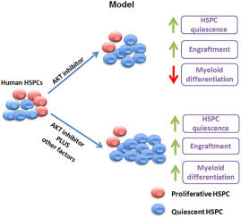
Dr. Lui and his coworkers wanted to examine the role of the signaling protein AKT (also known and protein kinase B) in UCB-HSC expansion in culture. To this end, they used silencing RNAs to knock-down AKT gene expression in cultured UCB-HSCs. AKT knock-down enhanced UCB-HSC quiescence and growth in culture. In a separate experiment, Lui and others treated human UCB-HSCs (so-called CD34+ cells) with a chemical that specifically inhibits AKT activity. Then they subjected these cells to a battery of tests in culture and in laboratory mice.

The results were astounding.  Treatment of human UCB-HSCs did not affect the identity of the HSCs and enhanced their ability to form isolated colonies in cell culture growth tests known as “replating assays.”  Additionally, the short-term inhibition of AKT with drugs also enhanced the ability of UBC-HSCs to repopulate the bone marrow of immunodeficient mice.

ubc-hsc-engraftment-improved-with-akt-inhibition

In summary, inhibition of AKT increases human UCB-HSC quiescence, growth potential, and engraftment in laboratory mice.

These interesting pre-clinical results suggest that AKT inhibitor can increase the expansion of UCB-HSCs in culture and potential increase their tendency of these cells to engraft in patients.

Factor From Umbilical Cord Blood Could Treat Harmful Inflammation


Umbilical cord blood turns out to have a factor that can potentially fight inflammation, according to scientists at the University of Utah School of Medicine. This study was published online Sept. 6, 2016, in The Journal of Clinical Investigation.

“We found something we weren’t expecting, and it has taken us to new strategies for therapy that didn’t exist before,” says Guy Zimmerman, M.D., a professor of internal medicine at the University of Utah School of Medicine, who was also the senior author of this work. Dr. Zimmerman collaborated with associate professor of pediatrics, Christian Con Yost, M.D., and their colleagues for this work.

Inflammation is well-known to anyone who has whacked their leg, been stung by a bee or a wasp, or anyone who over-stressed their muscles. The redness, heat pain, and swelling are signs that the body is cleaning up damaged cells and their debris, fighting invading microorganisms, and beginning the healing process. However, under certain circumstances, inflammation can go overboard and turn against us and seriously and chronically damage healthy tissues. Out-of-control inflammation is probably the culprit behind several different conditions ranging from rheumatoid arthritis to sepsis. In fact, the inflammatory overreaction to infections is one of the most common causes of hospital deaths.

Dr. Yost and his coworkers successfully isolated a cord blood factor, called “neonatal NET inhibitory factor” or nNIF. This name comes from the ability of this factor to inhibit “NETs” or neutrophil extracellular traps. NETs or neutrophil extracellular traps are composed of processed chromatin bound to granular and selected cytoplasmic proteins that are released by white blood cells called neutrophils. NETs seem to be a kind of last resort that neutrophils turn to in order to control microbial infections. Even though NETs usually help our bodies ward off infectious bacteria and viruses, they can also damage blood vessels and organs during sepsis.

nets

As physicians who have treated critically ill patients suffering from out-of-control inflammation, Drs. Zimmerman and Yost recognized the therapeutic potential of nNIF. “We knew we were onto something that could be very meaningful,” recalls Yost.

To test if this cord blood-based factor could control sepsis, Zimmerman and Yost and others treated groups of mice that suffered from laboratory-induced inflammatory disease. In the absence of treatment, only 20 percent of the mice survived longer than two to four days. However, 60% of those mice treated with nNIF survived after the same amount of time.

“Sepsis is a case where the body’s reaction to infection is lethal,” says Yost. “nNIF is offering insights into how to keep the inflammatory response within prescribed limits.” He adds that they will carry out additional studies to test the therapeutic properties of nNIF.

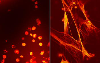
Anti-Inflammatory Agent Isolated From Umbilical Cord Blood Infection fighting cells from umbilical cord blood (left) and circulating blood three days after birth (right) from the same prematurely born baby. Umbilical cord blood has high levels of a factor, called neonatal NET inhibitory factor (nNIF), which inhibits a specific inflammatory response called NETs. Within two weeks after birth, nNIF levels drop and NETs can form. True to their name, they consist of a net-like substance that traps infectious agents like bacteria, as seen on the right. nNIF is showing promise as a potential therapy against harmful inflammation and sepsis.
Anti-Inflammatory Agent Isolated From Umbilical Cord Blood
Infection fighting cells from umbilical cord blood (left) and circulating blood three days after birth (right) from the same prematurely born baby. Umbilical cord blood has high levels of a factor, called neonatal NET inhibitory factor (nNIF), which inhibits a specific inflammatory response called NETs. Within two weeks after birth, nNIF levels drop and NETs can form. True to their name, they consist of a net-like substance that traps infectious agents like bacteria, as seen on the right. nNIF is showing promise as a potential therapy against harmful inflammation and sepsis.

nNIF seems to be present for just a brief window of time at the beginning of life. It circulates in cord blood and persists in the baby’s own bloodstream for up to two weeks after birth. However, after two weeks, nNIF disappears and is not found in older babies and is completely absent from the blood of adults. Scientists in Yost’s laboratory also discovered that the placenta also contains a similar, albeit less potent, anti-inflammatory agent. The evanescent nature of these factors possibly indicates that inflammation is under tight control during this time, since the fragility of young babies might make extensive amounts of inflammation deleterious to their health.

“The beginning of life is a delicate balance,” says Yost. “Our work is showing that it is important to have the right defenses, but they have to be controlled.”

Gamida Cell Announces First Patient with Sickle Cell Disease Transplanted in Phase 1/2 Study of CordIn™ as the Sole Graft Source


An Israeli regenerative therapy company called Gamida Cell specializes in cellular and immune therapies to treat cancer and rare (“orphan”) genetic diseases. Gamida Cell’s main product is called NiCord, which provides patients who need new blood-making stem cells in their bone marrow an alternative to a bone marrow transplant. NiCord is umbilical cord blood that has been expanded in culture. In clinical trials to date, NiCord has rapidly engrafted into patients and the clinical outcomes of NiCord transplantation seem to be comparable to transplantation of peripheral blood.

Gamida Cell’s two products, NiCord and CordIn, as well as some other products under development utilize the company’s proprietary NAM platform technology to expand umbilical cord cells. The NAM platform technology has the remarkable capacity to preserve and enhance the functionality of hematopoietic stem cells from umbilical cord blood. CordIn is an experimental therapy for those rare non-malignant diseases in which bone marrow transplantation is the only currently available cure.

Gamida Cell has recently announced that the first patient with sickle cell disease (SCD) has been transplanted with their CordIn product.  Mark Walters, MD, Director of the Blood and Marrow Transplantation (BMT) Program is the Principal Investigator of this clinical trial. The patient received their transplant at UCSF Benioff Children’s Hospital Oakland.

CordIn, as previously mentioned, is an experimental therapy for rare non-malignant diseases, including hemoglobinopathies such as Sickel Cell Disease and thalassemia, bone marrow failure syndromes such as aplastic anemia, genetic metabolic diseases and refractory autoimmune diseases. CordIn potentially addresses a presently unmet medical need.

“The successful enrollment and transplantation of our first SCD patient with CordIn as a single graft marks an important milestone in our clinical development program. We are eager to demonstrate the potential of CordIn as a transplantation solution to cure SCD and to broaden accessibility to patients with rare genetic diseases in need of bone marrow transplantation,” said Gamida Cell CEO Dr. Yael Margolin. “In the first Phase 1/2 study with SCD, the expanded graft was transplanted along with a non-manipulated umbilical cord blood unit in a double graft configuration. In the second phase 1/2 study we updated the protocol from our first Phase 1/2 study so that patients would be transplanted with CordIn as a standalone graft, which is expanded from one full umbilical cord blood unit and enriched with stem cells using the company’s proprietary NAM technology.”

Somewhere in the vicinity of 100,000 patients in the U.S suffer from SCD; and around 200,000 patients suffer from thalassemia, globally. The financial burden of treating these patients over their lifetime is estimated at $8-9M. Bone marrow transplantation is the only clinically established cure for SCD, but only a few hundred SCD patients have actually received a bone marrow transplant in the last ten years, since most patients were not successful in finding a suitable match. Unrelated cord blood could be available for most of the patients eligible for transplantation, but, unfortunately, the rate of successful engraftment of un-expanded cord blood in these patients is low. Therefore, cord blood is usually not considered for SCD patients. Without a transplant, these patients suffer from very high morbidity and low quality of life.

Eight patients with SCD were transplanted in the first Phase 1/2 study performed in a double graft configuration. This study is still ongoing. Preliminary data from the first study will be summarized and published later this year. A Phase 1/2 of CordIn for the treatment of patients with aplastic anemia will commence later this year.

Umbilical Cord Blood Mesenchymal Stem Cells do Not Cause Tumors in Rigorous Tests


Human umbilical cord blood mesenchymal stem cells (hUCB-MSCs) have the ability to self-renew and also can differentiate into a wide range of cell types. However, many clinicians and scientists fear that even these very useful cells might cause tumors.

To that end, Moon and colleagues from the Korean Institute of Toxicology have rigorously tested the tendency for hUBC-MSCs to cause tumors. They used a large battery of tests in living organisms and in culture. hUCB-MSCs were compared to MRC-5 and HeLa cells. MRC-5 cells are known to have no ability to cause tumors and HeLa cells have a robust ability to form tumors, and therefore, constitute negative and positive controls,

To evaluate the ability of cells to cause tumors, Moon and others examined the tendency of these cells to grow without being attached to a substratum. This is a hallmark of tumor cells and is called “anchorage-independent growth” (AIG). To assess AIG, the cells were grown in soft agar, which is a standard assay for AIG. hUCB-MSCs and MRC-5 cells formed few colonies in soft agar, but HeLa cells formed a greater number of larger colonies. This indicated that hUCB-MSCs and MRC-5 cells do not show AIG, a common trait of tumorigenic cells.

The next assay implanted these cells into live laboratory animals. hUCB-MSCs were implanted as a underneath the skin of BALB/c-nu mice (nasty creatures – they bite). All the mice implanted with hUCB-MSCs and NRC-5 cells showed any sign of tumors. Both gross and microscopic examination failed reveal any tumors. However, all mice transplanted with HeLa cells developed tumors that were clearly derived from the implanted cells.

These experiments, though somewhat mundane, rigorously demonstrate that hUCB-MSCs do not exhibit tumorigenic potential. This provides further evidence of these cells clinical applications.

The paper appeared in Toxicol Res. 2016 Jul;32(3):251-8. doi: 10.5487/TR.2016.32.3.251.

Genetic Switch to Making More Blood-Making Stem Cells Found


A coalition of stem cell scientists, co-led in Canada by Dr. John Dick, Senior Scientist, Princess Margaret Cancer Centre, University Health Network (UHN) and Professor, Department of Molecular Genetics, University of Toronto, and in the Netherlands by Dr. Gerald de Haan, Scientific Co-Director, European Institute for the Biology of Ageing, University Medical Centre Groningen, the Netherlands, have uncovered a genetic switch that can potentially increase the supply of stem cells for cancer patients who need transplantation therapy to fight their disease.

Their findings were published in the journal Cell Stem Cell and constitute proof-of-concept experiments that may provide a viable new approach to making more stem cells from umbilical cord blood.

“Stem cells are rare in cord blood and often there are not enough present in a typical collection to be useful for human transplantation. The goal is to find ways to make more of them and enable more patients to make use of blood stem cell therapy,” says Dr. Dick. “Our discovery shows a method that could be harnessed over the long-term into a clinical therapy and we could take advantage of cord blood being collected in various public banks that are now growing across the country.”

Currently, all patients who require stem cell transplants must be matched to an adult donor. The donor and the recipient must share a common set of cell surface proteins called “human leukocyte antigens” HLAs. HLAs are found on the surfaces of all nucleated cells in our bodies and these proteins are encoded by a cluster of genes called the “Major Histocompatibility Complex,” (MHC) which is found on chromosome six.

Map of MHC

There are two main types of MHC genes: Class I and Class II.

MHC Functions

Class I MHC contains three genes (HLA-A, B, and C). The three proteins encoded by these genes, HLA-A, -B, & -C, are found on the surfaces of almost all cells in our bodies. The exceptions are red blood cells and platelets, which do not have nuclei. Class II MHC genes consist of HLA-DR, DQ, and DP, and the proteins encoded by these genes are exclusive found on the surfaces of immune cells called “antigen-presenting cells” (includes macrophages, dendritic cells and B cells). Antigen-presenting cells recognize foreign substances in our bodies, grab them and, if you will, hold them up for everyone to see. The cells that usually respond to antigen presentation are immune cells called “T-cells.” T-cells are equipped with an antigen receptor that only binds antigens when those antigens are complexed with HLA proteins.

If you are given cells from another person who is genetically distinct from you, the HLA proteins on the surfaces of those cells are recognized by antigen-presenting cells as foreign substances. The antigen-presenting cells will them present pieces of the foreign HLA proteins on their surfaces, and T-cells will be sensitized to those proteins. These T-cells will them attack and destroy any cells in your body that have those foreign HLA proteins. This is the basis of transplant rejection and is the main reason transplant patients must continue to take drugs that prevent their T-cells from recognizing foreign HLA proteins as foreign.

When it comes to bone marrow transplantations, patients can almost never find a donor whose HLA surface proteins match perfectly. However, if the HLA proteins of the donor are too different from those of the recipient, then the cells from the bone marrow transplant attack the recipient’s cells and destroy them. This is called “Graft versus Host Disease” (GVHD). The inability of leukemia and lymphoma and other patients to receive bone marrow transplants is the unavailability of matching bone marrow. Globally, many thousands of patients are unable to get stem cell transplants needed to combat blood cancers such as leukemia because there is no donor match.

“About 40,000 people receive stem cell transplants each year, but that represents only about one-third of the patients who require this therapy,” says Dr. Dick. “That’s why there is a big push in research to explore cord blood as a source because it is readily available and increases the opportunity to find tissue matches. The key is to expand stem cells from cord blood to make many more samples available to meet this need. And we’re making progress.”

Umbilical cord blood, however, is different from adult bone marrow. The cells in umbilical cord blood are more immature and not nearly as likely to generate GVHD. Therefore, less perfect HLA matches can be used to treat patients in need of a bone marrow transplant. Unfortunately, umbilical cord blood has the drawback of have far fewer stem cells than adult bone marrow. If the number of blood-making (hematopoietic) stem cells in umbilical cord blood can be increased, then umbilical cord blood would become even more useful from a clinical perspective.

There has been a good deal of research into expanding the number of stem cells present in cord blood, the Dick/de Haan teams took a different approach. When a stem cell divides it produces a large number of “progenitor cells” that retain key properties of being able to develop into every one of the 10 mature blood cell types. These progenitor cells, however, have lost the critical ability to self-renew.

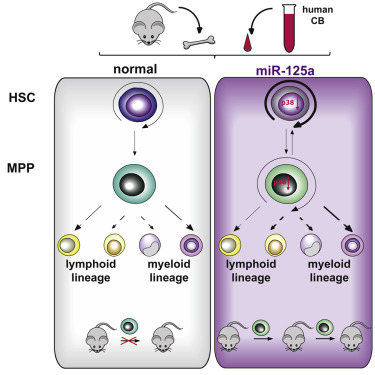
Dick and his colleagues analyzed mouse and human models of blood development, and they discovered that a microRNA called miR-125a is a genetic switch that is on in stem cells and controls self-renewal, but gets turned off in the progenitor cells.

“Our work shows that if we artificially throw the switch on in those downstream cells, we can endow them with stemness and they basically become stem cells and can be maintained over the long-term,” says Dr. Dick.

In their paper, Dick and de Haan showed that forced expression of miR-125 increases the number of hematopoietic stem cells in a living animal. Also, miR-125 induces stem cell potential in murine and human progenitor cells, and represses, among others, targets of the MAP kinase signaling pathway, which is important in differentiation of cells away from the stem cell fate. Furthermore, since miR-125 function and targets are conserved in human and mouse, what works in mice might very well work in human patients.

graphical abstract CSC_v9

This is proof-of-concept paper – no human trials have been conducted to date, but these data may be the beginnings of making more stem cells from banked cord blood to cure a variety of blood-based conditions.

Here’s to hoping.

Musashi-2 Protein Increases Number Hematopoietic Stem Cells in Umbilical Cord Blood


Umbilical cord blood infusions save the lives of many children and adults each year. Umbilical cord blood contains hematopoietic stem cells (HSCs) that can replace those lost to anticancer treatments, chemicals, or bone marrow collapse. However, despite their advantages for transplantation, the clinical use of umbilical cord blood is limited by the fact that HSCs in cord blood are found only in small numbers.

Small molecules that enhance hematopoietic stem and progenitor cell (HSPC) expansion in culture have been identified (see Boitano, A. E. et al. Science 329, 1345–1348 (2010), and Fares, I. et al. Science 345, 1509–1512 (2014). Unfortunately, the mechanisms of action or the nature of the pathways they impinge on are poorly understood.

Now a research team from McMaster University’s Stem Cell and Cancer Research Institute have discovered a key protein in the HSC/HSPC regenerative signaling pathway.

Kristin J. Hope and her team have elucidated the role of a protein called Musashi-2 in the function and development of HSCs.

Dr. Hope says that this discovery could help the tens of thousands of patients who suffer from blood-based disorders, including leukemia, lymphoma, aplastic anemia, sickle-cell disease, and more.

“We’ve really shone a light on the way these stem cells work,” she said. “We now understand how they operate at a completely new level, and that provides us with a serious advantage in determining how to maximize these stem cells in therapeutics. With this newfound ability to control over the regeneration of these cells, more people will be able to get the treatment they need.”

Only about five percent of all umbilical cord blood samples contain enough HSCs for a transplant, which is unfortunate because umbilical cord blood is less likely to be rejected by the immune system, because of the immaturity of the cells, and is also rather abundant.

Growing HSCs in culture is a possibility, but this remains a somewhat poorly understood and ill-defined procedure.

Musashi-2 is an RNA-binding protein in cells and was actually named for the Japanese samurai who fought using two swords.

In collaboration with researchers in Dr. Gene Yeo’s lab at the University of California San Diego, Dr. Hope’s lab has found that the Musashi-2 protein plays a pivotal role in controlling stem cell production in human cord blood HSCs. When Musashi-2 levels in HSCs are the knocked down, the cod blood HSCs were no longer able to regenerate the blood system. Conversely, when the levels of Musashi-2 were increased, the number of HSCs in the cord blood sample increased significantly.

The Hope’s group new discovery has identified a new way to tightly control on the development of HSCs. Essentially, Hope and her colleagues have discovered a new way to make more cord blood stem cells in a dish.

In the past, attempts to control HSC function and development has been approached at the level of transcriptional factors. The Hope lab’s approach of directing stem cell function through manipulation of an RNA-binding protein is somewhat novel, and represents a paradigm shift in the way we think about stem cell biology.

“This discovery really highlights the underappreciated role that RNA-binding protein-mediated control has on the properties of stemness in the blood system,” explained Dr. Hope.

This paradigm shift provides new targets for pharmaceuticals that may be able to expand these cells in a safe and targeted manner.

These findings represent an important step forward in surmounting the obstacles associated with stem cell transplants. According to Dr. Hope, the ability to increase the number of available cord blood stem cells has the potential to “mitigate a lot of the problems that arise post-transplantation.” Elaborating further, Dr. Hope explained that stem cells from cord blood are a “safer and more efficient transplant product,” and detailed how their use could reduce the number of patient follow-up visits and treatments required post-transplantation. Streamlining the transplantation process could help to alleviate the stress on the healthcare system and open up space for more transplant patients.

Making Cartilage from Umbilical Cord Stem Cells Without Growth Factors


Loïc Reppel and his colleagues at CNRS-Université de Lorraine in France have found that mesenchymal stem cells from human umbilical cord can not only be induced to make cartilage, but that these remarkable cells can make cartilage without the use of exogenous growth factors.

Mesenchymal stromal/stem cells from bone marrow (BM-MSC) have, for some time, been the “all stars” for cartilage regeneration. In fact, a very innovative clinic near Denver, CO has pioneered the use of BM-MSCs for patients with cartilage injuries. Chris Centeno, the mover and shaker, of this clinic has carefully documented the restoration of articular cartilage in many patients in peer-reviewed articles.

However, there is another “kid’ on the cartilage-regeneration block; mesenchymal stromal/stem cells from Wharton’s jelly (WJ-MSC). The advantages of these cells are their low immunogenicity and large cartilage-making potential. In this paper, which was published in Stem Cell Research and Therapy, Reppel and others evaluated the ability of WJ-MSCs to make cartilage in three-dimensional culture systems.

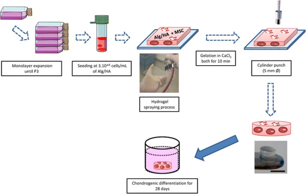
Reppell and his coworkers embedded WJ-MSCs isolated from the umbilical cords of new-born babies in alginate/hyaluronic acid hydrogel and grew them for over 28 days. These hydrogels were constructed by the spraying method. The hydrogel solution (for those who are interested, it was 1.5 % (m/v) alginate and hyaluronic acid (ratio 4:1) dissolved in 0.9 % NaCl) was sprayed an airbrush connected to a compressor. The solution was seeded with WJ-MSCs and then sprayed on a sterile glass plate. The hydrogel was made solid (gelation) in a CaCl 2 bath (102 mM for 10 minutes). Then small cylinders were cut (5 mm diameter and 2 mm thickness) with a biopsy punch. Then Reppel and others compared the chondrogenic differentiation of WJ-MSC in these three-dimensional scaffolds, without adding growth factors with BM-MSC.

Illustration of protocol steps used to perform scaffold construct and chondrogenic differentiation. After monolayer expansion, MSC were seeded at 3× 10 6 cells/mL of Alg/HA hydrogel. Hydrogel was sprayed, gelated, and cut into 5 mm diameter cylinders; scale bar = 5 mm. Scaffolds were cultivated in a 48-well plate in differentiation medium for 28 days. Alg/HA alginate/hyaluronic acid, MSC mesenchymal stromal/stem cells, P3 passage 3

After 3 days in culture, WJ-MSCs seemed nicely adapted to their new three-dimensional culture system without any detectable damage. From day 14 – 28, the proportion of WJ-MSC cells that expressed all kinds of cell surface proteins characteristic of MSCs (i.e., CD73, CD90, CD105, and CD166) decreased significantly. This suggests that these cells were differentiating into some other cell type.

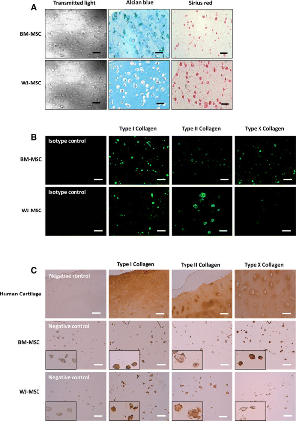
After 28 days in this scaffold culture, both WJ-MSCs and BM-MSCs showed strong upregulation of cartilage-specific genes. However, WJ-MSCs exhibited greater type II collagen synthesis than BM-MSCs, and these differences were evident at the RNA and protein levels. Collagen II is a very important molecule when it comes to cartilage synthesis because chondrogenesis, otherwise known as cartilage production, occurs when MSCs differentiate into cartilage-making cells known as chondroblasts that begins secreting aggrecan and collagen type II that form the extracellular matrix that forms cartilage. Unfortunately, in order to complete the run to mature cartilage formation, the chrondrocytes must enlarge (hypertrophy), and express the transcription factor Runx2 and secrete collagen X. Unfortunately, WJ-MSCs expressed Runx2 and type X collagen at lower levels than BM-MSCs in this culture system.

Matrix synthesis detected after 28 days of chondrogenic induction. Proteoglycans and total collagen were stained by Alcian blue and Sirius red (a), respectively. To explore the synthesis of various collagens in depth, immunofluorescence (b) and immunohistochemistry staining (c) were performed and detected using fluorescence microscopy and light microscopy, respectively; scale bar = 100 μm. BM-MSC bone marrow-derived mesenchymal stromal/stem cells, WJ-MSC Wharton’s jelly-derived mesenchymal stromal/stem cells

These experiments only examined cells in culture, which is not the same as placing cells in a living animal, but it is a start. Thus, when they are seeded in the hydrogel scaffold, WJ-MSCs and BM-MSCs, after 4 weeks, were able to adapt to their environment and express specific cartilage-related genes and matrix proteins in the absence of growth factors. In order to properly make cartilage in clinical applications, WJ-MSCs must go the full way and express high levels of Runx2 and collagen X. However, these experiments show that WJ-MSCs, which in the past were medical waste, are a potential alternative source of stem cells for cartilage tissue engineering.

Reppel and his colleagues note in their paper that to improve cartilage production from WJ-MSCs, it might be important to mimic the physiological environment in which chondrocytes normally find themselves. For example, they could apply mechanical stress or even a low-oxygen culture system. Additionally, Reppel and others could apply stratified cartilage tissue engineering. Reppel thinks that they could adapt their spraying method to design new stratified engineered tissues by applying progressive cells and spraying hydrogel layers one at a time.

All in all, cartilage repair based with WJ-MSC embedded in Alginate/Hyaluronic Acid hydrogel will hopefully be tested in laboratory animals and then, perhaps, if all goes well, in clinical trials.

Stem Cell-Based Cartilage Regeneration Could Decrease Knee and Hip Replacements


Work by Chul-Won Ha, director of the Stem Cell and Regenerative Medicine Institute at Samsung Medical Center and his colleagues illustrates the how stem cell treatments might help regrow cartilage in patients with osteoarthritis or have suffered from severe hip or knee injuries.

A 2011 report from the American Academy of Orthopedic Surgeons showed that approximately one million patients in the US alone (645,000 hips and 300,000 knees) have had joint replacements in the U.S. alone. Most joint replacements occur with few complications, artificial joints can only last for a certain period of time and some will even eventually require replacement. Also these procedures require extensive rehabilitation and are, in general, quite painful. A goal for regenerative medicine is the regenerate the cartilage that was worn away to prevent bones from eroding each other and obviate the need for artificial joint replacement procedures.

Extensive research from the past two decades from a whole host of laboratories in the United States, Europe, and Japan have shown that mesenchymal stem cells (MSCs) have the ability to make cartilage, and might even have the capability to regenerate cartilage in the joint of a living organism. MSCs have the added benefit of suppressing inflammation, which is a major contributor to the pathology of osteoporosis. Additionally, MSCs are also relatively easy to isolate from tissues and store.

“Over the past several years, we have been investigating the regeneration potential of human umbilical cord blood- derived MSCs in a hyaluronic acid (HA) hydrogel composite. This has shown remarkable results for cartilage regeneration in rat and rabbit models. In this latest study we wanted to evaluate how this same cell/HA mixture would perform in larger animals,” said Ha.

Ha collaborated with researchers from Ajou University, which is also in Seoul, and Jeju University in Jeju, Korea. Ha and his team used pigs as their model system, which is a better system than rodents for such research.

The stem cells for this project were isolated from human umbilical cord blood that was obtained from a cord blood bank. They isolated MSCs from the umbilical cord blood and grew them in culture to establish three different human Umbilical Cord Blood MSC lines. Then they pelleted the cells and mixed them with the HA solution and applied them to the damaged knee joints of pigs.

“After 12 weeks, there was no evidence of abnormal findings suggesting rejection or infection in any of the six treated pigs. The surface of the defect site in the transplanted knees was relatively smooth and had similar coloration and microscopic findings as the surrounding normal cartilage, compared to the knees of a control group of animals that received no cells. The borderline of the defect was less distinct, too,” said the study’s lead investigator, Yong-Beom Park, who is a colleague of Ha’s at the SungKyunKwan University’s Stem Cell and Regenerative Medicine Institute.

“This led us to conclude that the transplantation of hUCB-MSCs and 4 percent HA hydrogel shows superior cartilage regeneration, regardless of the species. These consistent results in animals may be a stepping stone to a human clinical trial in the future,” Dr. Ha noted.

“These cells are easy to obtain, can be stored in advance and the number of potential donors is high,” said Anthony Atala, M.D., Editor of STEM CELLS Translational Medicine and Director of the Wake Forest Institute for Regenerative Medicine. “The positive results in multiple species, including the first study of this treatment in large animals, are certainly promising for the many patients requiring treatments for worn and damaged cartilage.”

Next-Generation Cell Therapy for Graft-Versus-Host Disease


Endonovo Therapeutics, Inc has announced its development of a cell-based treatment for Graft-versus-Host Disease (GvHD). This treatment utilizes umbilical cord blood stem cells that have been grown and enhanced by specific treatments.

GVHD occurs when newly transplanted donor cells attack the recipient’s body. It can occur after a bone marrow or stem cell transplant if the cells have not been properly matched or even if the donor and recipient are relatively well matched. The chances of suffering GVHD are around 30 – 40% if the donor and recipient are genetically related and close to 60 – 80% when the donor and recipient are not related.

GVHD can be either acute or chronic and the symptoms of GvHD can be either mild or severe. Typically, acute GVHD comes on within the first 6 months after a transplant. Common acute symptoms include: Abdominal pain or cramps, nausea, vomiting, and diarrhea, Jaundice (yellow coloring of the skin or eyes) or other liver problems, skin rash, itching, redness on areas of the skin. Chronic GVHD usually starts more than 3 months after a transplant, and can last for the lifetime or the patient. The symptoms of chronic GvHD include: dry eyes or vision changes, dry mouth, white patches inside the mouth, and sensitivity to spicy foods, fatigue, muscle weakness, and chronic pain, joint pain or stiffness, skin rash with raised, discolored areas, as well as skin tightening or thickening, shortness of breath, weight loss.

Endonovo uses a novel method to enhance stem cells. Their so-called “Cytotronics platform” utilizes Time-Varying Electromagnetic Field (TVEMF) technology to expand and enhance the therapeutic properties of stem cells and other types of cells for regenerative treatments and tissue engineering. This platform can potentially optimize cell-based therapies so that they have greater therapeutic potential than they had prior to their treatment.

The Cytotronics™ platform dates back to experiments conducted at NASA to expand stem cells in culture. NASA’s goal was to create stem cell therapies that could be used to treat astronauts during long-term space exploration. NASA scientists showed that Time-Varying Electromagnetic Fields (TVEMF) could stimulate the expansion of stem cells in the lab. Additionally, TVEMF increased the expression of dozens of genes related to cell growth, tumor suppression, cell adhesion and extracellular matrix production.

By testing and tweaking this technology over a period of 15 years, Endonovo scientists created a novel protocol for augmenting the therapeutic properties of cells in culture through physics rather than genetic engineering. The Cytotronics™ platform seems to be able to make stem cells that express higher levels of key genes necessary for tissue healing and regeneration.

As an example of the efficacy of this technology. Endonovo scientists have shown that Cytotronic™ expansion of peripheral blood stem cells resulted in an over 80-fold expansion of CD34+ cells in as little as 6 days.

Endonovo is using the Cytotronic platform to enhance the regenerative properties of mesenchymal stem cells (MSCs), which have the capacity to staunch inflammation in patients with GvHD and other inflammatory diseases.

However, despite their promise, MSC-based therapies suffer from poor engraftment and short-term survival when transplanted into sick patients. These remain major limitations to the effective therapeutic use of MSCs. If there was a safe and effective way to beef up the survival and regenerative properties of MSCs, such a technique would be indispensable.  This makes MSCs prime candidates for the Cytotronic Platform.

Dr. Donnie Rudd, Chief Scientist & Director of Intellectual Property at Endonovo, said: “Our Cytotronics platform is particularly suited to address many of the issues that have plagued stem cell therapies that have recently failed, such as their loss of potency and self-renewal when expanded ex vivo, their poor engraftment and their limited ability to survive when transplanted.”

Earlier this year, Endonovo announced a protocol for the creation of a cell mixture from a portion of the human umbilical cord co-cultured with adipose-derived stem cells. This resulting cell mixture contains a rich source of highly-proliferative, immunosuppressive cells that are not recognized by the patients immune system, since they contain neither of the major histocompatibility markers (HLA double negative). These cells are “immune privileged,” which means that are not recognized as foreign cells by the patient’s immune system, and therefore are a significant source of cells for MSC-based therapies.

Endonovo Therapeutics has used this new technology to create a biologically potent, off-the-shelf, allogeneic treatment for Graft-Versus-Host disease and a wide-array of other conditions. They would like to test these products in clinical trials eventually.

Endonovo hopes that stem cells enhanced by the Cytotronics™ platform will become a major innovation in the regenerative medicine market.

“We are very excited to be a leader in the development of next-generation, ex vivo enhanced cells for regenerative medicine,” stated Endonovo CEO, Alan Collier. “We have seen several stem cell therapies fail in clinical trials over the last couple of years, which points to a critical need for the development of methods to increase the biological and therapeutic properties of stem cells.”

“We believe that enhancing the biological and therapeutic properties of stem cells using bioelectronics is the future of cell-based therapies,” concluded Mr. Collier.

Partial Repair of Full-Thickness Rotator Cuff Tears By Guided Application of Umbilical Cord Blood Mesenchymal Stem Cells


Baseball players, weight lifters, tennis players, basketball players, and other athletes have experienced the pain and frustration of a rotator cuff injury. The rotator cuff is the capsule that surrounds the shoulder joint, in combination with the fused tendons that support the arm at the shoulder joint. A tear in any of these tendons constitute a rotator cuff tear, and it is painful, and debilitating. Furthermore, rotator cuff tears are notoriously slow healing, if they heal at all.

The main option for a rotator cuff tear is microsurgical repair of the tendon. However, as Christopher Centeno at the Regenexx blog points out, sewing together atrophied tissue does not make a lot of sense, and consequently, rotator cuff repairs by means of microsurgery can have a high percentage of re-tearing. Is there a better way?

In the journal Stem Cells and Translational Medicine, Dong Rak Kwon and his two colleagues, Gi-Young Park and Sang Chui Lee, from the Catholic University of Daugu School of Medicine in Daegu, Korea have reported the results of treating whole-thickness rotator cuff tears in rabbits with human umbilical cord blood mesenchymal stem cells (UCB-MSCs). The results are quite interesting.

Kwon and his colleagues broke a colony of New Zealand White rabbits into three groups and surgically subjected all animals to full-thickness tears in the subscapularis tendon. Because rabbits are four-legged creatures, such tears severely compromise their ability to walk, and Kwon and his team measured the ability of these rabbits to walk and the speed at which they walked. All three groups of rabbits showed about the same ability to walk: they walked at about the same speed at for the same distance before giving up.

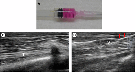
Human umbilical cord blood-derived mesenchymal stem cell (MSC) and ultrasound images. (A): Human umbilical cord blood-derived MSCs. (B): Injection was made in the left shoulder subscapularis (SCC) full-thickness tears under ultrasound guidance. (C): Longitudinal ultrasound image showed the needle (arrows) in the left shoulder SCC of the rabbit. Abbreviations: S, mesenchymal stem cell; T, tendon.

The first group of rabbits received injections of UCB-MSCs into their rotator cuffs. These injections were guided by ultrasound so that Kwon and his colleagues were able to place the stem cells directly on the damaged tendons. The second group of rabbits received injections of hyaluronic acid (HA), which is a component of connective tissue and the synovial fluid within bursal sacs that surround and lubricated some our joints. The third group received injections of sterile saline into their joints. The animals were then examined four weeks later.

shoulder-joint

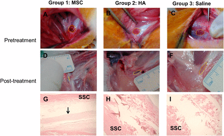
The HA- and saline-injected animals showed few changes, but the UCB-MSC-injected animals were able to walk almost twice as far as the other rabbits and almost twice as fast. When the joint tissue of these animals was examined in detail, the HA and saline-injected animals still had full-thickness rotator cuff tears, although the HA-injected animals showed more healing that then the saline-injected rabbits. When the UCB-MSC-injected animals were examined, seven of the ten animals have rotator cuffs that had healed so that the tears could be classified as partial-thickness tears rather than full-thickness tears. Furthermore, a more detailed examination of these joint revealed that they showed regeneration of the tendon and the production of tough, high-quality collagen I.

Gross morphological (A–F) and histological (G–I) findings of the subscapularis tendons in groups 1, 2, and 3. The polygon in each of the first six images depicts the area of the full-thickness subscapularis tendon tear. (A–C): Pretreatment images. (D–F): Posttreatment images. (G): Parallel arrangement of hypercellular fibroblastic bundles (arrow) was noted in group 1. (H, I): Histological findings in groups 2 and 3 showed absence of fiber bundles. Group 1 received a 0.1-ml injection of MSCs; group 2, 0.1 ml of HA; group 3, 0.1 ml of saline. Hematoxylin-and-eosin stain, ×40. Abbreviations: MSC, human umbilical cord blood-derived mesenchymal stem cell; HA, hyaluronic acid; SSC, subscapularis muscle.

Collagen I is the tough material that makes tendon. When rotator cuff surgeries fail, it can be for a variety of reasons, such as poor blood supply, intrinsic tendon degeneration, fatty infiltration, or muscle atrophy (see UG Longo, et al., British Medical Bulletin 2011, 98:31-59).

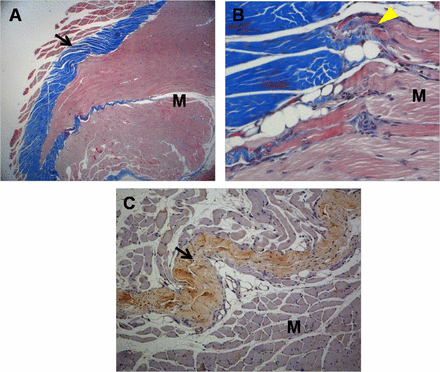
Histological micrographs of tissue from group 1 rabbits. (A): Newly regenerated tendons are shown in the blue-stained fibers (black arrow; Masson’s trichrome stain; magnification, ×12.5). (B): Regenerated tendon fibers (yellow arrowhead; Masson’s trichrome stain; magnification, ×250) are connected to adjacent M fibers. (C): The regenerated tendon fibers (black arrow) stained with anti-type 1 collagen antibody. The defect was reconstructed with human umbilical cord blood-derived mesenchymal stem cells (magnification, ×100). Abbreviation: M, muscle.

However, tendon failures after surgery usually result from the production of collagen III, which is mechanically weaker than collagen I, instead of collagen I (see MF Pittenger, et al., Science 1999, 284: 143-147; V Rocha, et al., New England Journal of Medicine 2000, 342: 1846-1854). None of the animals in the other groups showed any sign of collagen I production.

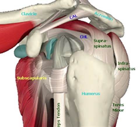
This experiment shows that full thickness tears in the subscapularis tendon of the rotator cuff of rabbits, which is functionally similar to the supraspinatus in humans (see figure below), can be partially healed by the ultrasound-guided infusion of UCB-MSCs.

th48RY4PHI

If larger numbers of UCB-MSCs were implanted, it is possible that the tears would have been completely repaired. Also, it is possible that partial tears can be completely repaired by this procedure, but clearly more work is required.

Other questions also remain besides the optimal dose of the cells. What sized tears can be regenerated by this procedure? What immobilization procedures are appropriate after the stem cell injections and for how long? What are the most effective rehabilitation techniques after the surgery? These are all questions that are amenable to research so take heart athletes; a better cure is slowly, but surely on its way.

Combining Umbilical Cord Cells with Hyaluronic Acid Improves Heart Repair After a Heart Attack


Umbilical cord blood cells have an advantage over bone marrow or peripheral blood cells in that aging, systemic inflammation, and stress or damage caused by cell processing procedures can potentially compromise and diminish the regenerative capability of these cells. This problem is particularly acute in the case of treating patients who have recently suffered a heart attack, since transplanted cells experience a rather hostile environment that kills off most cells. Additionally, blood flow through the heart tends to wash out infused cells, which further decreases any regenerative activities the cells might have otherwise exerted.

With this in mind, Patrick Hsieh and his colleagues at the Academia Sinica, in Taipei, Taiwan tested if ability of human cord blood mononuclear cells (CB-MNCs) injected into the heart in combination with a hyaluronan (HA) hydrogel could extend the regenerative abilities of these cells in a pig model. HA is a common component of connective tissue, and, in general, it is very well tolerated by patients and implanted cells. Furthermore, it has the added bonus of shielding cells from a hostile environment and preventing them from being washed out of the heart.

Hsieh used a total of 34 minipigs and divided them into five different groups. One group was the sham operation group in which minipigs received surgical incisions but no heart attack was induced. The second group had heart attacks surgically induced and received infusions of normal saline solutions. The third group of minipigs also experienced heart attacks, and had HA injected into the heart walls. The fourth group also suffered heart attacks and received injections of human umbilical cord stem cells into their heart walls. The fifth group experienced heart attacks and received injections of both HA and human umbilical cord blood cells. The animals were kept and examined two months after surgery.

Two months after the surgery, the minipigs that received injections of human umbilical cord blood cells plus HA showed the highest left ventricle ejection fraction (51.32% ± 0.81%). This is significant when compared to 42.87% ± 0.97%, for the group that received injections of normal saline, 44.2% ± 0.63% for the group that received injections of HA alone, and 46.17% ± 0.39% for the group that received injections of umbilical cord blood cells only. Additionally, hearts from minipigs that received cord blood cells plus HA improved the systolic and diastolic function significantly better than the other experimental groups. Injections of either cord blood cells alone or in combination with HA significantly decreased the scar area and promoted the formation of new blood vessels in the infarcted region. In general, this study suggests that combined infusion of umbilical cord blood cells and HA improves the function of the heart after a heart attack and might prove to be a promising treatment option of heart attack patients.

This is a preclinical study, but it is a preclinical study in a larger animal model system. Umbilical cord blood cells have a demonstrated ability to induce healing in the heart after a heart attack. However, the combination of these cells with HA almost certainly significantly increases cell retention in the heart, thereby significantly improving cardiac performance, and preventing cardiac remodeling. Therefore, using healthy cells donated from another source to replace damaged or moribund cells may be a better option to treat a heart patient and repair their sick heart.

This work appeared in Stem Cells Trans Med November 2015, doi: 10.5966/sctm.2015-0092

Blocking Differentiation is Enough to Turn Mature Cells into Stem Cells


Hiroshi Kawamoto led a collaboration between the RIKEN Center for Integrative Medical Science and other institutions in Japan and Europe that examined the possibility that adult cells can be maintained in a stem cell-like state where they can proliferate without undergoing differentiation. They discovered that in immune cells, blocking the activity of one transcription factor can maintain the cells in a stem cell-like state where they continue to proliferate and still have the capacity to differentiate into different mature cell types.

Kawamoto and his team genetically engineered hematopoietic progenitor cells from mice to overexpress the Id3 protein. Id3, or inhibitor of DNA binding 3, is an inhibitory protein that forms nonfunctional complexes with other transcription factors. In particular, Id3 inhibits so-called “E-proteins,” (such as TCF3) which drive the progenitor cells to differentiate into immune cells.

Overexpression of Id3, in addition to soaking the cells in a cocktail of cytokines, cause the cells to continue to divide as stem cells. However, when the cytokines were withdrawn, the cells differentiated into various types of immune cells.

Next, Kawamoto and his collaborators infused these engineered hematopoietic progenitors into mice that had been depleted of white blood cells. They discovered that their Id3-overexpressing cells could expand and replenish the white blood cell population of these.

In a follow-up experiment, Kawamoto and his crew recapitulated this experiment using human umbilical cord blood hematopoietic progenitors. Just like their mouse counterparts, these umbilical cord cells could be maintained in culture, and then, upon change of culture conditions, could differentiate into blood cells.

Because these cells can be kept in an undifferentiated state and can extensively proliferate, this culture system provides a model for studying the genetic and epigenetic basis of stem cell self-renewal. And it might also allow scientists to inexpensively grow large quantities of immune cells for regenerative medicine or immune therapies.

This work was published in Stem Cell Reports, October 2015 DOI: 10.1016/j.stemcr.2015.09.012.

Cord Blood Cells As a Potential Treatment for Alzheimer’s Disease


Jared Ehrhart from the University of South Florida, who also serves as the Director of Research and Development at Saneron CCEL Therapeutics Inc, and his coworkers have shown that cells from umbilical cord blood can not only improve the health of mice that have an experimental form of Alzheimer’s disease (AD), but these can also be administered intravenously, which is safer and easier than other more invasive procedures.

Laboratory mice can be engineered to harbor mutations that can cause a neurodegenerative disease that greatly resembles human AD. One such mouse is the PSAPP mouse that harbors two mutations that are known to cause an inherited, early-onset form of AD in humans. By placing both mutations in the same mouse, the animal forms the characteristic protein plaques more rapidly and shows significant AD symptoms and brain pathology.

Ehrhart used PSAPP mice to test the ability of human umbilical cord blood to ameliorate the symptoms of AD. He injected one million Human Umbilical Cord Blood Cells (HUCBCs) into the tail veins of PSAPP mice and 2.2 million into the tail veins of Sprague-Dawley rats. Then he harvested their tissues at 24 hours, 7 days, and 30 days after injection. Then Ehrhart and his team used a variety of techniques to detect the presence of the HUCBCs.

Interestingly, the HUCBCs were able to cross the blood-brain barrier and take up residence in the brain. The cells remained in the brain and survived there for up to 30 days and did not promote the growth of any tumors.

Several studies have shown that the administration of HUCBCs to mice with a laboratory form of AD can improve the cognitive abilities of those mice (see Darlington D, et al., Cell Transplant. 2015;24(11):2237-50; Banik A, et al., Behav Brain Res. 2015 Sep 15;291:46-59; Darlington D, et al., Stem Cells Dev. 2013 Feb 1;22(3):412-21). However, in such cases it is essential to establish that the administered cells actually found their way to the site of damage and exerted a regenerative response.

Even though Ehrhart and his troop found that the intravenously administered HUCBCs were widely distributed throughout the bodies of the animals, they persisted in the central nervous system for up to one month after they were injected. In the words of this publication, which appeared in Cell Transplantation, the HUCBCs were “broadly detected in both in the brain and several peripheral organs, including the liver, kidneys, and bone marrow.”. The fact that such a minimally invasive procedure like intravenous injection can effectively introduce these cells into the bodies of the PSAPP mice and still produce a significant therapeutic effect is a significant discovery.

Ehrhart and his colleagues concluded that HUCBCs might provide therapeutic effects by modulating the inflammation that tends to accompany the onset of AD. Furthermore, these cells do not need to be delivered by means of an invasive procedure like intracerebroventricular injection. Furthermore, even though HUCBCs were detected in other organs, their numbers in those places was not excessive and the ability of the HUCBCs to cross the blood-brain barrier suggests that these cells might serve as safe, effective therapeutic agents for AD patients some day.

Umbilical Cord Blood Contains c-kit+ Cells that Can Differentiate into Heart-like Cells


Bone contains a wide variety of stem cells whose potential are only beginning to be tapped. One cell population possesses a cell surface protein called c-kit, and these c-kit+ progenitor cells seem to support myocardial regeneration. Do c-kit+ cells from umbilical cord blood have the same capacity?

Luciana Teofili from the Catholic University of the Sacred Heart in Rome, Italy and her colleagues purified c-kit+ cells from umbilical cord blood by means of magnetic beads that were coated with c-kit-binding antibodies. Teofili and others induced heart muscle differentiation in these cells with several different protocols. Then the expression of cardiac markers (GATA4, GATA6, β-myosin heavy chain, α-sarcomeric actin and cardiac Troponin T) was investigated, and whole-cell current and voltage-clamp recordings were performed.

The c-kit+ cells from umbilical cord blood showed a rather immature gene profile, and by themselves, they did not differentiate into heart muscle-like cells in culture. In contrast, if whole mononuclear cells from umbilical cord blood were subjected to the same treatment, several if the employed protocols produced large, adherent cells that expressed several heart muscle-specific genes and exhibited an excitability much like that of heart muscle cells.

Formation of these heart muscle-like cells was prevented if the c-kit+ cells were removed from the other cells. Tracking studies showed that the c-kit+ cells were the ones that differentiated into heart muscle-like cells, but they only did so when they were together with c-kit– cells.

Thus umbilical cord blood contains progenitors endowed with the ability to differentiate into heart muscle-like cells. The cells with this potential reside in the c-kit+ fraction but they require the presence of abundant accessory cells to differentiate properly.

These preliminary observations suggest that it is a good idea to consider the storage of the umbilical cord blood of patients with prenatal diagnosis of congenital heart diseases. Such conditions might be potentially amenable to myocardial regenerative therapies with umbilical blood-based stem cells.

This paper was published in the journal Cytotherapy, but it must be said that the evidence that these cells differentiated into heart muscle cells was not completely convincing.

Gamida Cell Phase 3 Study Design Outline Approved by FDA and EMA


Gamida Cell, a cell therapy company based in Jerusalem, Israel, has reached agreements with the US Food and Drug Administration (USFDA) and the European Medicines Agency (EMA) with regards to a Phase III study design outline for testing their NiCord product. NiCord is a blood cancer treatment derived from a single umbilical cord blood until expanded in culture and enriched with stem cells by means of the company’s proprietary NAM technology.

Gamida Cell is moving forward now with plans to commence an international, multi-center, Phase III study of NiCord in 2016. Phase I/II data of 15 patients are expected in the fourth quarter of 2015. NiCord is in development as an experimental treatment for various types of blood cancers including Acute Myeloid Leukemia (AML), Acute Lymphoblastic Leukemia (ALL), Myelodysplastic Syndrome (MDS), and Chronic Myelogenous Leukemia (CML).

NiCord® is derived from a single cord blood unit which has been expanded in culture and enriched with stem cells using Gamida Cell’s proprietary NAM technology. NAM technology proceeds from the observation that nicotinamide, a form of vitamin B3, inhibits the loss of functionality that usually occurs during the culture process of umbilical cord blood stem cells, when added to the culture medium. Pre-clinical studies have also shown that the expanded cell grafts manufactured using NAM technology demonstrate improved functionality following infusion in a living animal. These stem cells show improved movement, home to the bone marrow, and show higher rates of engraftment, or durable retention in the bone marrow. Based on these results, Gamida Cell is currently testing in clinical trials (in patients) cells expanded in culture with the NAM platform to determine their safety and effectiveness as a treatment for blood cancers, sickle-cell anemia and thalassemia. NiCord is intended to fill the crucial clinical need for a treatment for the vast majority of blood cancer patients indicated for bone marrow transplantation, with insufficient treatment options. This segment has a multi-billion dollar market potential.

“The FDA and EMA feedback is a major regulatory milestone for NiCord. NiCord is a life-saving therapy intended to provide a successful treatment for the large number of blood cancer patients who do not have a family related matched donor. Gamida Cell is dedicated to changing the paradigm in transplantation by bringing this therapy to market as soon as possible,” said Dr. Yael Margolin, president and CEO of Gamida Cell.

“The positive regulatory feedback confirms that Gamida Cell’s NiCord program is on a clear path to approval both in the U.S. and EU. We look forward to continuing the development of this very important product in cooperation with sites of excellence in cord blood transplantation worldwide,” said Dr. David Snyder, V.P. of Clinical Development and Regulatory Affairs at Gamida Cell.

The Phase III study will be a randomized, controlled study of approximately 120 patients. It will compare the outcomes of patients transplanted with NiCord to those of patients transplanted with un-manipulated umbilical cord blood.


Cartilage Repair Using Human Umbilical Cord Blood-Derived Mesenchymal Stem Cells Embedded in Hyaluronic Acid Hydrogel in a Minipig Model


Cartilage shows lousy regenerative capabilities. Fortunately, it is possible to regenerate cartilage with human umbilical cord blood-derived mesenchymal stem cells (hUCB-MSCs) that have been embedded in a hyaluronic acid (HA) hydrogel composite. In fact, such a combination has shown remarkable results in rat and rabbit models.

In this present study, published in Stem Cells Translational Medicine, Yong-Geun Park and his colleagues from SungKyunKwan University School of Medicine, in Seoul, South Korea sought to confirm the efficacy of this protocol in a in a pig model using three different hUCB-MSC cell lines.

Park and his coworkers generated full-thickness cartilage injuries in the trochlear groove of each knee in 6 minipigs. Three weeks later, an even larger cartilage defect, 5 mm wide by 10 mm deep, was created, followed by an 8-mm-wide and 5-mm-deep boring. In short, the knee cartilages of these minipigs were very messed up.

Trochlear-groove

To these knee cartilages, a mixture (1.5 ml) of hUCB-MSCs (0.5 × 10⁷ cells per milliliter) and 4% HA hydrogel composite were troweled into was then cartilage defects of the right knee. The left knee served as an untreated control. Each cell line was used in two minipigs.

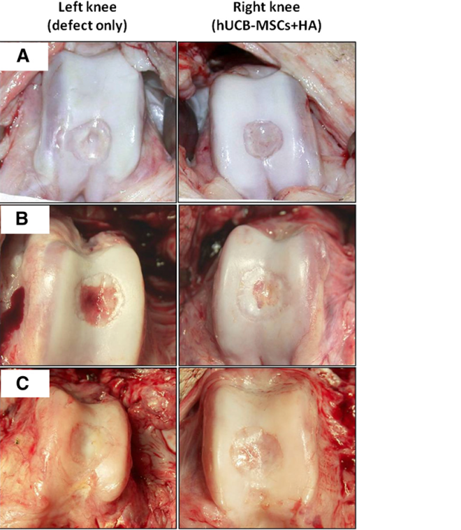
Macroscopic findings of the osteochondral defects of the porcine knees. At 12 weeks postoperatively, the defects of both knees had produced regenerated tissues that were pearly white and firm. These new tissues, which resembled articular cartilage, appeared adherent to the adjacent cartilage and had restored the contour of the femoral condyles (smooth articular surfacewithout depression). The regenerated tissue of the control knee (left knee) looked fibrillated. Grossly, no differencewas seen in the quality of the repaired tissue in the transplanted knee (right knee) among the three groups with different cell lines. (A): Group A. (B): Group B. (C): Group C. Abbreviations: HA, hyaluronic acid; hUCB-MSCs, human umbilical cord blood-derived mesenchymal stem cells.
Macroscopic findings of the osteochondral defects of the porcine knees. At 12 weeks postoperatively, the defects of both
knees had produced regenerated tissues that were pearly white and firm. These new tissues, which resembled articular cartilage, appeared adherent to the adjacent cartilage and had restored the contour of the femoral condyles (smooth articular surface without depression). The regenerated tissue of the control knee (left knee) looked fibrillated. Grossly, no difference was seen in the quality of the repaired tissue in the transplanted knee (right knee) among the three groups with different cell lines. (A): Group A. (B): Group B. (C): Group C. Abbreviations: HA, hyaluronic acid; hUCB-MSCs, human umbilical cord blood-derived mesenchymal stem cells.

12 weeks after surgery, the pigs were sacrificed, and the degree of subsequent cartilage regeneration was evaluated by gross and more detailed microscopic analysis of the knee tissue. The transplanted knee showed superior and more complete joint-specific (hyaline) cartilage regeneration compared with the control knee. The microscopic characteristics of the knee cartilage showed that those animals that received the hUCB-MSCs had greater rates of cell proliferation and cells that differentiated into cartilage-making cells.

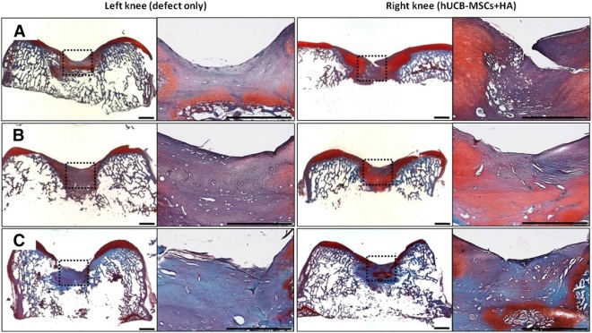
Microscopic findings of the regenerating osteochondral defects on porcine articular cartilage (safranin O and fast green staining). At 12 weeks postoperatively, the surface of the repairing tissue in the control knee (left knee) was poorly stained for glycosaminoglycan. In the transplanted knee (right knee), both the regenerated tissue and the adjacent cartilage to which it had become adherent exhibited the normal orthochromatic staining properties with safranin O. (A): Group A. (B): Group B. (C): Group C. Scale bars = 2 mm. Abbreviations: HA, hyaluronic acid; hUCB-MSCs, human umbilical cord blood-derived mesenchymal stem cells.

These data show consistent cartilage regeneration using composites of hUCB-MSCs and HA hydrogel in a large animal model. These experiments could be a stepping stone to a human clinical trial in the future that treats osteoarthritis of the knees with hUCB-MSCs embedded in HA hydrogel.

Using Silk to Grow Salivary Glands in the Laboratory


Colloquially, we use the word “spit” to describe saliva, which is secreted by our salivary glands. Saliva is a complex combination of water, salts, proteins, small molecules, and other components that lubricate our throats and mouths to facilitate swallowing, coat our gums and teeth to maintain good gum health and keep tooth decay at bay, and keep our breath fresh. Insufficient salivation production increases tooth decay rates, causes chronic halitosis (bad breath), and can swallowing difficult. Additionally, there are no treatments for poorly-functioning salivary glands, and these glands have poor regenerative capabilities.

Patients who suffer from head/neck cancers and have been treated with radiation suffer from “xerostoma” or dry mouth. Certain medications can also cause dry mouth as can old age. 50% of older Americans suffer from xerostoma.

If that isn’t bad enough, salivary glands are notoriously hard to grow in the laboratory.  So they slow down when we grow old, do not regenerate and grow poorly, in at all, in the laboratory.  Is there any good news about salivary glands?

Make that a yes!  A research team from the University of Texas Health Science Center, led by Chih-Ko Yeh has discovered a process that may lead to the growth of salivary glands in cell culture.  Yeh and his team used purified silk fibers that had many of their contaminants removed to grow salivary stem cells from rat salivary glands.  These cells grew in the laboratory and after several weeks in culture, generated a three-dimensional matrix that covered the silk scaffolds and shared many characteristics with the salivary glands that grow in the mouth.

Yeh underscored the importance of this discovery: “Salivary gland stem cells are some of the most difficult cells to grow in culture and retain their function.”  This work in Yeh’s laboratory have is the first time that salivary gland stem cells have been grown in cell culture while retaining their salivary gland properties.

Yeh continued, “The unique culture system has great potential for future salivary gland research and for the development of new cell-based therapeutics.”

Silk, contrary to what you might think, is an excellent choice for stem cell scaffolding because it is natural, biodegradable, flexible, porous material that provides cells easy access to oxygen and nutrition.  Silk also does not cause inflammation, which is a problem with other types of stem cell scaffolds.

Since there are so few salivary gland stem cells in the human mouth, Yeh and his group plan to continue using the rat model to refine their techniques.  Eventually, Yeh and others would like to use stem cells derived from bone marrow or umbilical cord blood to regenerate salivary glands in human patients.

In fact, Yeh and his coworkers have pioneered protocols for harvesting large numbers of bone marrow stem cells from bone marrow and human umbilical cord blood and growing them in culture.  These stem cells are abundant and can be differentiated into different cell types by means of tissue engineering technologies.

Yeh hopes that by the next decade, human salivary stem cells or tissue engineered artificial salivary gland will be used to initiate salivary gland regeneration in human patients.

This research was published in Tissue Engineering part A 2015; 21(9-10).

Wound Healing and Human Umbilical Cord Mesenchymal Stem Cells


Previous studies have shown that human bone marrow–derived mesenchymal stromal cells have potential to accelerate and augment wound healing. However, in the clinic, it is difficult to properly culture and then use bone marrow stem cells. Human umbilical cord blood–derived mesenchymal stromal cells (hUCB-MSCs) recently have been commercialized for cartilage repair as a cell-based therapy product that uses allogeneic stem cells.

Presently, current cell therapy products for wound healing utilize fibroblasts. Is it possible that hUCB-MSCs are superior to fibroblasts for wound healing? Seung-Kyu Han and his colleagues from the Department of Plastic Surgery at the Korea University College of Medicine in Seoul, South Korea used a cell culture system to compare the ability of hUCB-MSCs and fibroblasts to heal wounds.

For their study, Han and others used diabetic mice and isolated fibroblasts from normal and diabetic mice. Then they tested the ability of these cells to heal skin wounds in the very mice from which they were isolated. A third group of diabetic mice with skin wounds were treated with hUCB-MSCs. A comparison of all three groups examined the cell proliferation, collagen synthesis and growth factor (basic fibroblast growth factor, vascular endothelial growth factor and transforming growth factor-β) production and compared them among the three groups.

The results showed that hUCB-MSCs produced significantly higher amounts of vascular endothelial growth factor and basic fibroblast growth factor in comparison to both fibroblast groups. Human UCB-MSCs were better than diabetic fibroblasts but healthy fibroblasts in collagen synthesis, and there were no significant differences in cell proliferation and transforming growth factor-β production. Human UCB-MSCs produced significantly higher amounts of VEGF and bFGF when compared with both fibroblasts.

These results suggest that Human UCB-MSCs might be a better source for diabetic wound healing than either allogeneic or autologous fibroblasts. Larger animal studies will be needed, but this particular study seems like a good start.

Umbilical Cord Stem Cells Improve Heart Function after a Heart Attack


The umbilical cord connects the baby to the placenta and contains umbilical arteries, umbilical veins, and a gooey material between the umbilical vessels called Warton’s jelly. Warton’s Jelly (WJ), besides being rich in extracellular matrix molecules also contains a mesenchymal stem cell population that is rather primitive. These WJ mesenchymal stem cells or WJMSCs have excellent potential for therapeutic strategies.

Lian Gao and her colleagues from the Navy General Hospital in Beijing, China, in collaboration with coworkers from the Shenzhen Beike Cell Engineering Research Institute in Shenzhen, China conducted a clinical trial that examined the use of these WJMSCs in human patients who had suffered a heart attack.  The results are as interesting as they are suggestive and were published in the journal BMC Medicine.

First we must consider the design of the study. Gao and others recruited 160 heart attack patients who were no younger than 18 and no older than 80-years old. All patients had to be free of liver or kidney disease, cancer or some other terminal illness. They were admitted to 11 hospitals in China between February 2011 and January 2012 and had suffered from a documented heart attack as defined by symptoms and their EKG (ST elevation). All patients has also been treated with the implantation of a stent within 12 hours of their heart attack and still retained a respectable amount of movement of the heart wall in the left ventricle. If patients were outside these parameters, they were excluded from the study.

Of the 160 patients who were recruited for the study, 44 were excluded, either because they did not fit within the exclusion criteria, did not wish to participate in the trial, or opted out for undisclosed reasons. This left 116 patients who were randomly assigned to the placebo group or the experimental group (58 in each group). Of these two groups, the placebo group had one patient discontinue the study because of a bout with stomach cancer. The experimental group had one patient die ten days into the trial, another was lost because they moved and a third patients withdrew because of leukemia. This left 57 subjects for the placebo group and 55 for the experiment group who went through all 18 months of follow-up after their respective procedures.

There were two end points for this clinical trial after patients were observed for 18 months after the procedure. The first was safety and this was measured by examining the number of adverse effects (AEs) within these 18 months. Such AEs include things like death, hospitalization for worsening heart function, severe arrhythmias, repeated coronary intervention, blood clots forming in the stents (stent thrombosis), coronary artery obstruction, and the growth of extra tissue in the heart that does not belong there, disorders of the immune system and so on. The second end pointy was efficacy of the implanted cells. To ascertain this, the function of the heart was measured using positron emission computer tomography (PET), and single-photo-emission computer tomography or SPECT. These imaging procedures allow cardiologists to take very precise snapshots of the heart and determine with a good deal of accuracy the performance of the heart.

The WJMSCs were acquired from umbilical cords that were donated from healthy mothers who had delivered healthy babies by means of Caesarian section. 21 of these umbilical cords had their blood vessels removed and then the gelatinous tissue surrounding the vessels was removed, sliced up, and cultured. The MSCs in the gelatinous tissue, which is Warton’s Jelly, migrated from the WJ to the culture dishes. After three passages in the culture dishes, he cells were harvested, concentrated, and tested for viruses, toxins, and cell viability. All cells were negative for viruses and toxins and other contaminants, and were also clearly MSCs, based on the ensemble of cell surface proteins that presented on their membranes, and showed high degrees of viability.

In infuse the cells into the hearts of the patients, six million WJMSCs were delivered into the coronary arteries using the usual over-the-wire techniques that are used to place stents, except that instead of placing stents, WJMSCs were slowly released into the coronary arteries. The cells will home to the damaged heart tissue and are able to pass through the blood vessels into the area of the infarct. Patients receiving the placebo, only received infusions of physiological saline solution, which was used to resuspend the WJMSCs.

The results are very encouraging. With respect to safety, the number of AEs was approximately the same for both groups. In the words of the study, “The groups did not differ in occurrences of MACEs (major adverse cardiac events), including death, recurrences of AMIs (acute myocardial infarctions) and re-hospitalization due to heart failure, during the course of treatment and the 18-month follow-up period.” There were no indications of cancer or the increase in tumor-specific molecules in the blood of the patients from either group. No biochemical or immune abnormalities were observed in any pf the patients either. The stomach cancer in one patient in the placebo group and leukemia in a patient from the experimental group were shown to be unrelated to the procedures. Therefore, at 18 months after the procedure, the infusion of these cells appears to be safe.

As to the efficacy of the procedure, there were significant improvements in the heart function of patients who had received the WJMSCs over those who had received placebo. First of all, the baseline heart function of patients in both groups was approximately the same on the average, except that the patients in the experimental group had slightly better heart parameter than those in the placebo group. Therefore, the efficacy of this procedure was determined by measuring the change in heart performance after the procedure. Patients who had received the placebo had about a 3% increase in the uptake of the F18-labeled sugar molecule after 4 months. The uptake of this marker indicates the presence of live cells. An increase in uptake of the modified sugar molecule shows that some new heart tissue has been produced, probably by the resident stem cell population in the heart. The experimental group, however, after 4 months showed an approximate 7% increase in PET signal intensity. This shows that a good deal more heart cells are being formed in the WJMSC-treated hearts that in the placebo-treated hearts. The SPECT imaging assays the “perfusion” of the heart tissue or the degree to which the heart tissue is being fed by blood vessels. After a heart attack, the dead area of the heart lacks blood vessels and its poor perfusion can affect nearby areas. The placebo-treated patients had a roughly 4% increase in SPECT signal, whereas the WJMSC-treated group had a 7% increase. Thus, the WJMSC-treated hearts had more blood vessels to feed the blood, oxygen and nutrients to the heart muscle and therefore, better perfusion.

Finally, the percentage of blood ejected by the heart during each contraction increase about 3% in the placebo group, but increase by about 8% in the WJMSC-treated group after 18 months. This parameter of heart function, the ejection fraction, is a very important measure of heart function and the fact that it significantly increased in the WJMSC-treated patients over the placebo-treated patients is an important finding.

This was a double-blinded, placebo-controlled study that determined the safety and efficacy of infusions of WJMSCs into the hearts of patients who had recently suffered from a heart attack. In animal experiments, these cells have been shown to increase heart function, increase blood vessel density in the hearts of animals, and increase resident heart-specific stem cell activity in the heart (see Lupu and others, Cell Physiol Biochem 2011; 28:63-76; Gao and others, Cell Transplant 2013; 22:1883-1900; Lopez Y, and others, Current Stem Cell Res Ther 2013;8:46-59). This clinical trial suggests that those benefits documented in laboratory animals might translate to human patients.

This is not a perfect study. These patients will need to be followed for several years to establish that these benefits are long-term and not short-term. Also, there is no indication that patients were given a 6-minute walking test to determine if the improvements in cardiac function translated to improvements in basic activities. However, it is an interesting study and it suggests that banking WJMSCs in addition to cord blood might be a good idea for use in trials like this one and maybe, someday for treatments of heart attack patients.