Minneapolis Heart Institute Foundation Tests Stem Cell Combination in Heart Attack Patients


The Minneapolis Heart Institute Foundation has announced a new clinical trial that will examine the ability of a stem cell combination to treat patients with ischemic heart failure.

In patients who have suffered from former heart attacks, clogged coronary blood vessels and heart muscle that hibernates can result in a heart that no longer works well enough to support the life of the patient. The lack of blood flow to vital parts of the heart and an increasing work load can result is so-called “Ischemic heart failure.” Such heart failure after a previous heart attack is one of the leading cause of death and morbidity in the world. According to the World Health Organization, ischemic heart disease affects more than 12% of the world’s population.

Stem cell therapy has been tested as a potential treatment for ischemic heart disease. Despite flashes of remarkable success, the overall efficacy of these treatments has been relatively modest. Most clinical trials have used the patient’s own bone marrow cells. In this case, the cell population is very mixed and it might not even be stem cell populations in the bone marrow that are eliciting recovery. Also, the quality of each patient’s bone marrow is probably quite varied, which makes standardizing such experiments remarkably difficult. Other clinical trials have used bone marrow derived mesenchymal cells [MSCs]. Several clinical trials with MSCs have seen some improvement in patients. MSCs seem to induce the formation of new blood vessels and also seem to induce endogenous stem cell populations in the heart to come to life and fix the heart. Other trials have used cardiac stem cells (CSCs) that were derived from biopsies of the heart. Even though fewer clinical trials have tested the efficacy of CSCs in human patients, the trials that have been conducted suggest that these cells can truly regenerate damaged heart tissue.

The Minneapolis Heart Institute Foundation® (MHIF) has announced a new clinical trial which will examine the combination of MSCs with CSCs to treatment patients with ischemic heart failure. This clinical trial, the CONCERT study, will be led by Principal Investigator Jay Traverse, MD. The CONCERT study will implant MSC’s and CSC’s in order to determine if the combination would be more successful than using either alone based on pre-clinical studies in swine demonstrating an enhanced synergistic effect of the combination.

CONCERT is sponsored by the National Institutes of Health and the Cardiovascular Cell Therapy Research Network (CCTRN), of which MHIF is a charter member. This will be a phase II clinical trial, which means that the focus of this leg of the study is to assess the relative safety of CSCs and MSCs, delivered either alone, or in combination, in comparison to placebo, and to measure the efficacy of the stem cell cocktail as well. To that end, researchers will measure and note any change or improvement in left ventricular (LV) function by cardiac MRI as well as changes in various clinical outcomes (survival, 6-minute walking, blood pressure, etc.), and quality of life.

This phase II study is a randomized, blinded, placebo-controlled study that will enroll 160 subjects at seven different CCTRN sites throughout the U.S. All recruited subjects will have ischemic cardiomyopathy and an ejection fraction 5%). This is significant, because some work in animals suggests that CSCs can make new heart muscle tissue that can shrink the heart scar. The first 16 patients were recently enrolled in a FDA-required safety run-in phase, but the remaining patients will be enrolled in the fall after a three-month safety analysis is performed. Incidentally, this is the first cardiac stem cell trial to perform MRIs on patients with defibrillators and pacemakers

“This combination of cells represents the most potent cell therapy product ever delivered to patients,” said Dr. Traverse. “Confirming that both types of stem cells together work better than either individual cell type could lead to improved patient outcomes and better quality of life for ischemic heart failure patients.”
Dosing Recent Heart Attack Patients with G-CSF Doesn’t Seem To Work


Granulocyte-Colony Stimulating Factor (G-CSF)is a small protein that stimulates the bone marrow to produce more of a particular class of white blood cells called granulocytes and release them into the bloodstream. A commercially available version of G-CSF called Filgrastim (Neupogen) is used to boost the immune system of cancer patients whose immune systems have taken a beating from chemotherapy.

Because several clinical trials have shown that implanting bone marrow mononuclear fractions into the hearts of heart attack patients can improve the heart health of some heart attack patients, clinicians have supposed that injecting heart attack patients with drugs like filgrastim, which moves many bone marrow-derived cells into the bloodstream might also provide some relief for heart attack patients.

Nice idea, but it does not seem to work. Two clinical trials, STEMMI and REVIVAL-2, have given G-CSF to heart attack patients at different times after their heart attacks. Unfortunately both studies have failed to show a difference from the placebo.

In the REVIVAL-2 study, 114 patients were enrolled, and 56 received 10 micrograms per kilogram body weight G-CSF for five days, and the remaining patients received a placebo treatment.  G-CSF and the placebo were administered to patients five days after the hearts were successfully reperfused by percutaneous coronary intervention (this is a fancy way of saying stenting).  This study was double-blinded, placebo-controlled and well designed.  Unfortunately, when patients were studied seven years after treatment, there were no statistically significant differences between the treatment and the placebo groups when it came to the number of deaths, heart attacks, and strokes.  Thus, the authors conclude that G-CSF administration did not improve clinical outcomes for patients who had a heart attack (see Birgit Steppich, et al, Atherosclerosis and Ischemic Disease 115.4, 2016).

A second clinical trial, the STEMMI trial, was a prospective trial in which G-CSF treatment was begun 10-65 hours after reperfusion.  Here again, there were no structural differences between the placebo group and the G-CSF-treated group six months after treatment and a five-year follow-up analysis of 74 patients revealed no differences in the occurrence of major cardiovascular incidents between the two treatment groups (R.S. Ripa, and others, Circulation 2006; 113: 1983-1992).

The STEM-AMI clinical trial also showed no differences in clinical outcomes after G-CSF treatment as compared to placebo in 60 patients after three years (F. Achilli, and others, Heart 2014, 100: 574-581).

Why does this technique fail?  It is possible that the white blood cells that are mobilized by G-CSF are low-quality and do not express particular genes.  A study in rats has shown that G-CSF infusion increases the number of progenitor cells in the bloodstream, but fails to increase the number of progenitor cells in the heart after a heart attack (D. Sato, and others, Experimental Clinical Cardiology, 2012; 17:83-88).  In order for cells to home to the infarcted heart, they must express particular proteins on their surfaces.  For example, the cell surface protein CXCR4 is known to play an integral role in progenitor cell homing, along with several other proteins (see Taghavi and George, American Journal of Translational Research 2013; 5:404-411; Shah and Shalia, Stem Cells International 2011;2011:536758; Zaruba and Franz, Expert Opinion in Biological Therapy 2010; 10:321-335).  Indeed, Stein and others have shown that progenitor cells mobilized with G-CSF in human patients lack CXCR4 and other cell adhesion proteins thought to play a role in homing to the infarcted heart (Thromb Haemost 2010;103:638-643).

Therefore, even though all of these studies have not uncovered a risk in G-CSF treatment, the consensus of the data seems to be there no clinical benefit is conferred by treating heart attack patients with G-CSF.

Enrollment Completed in Phase 2 ALLSTAR Cardiac Clinical Trial


Capricor Therapeutics Inc. has announced the completion of patient enrollment in their Phase 2 ALLSTAR clinical trial.  ALLSTAR stands for ALLogeneic Heart STem Cells to Achieve Myocardial Regeneration, and this trial will test Capricor’s CAP-1002 product in patients suffering from cardiac dysfunction following a heart attack.

CAP-1002 cells are cardiosphere derived cells (CDCs) that were isolated from donors.  This investigational therapy is an off-the-shelf “ready to use” cardiac cell therapy that comes from donor heart tissue.  CAP-1002 cells are made to be directly infused into a patient’s coronary artery during a catheterization procedure.

These CDCs were tested in the CADUCEUS clinical trial, in which they were shown to decrease scar size and increase viable heart tissue when implanted into the hearts of heart attack patients.  One-year follow-up examinations of these confirmed the earlier results.

ALLSTAR will study a population similar to the one in the CADUCEUS study (patients who had experienced a heart attack 30-90 days earlier), except that ALLSTAR will treat patients 91-365 days after suffering a heart attack.  The extension of the patient pool was to see if the indication window for CAD-1002 could be extended.

The Capricor CEO Linda Marbàn said, “With the last patient in ALLSTAR having been dosed on September 30, we expect to report top-line 12-month primary efficacy outcome results in the fourth quarter of 2017.”

ALLSTAR is being sponsored by Capricor and is led by Drs. Timothy Henry and Rajendra Makkar of the Cedars-Sinai Heart Institute.  The trial is being conducted at approximately 25-40 sites across the U.S.

The Phase I portion of the trial was funded in part by the National Institutes of Health and completed enrollment in December 2013, and the Phase II portion of the trial is supported in large part by the California Institute for Regenerative Medicine (CIRM).

Patient-Specific Heart Muscle Cells Before the Baby Is Born


Prenatal ultrasound scans can detect congenital heart defects (CHDs) before birth. Some 1% of all children born per year have some kind of CHD. Most of these children will require some kind of rather invasive, albeit life-saving surgery but an estimated 25% of these children will die before their first birthday. This underscores the need for netter therapies of children with CHDs.

To that end, Shaun Kunisaka from C.S. Mott Children’s Hospital in Ann Arbor, Michigan and his colleagues have used induced pluripotent stem cell (iPSC) technology to make patient-specific heart muscle cells in culture from the baby’s amniotic fluid cells. Because these cells can be generated in less than 16 weeks, and because the amniotic fluid can be harvested at about 20-weeks gestation, this procedure can potentially provide large quantities of heart muscle cells before the baby is born.

In this paper, which was published in Stem Cells Translational Medicine, Kunisaki and others collected 8-10 milliliter samples of amniotic fluid at 20 weeks gestation from two pregnant women who provided written consent for their amniocentesis procedures. The amniotic fluid cells from these small samples were expanded in culture, and between passages 3 and 5, cells were selected for mesenchymal stem cell properties. These amniotic fluid mesenchymal stem cells were then infected with genetically engineered non-integrating Sendai viruses that caused transient expression of the Oct4, Sox2, Klf4, and c-Myc genes in these cells. The transient expression of these four genes drove the cells to dedifferentiate into iPSCs that were then grown and then differentiated into heart muscle cells, using well-worked out protocols that have become rather standard in the field.

Not only were the amniotic fluid mesenchymal stem cells very well reprogrammed into iPSCs, but these iPSCs also could be reliably differentiated into cardiomyocytes (heart muscle cells, that is) that had no detectable signs of the transgenes that were used to reprogram them, and, also, had normal karyotypes. Karyotypes are spreads of a cell’s chromosomes, and the chromosome spreads of these reprogrammed cells were normal.

As to what kinds of heart muscle cells were made, these cells showed the usual types of calcium cycling common to heart muscle cells. These cells also beat faster when they were stimulated with epinephrine-like molecules (isoproterenol in this case). Interestingly, the heart muscle cells were a mixed population of ventricular cells that form the large, lower chambers of the heart, atrial cells, that form the small, upper chambers of the heart, and pacemaker cells that spontaneously form their own signals to beat.

This paper demonstrated that second-trimester human amniotic fluid cells can be reliably reprogrammed into iPSCs that can be reliably differentiated into heart muscle cells that are free of reprogramming factors. This approach does have the potential to produce patient-specific, therapeutic-grade heart muscle cells for treatment before the child is even born.

Some caveats do exist. The use of the Sendai virus means that cells have to be passaged several times to rid them of the viral DNA sequences. Also, to make these clinical-grade cells, all animal produces in their production must be removed. Tremendous advances have been made in this regard to date, but those advancements would have to be applied to this procedure in order to make cells under Good Manufacturing Practices (GMP) standards that are required for clinical-grade materials. Finally, neither of these mothers had children who were diagnosed with a CHD. Deriving heart muscle cells from children diagnosed with a CHD and showing that such cells had the ability to improve the function of the heart of such children is the true test of whether or not this procedure might work in the clinic.

Intravenous Preconditioned Mesenchymal Stem Cells from Donors Improve the Heart Function of Heart Failure Patients


CardioCell is a global biotechnology company that was founded in 2013 in San Diego, California. CardioCell specializes in ischemia-tolerant mesenchymal stem cells (itMSCs). These stem cells are derived from bone marrow-derived mesenchymal stems extracted from healthy donors. However, after isolation, these cells are grown in low-oxygen conditions, which induces the expression of genes that allow cells to adapt to stressful, oxygen-poor conditions.

Non-ischemic dilated cardiomyopathy (NIDCM) is a progressive disorder with no current cure, often culminating in heart transplantation. Because the heart has enlarged, there are areas where the blood supply of the heart fails to properly provide oxygen to the tissues. Without proper muscular support, the walls of the heart begin to thin and the blood supply becomes less and less adequate to the task of feeding the heart muscle. Also, the heart of a patient experiencing chronic heart failure also seems to have some low-level of inflammation that slowly damage the heart (Circ Res. 2016;119(1):159-76). Stem cell treatments might help ameliorate the physiological quandary in which the heart finds itself, but these oxygen-poor areas of the heart are inimical to stem cell survival and flourishing. Therefore, itMSCs stand a better chance of surviving when implanted into a damaged heart than non-conditioned stem cells. Experiments in laboratory animals have confirmed that itMSCs show a greater ability to seek out and find the damaged heart and engraft into the heart at higher rates than MSCs grown under normal culture conditions (see PLoS One. 2015 Sep 18;10(9):e0138477; Stem Cells. 2015 Jun;33(6):1818-28). These itMSCs also secrete higher levels of growth factors and angiogenic factors than normal MSCs. On the strength of these laboratory and animal-based studies itMSCs are now in the process of being tested as a treatment for heart attack patients.

CardioCell has sponsored a single-blind, placebo-controlled, crossover, randomized phase II-a trial of patients with NIDCM who have an ejection below 40% (the ejection fraction refers to the average percentage of blood pumped from the left ventricle at each contraction. The average ejection from for a healthy individual is about 65% or so).  The results of this study were published in the journal Circulation Research (;

Patients who volunteered for this study were randomly assigned to group I or group II. Group I patients received intravenous infusions of one and a half-million itMSCs per kilogram body weight. Group II received the placebo. There were 22 patients in all, and 10 received the itMSCs and 10 received the placebo. Since this was a crossover trial, after 90 days, patients in group I received he placebo and group II received the intravenous itMSCs. After crossover, safety and efficacy data were available for all 22 itMSC patients.

With respect to safety issues, there were no major differences in the number of deaths, hospitalizations, or serious adverse events between the two treatments. With respect the efficacy, the data is but more difficult to analyze. In the first place, when it comes to changes in the ejection fraction of the left ventricle from the originally measured baseline, there were no statistically significant changes between the two treatments. The same could be said for the volume of the left ventricle. This is an unfortunately finding, since heart failure includes a decrease in the ejection fraction of the heart and stretching and dilation of the ventricles. Stem cell treatments, if they are to properly treat heart failure, should increase the ejection fraction of the heart and reduce the dilation of the left ventricle. However, there might be more to these data than originally meets the eye. When it came to patient performance, the data was much more hopeful. Compared to patients who received the placebo, patients who received the itMSCs significantly increased the distance they were able to walk during 6-minutes. Patients who had received the itMSCs walked an average of 36.47 longer meters than patients who had received the placebo. Additionally, patients were also given a commonly-used survey, called the Kansas City Cardiomyopathy clinical summary. This survey is a 23-item, self-administered instrument that quantifies physical function, symptoms (frequency, severity and recent change), social function, self-efficacy and knowledge, and quality of life. Administration of this survey to both sets of patients revealed that patients who had received the itMSCs consistently and statistically significantly scored higher on this survey than those patients who received the placebo. The same was also demonstrated for particular functional status tests. Therefore, when it came to how well patients felt and well they functioned, itMSC treatments seemed to excel significantly better than placebo.

Given the ability of MSCs to suppress inflammation, and given the tendency for patients with heart failure to suffer from chronic inflammation of the heart, individual patients were measured for their degree of inflammation. There was an inverse relationship between the degree of inflammation in a patient and their ejection fraction; the lower their level of inflammation, the higher their ejection fraction.

Thus this study seems to suggest that treatment of heart failure with itMSCs is indeed safe. These treatments also did reduce inflammation in heart failure patients and these reductions in inflammation were also associated with improvements in health status and functional capacity.

Capricor Therapeutics Enrolls Patients in HOPE Clinical Trial


The Beverly Hills-based biotechnology company Capricor Therapeutics, Inc. (CAPR) has announced the enrollment of 25 patients for their randomized Phase 1/2 HOPE-Duchenne clinical trial.

“HOPE” stands for “Halt cardiomyOPathy progrEssion in Duchenne” Muscular Dystrophy. The HOPE trial will evaluate the company’s CAP-1002 investigational cardiac cell therapy in patients suffering from Duchenne muscular dystrophy (DMD)-associated cardiomyopathy. If all goes as planned, CAPR expects to the first data points from this trial in six months (first quarter of 2017).

DMD most seriously affects skeletal muscle, but the disease can also devastate heart muscle. In fact, the most common cause of death from DMD results from the consequences of the disease on heart muscle.

The HOPE trial will assess the safety and efficacy of CAP-1002 in these 25 patients.

In DMD patients, scar tissue gradually accumulates in the heart, which leads to a deterioration of cardiac function.

CAP-1002 consists of cells donated from the hearts of healthy volunteers. These “cardiosphere-derived cells” or CDCs, have been shown by work in the laboratory of Dr. Eduardo Marbán, Director of the Heart Institute at Cedars-Sinai Medical Center, to reduce scar tissue in damaged hearts and improve heart function in studies with laboratory animals. Furthermore, a clinical study with CDCs, the CADUCEUS study, showed that the reduction of heart scar tissue in patients given infusions of CDCs. Therefore CAD-1002 might be the only therapeutic agent that can potentially reduce scar tissue in the damaged heart.

The HOPE trial enrolled 25 boys with DMD who were at least 12 years of age at the time of screening and who show signs of DMD-associated cardiomyopathy. These boys all have significant scar tissue in at least four left ventricular segments, according to magnetic resonance imaging (MRI) scans.

Of these 25 subjects, 13 subjects were randomly assigned to receive CAP-1002 by means of intracoronary infusion into each of the three main coronary arteries in a single procedure.

The 12 subjects randomized to the control arm received usual care and received no such infusion.

Efficacy of CAD-1002 will be assessed by means of specified secondary outcome measures that include absolute and relative changes in cardiac scar tissue and cardiac function as measured by MRI, performance on the Six-Minute Walk Test (6MWT) and the Performance of the Upper Limb (PUL), and scoring on the Pediatric Quality of Life Inventory (PedsQL).

The HOPE trial is a multicenter study; it is being conducted at Cincinnati Children’s Hospital Medical Center in Cincinnati, Ohio, Cedars-Sinai Heart Institute in Los Angeles, Calif., and the University of Florida in Gainesville, Fla.

DMD is a genetically inherited condition. The dystrophin gene that is abnormal in DMD patients is on the X chromosome, and therefore, the vast majority of DMD patients are male. DMD afflicts approximately 20,000 boys and young men in the U.S. The dystrophin complex is a structural component of muscles, integral to the integrity of muscle fibers. Abnormalities in dystrophin leads to chronic skeletal and cardiac muscle damage.

Induced Pluripotent Stem Cell-Based Model System of Hypertrophic Cardiomyopathy Provides Unique Insights into Disease Pathology


A research team at the Icahn School of Medicine at Mount Sinai led by Bruce Gelb created a model of hypertrophic cardiomyopathy (HCM) by using human induced pluripotent stem cells.

Patients who suffer from an extreme thickening of the walls of the heart exhibit HCM. This excessive heart thickening is associated with a several rare and common illnesses. There is a strong genetic component to the risk for developing HCM. Can stem cell-based model system be used to study the genetics of HCM?

The answer to this question seems to be yes, since laboratory-generated induced pluripotent stem cells lines that have been differentiated into heart cells that, in many cases, closely resemble human heart tissue. Studies with such stem cell-based model systems have reaped useful insights into disease mechanisms (see F Kamdar, et al., J Card Fail. 2015 Sep;21(9):761-70; Lee YK, Ng KM, Tse HF. J Biomed Nanotechnol. 2014 Oct;10(10):2562-85).

In this paper, Bruce Gelb and his colleagues examined a genetic disorder called cardiofaciocutaneous syndrome (CFC). CFC is caused by mutations in a gene called BRAF. It is a rare condition that affects fewer than 300 people worldwide, and causes head, face, skin, and muscular abnormalities, including abnormalities of the heart.

Gelb and his coworkers isolated skin cells from three CFC patients and reprogrammed them into induced pluripotent stem cells, which were then differentiated into heart cells. In this disease model system, the heart muscle cells enlarged, but this seemed to be due to the interaction of the heart muscle cells with heart-specific fibroblasts. Fibroblasts constitute a significant portion of total heart tissue, even though the heart muscle cells are responsible for the actual pumping activity of the heart. In their model system, Gelb and others observed that these fibroblast-like cells produce an excess of a protein growth factor called TGF-beta, which causes the cardiomyocytes to undergo hypertrophy or abnormal enlargement.

This model system has relevance for research on several related and more common genetic disorders, including Noonan syndrome, which is characterized by unusual facial features, short stature, heart defects, and skeletal malformations.

There is no cure for HCM in patients with these related genetic conditions, but if these findings are correct, then scientists might be able to treat HCM by blocking specific cell signals. This is something that scientists already know how to do. Approximately 40 percent of patients with CFC suffer from HCM (two of the three participants in this study had HCM). This suggests a pathogenic connection, though the link has never been adequately researched.

“We believe this is the first time the phenomenon has been observed using a human induced pluripotent stem cell model of the disease,” said Bruce Gelb.

Please see Rebecca Josowitz et al., “Autonomous and Non-Autonomous Defects Underlie Hypertrophic Cardiomyopathy in BRAF-Mutant hiPSC -Derived Cardiomyocytes,” Stem Cell Reports, 2016; DOI: 10.1016/j.stemcr.2016.07.018.

Computer Simulations of MSC-Heart Muscle Interactions Identify A Family of MSCs that Produce Few Side Effects


A research team at the Icahn School of Medicine at Mount Sinai has utilized a mathematical modeling to simulate the delivery of human mesenchymal stem cells to a damaged heart. In doing so, they found that a particular subset of harvested MSCs minimizes the risks associated with this therapy. This study represents a development that could lead to novel strategies to repair and regenerate heart muscle and might even improve stem cell treatments for heart attack patients.

In the United States alone, one person suffers a myocardial infarction or heart attack every 43 seconds (on the average). The urgency of this situation has motivated stem cells scientists and cardiologists to develop novel therapies to repair and regenerate heart muscle. One of these therapies includes the implantation of human mesenchymal stem cells (hMSCs). However, in clinical trials the benefits of hMSC implantation have often been modest and even transient. This might reflect our understanding of the mechanism by which hMSCs influence cardiac function.

Kevin D. Costa and his colleagues at the Icahn School of Medicine have used mathematical modeling to simulate the electrical interactions between implanted hMSCs and endogenous heart cells. They hoped to eventually understand the possible adverse effects of hMSC transplantation and new methods for reducing some potential risks of this therapy.

Implanted hMSCs can disrupt the electrical connections between heart muscle cells and can even cause the heart to beat irregularly; a condition called “arrhythmias.” One particular type of hMSCs, however, did not express an ion channel called EAG1 (which stands for “ether-a-go-go”). The EAG1-less hMSCs did not cause arrhythmias at nearly the rate as the EAG1-containing hMSC, in computer simulations run by Costa’s group.

These EAG1-less hMSCs, also known as “Type C” MSCs, minimized electrochemical disturbances in cardiac single-cell and tissue-level electrical activity. The benefits of these EAG1-less hMSCs may enhance the safety of hMSC treatments in heart attack patients who receive stem cell therapy. This advance could therefore lead to new clinical trials and future improvements in treatment of patients with heart failure.

Costa’s study might provide a template for future computational studies on mesenchymal stem cells. It also provides novel insights into hMSC-heart cell interactions that can guide future experimental studies to understand the mechanisms that underlie hMSC therapy for the heart.

This work was published in Joshua Mayourian et al., “Modeling Electrophysiological Coupling and Fusion between Human Mesenchymal Stem Cells and Cardiomyocytes, PLOS Computational Biology, 2016; 12 (7): e1005014. DOI: 10.1371/journal.pcbi.1005014.

Patient-Specific Heart Cells Made from Amniotic Fluid Cells Before a Baby is Born


The dream of cardiologists is to have stockpiles of cardiac progenitor cells that could be transplanted into a sick heart and regenerate it. Even more remarkable would be a source of heart cells for newborn babies with congenital heart problems. What about making these cells before they are born? Science fiction?

Probably not. Dr. Shaun M. Kunisaki from Mott Children’s Hospital and the University of Michigan School of Medicine and his colleagues made heart progenitor cells from Amniotic Fluid Cells. These cells were acquired from routine amniocentesis procedures, with proper institutional review board approval.

These amniotic fluid specimens (8–10 ml), which were taken from babies at 20 weeks gestation, were expanded in culture and then reprogrammed toward pluripotency using nonintegrating Sendai virus (SeV) vectors that expressed the four commonly-used reprogramming genes; OCT4, SOX2, cMYC, and KLF4. The resulted induced pluripotent stem cell (iPSC) lines were then exposed to cardiogenic differentiation conditions in order to generate spontaneously beating amniotic fluid-derived cardiomyocytes (AF-CMs). AF-CMs were formed with high efficiency.

After 6 weeks, Kunisaki and his team subjected their AF-CMs to a battery of quantitative gene expression experiments. They discovered that their AF-CMs expressed high levels of heart-specific genes (including MYH6, MYL7, TNNT2, TTN, and HCN4). However, Kunisaki and others also found that their AF-CMs consisted of a mixed population of differentiated atrial, ventricular, and nodal cells, as demonstrated by various genes expression profiles.

All AF-CMs were chromosomally normal and had no remnants of the SeV transgenes. Functional characterization of these AF-CMs showed a higher spontaneous beat frequency in comparison with heart cells made from dermal fibroblasts. The AF-CMs also showed normal calcium currents and appropriately responded to neurotransmitters that usually speed up the heart, like norepinephrine.

Collectively, these data suggest that human amniotic fluid-derived cells can be used to produce highly scalable sources of functional, transgene-free, autologous heart cells before child is born. Such an approach may be ideally suited for patients with prenatally diagnosed cardiac anomalies.

Mesenchymal Precursor Cells Reduce Cardiac Scar in Heart Failure Patients


Heart failure is a life-limiting condition that affects over 40 million patients worldwide. Fortunately, people who suffer from heart disease now may have new hope. A new study suggests that damaged tissue can be regenerated by means of a stem cell treatment that was injected into the heart during surgery.

This small-scale study was published in the Journal of Cardiovascular Translational Research. It treated and then followed 11 patients who, during coronary artery bypass graft surgery, had stem cells directly injected into their heart muscle near the site of the tissue scars that had resulted from previous heart attacks.

The most common cause of heart failure is “Ischemic cardiomyopathy” or ICM. ICM occurs when the heart has enlarged to such a degree that the vasculature can no longer supply the heart with adequate blood. ICM can also result from multiple sites of blockage in the coronary arteries of the heart that prevent adequate circulation in the heart.

In this study, researchers delivered a novel stem cell mesenchymal precursor cell type (iMP) during coronary artery bypass surgery (CABG) in patients with ICM whose ejection fractions were below 40%. The iMP cells are derived from what seem to be very young mesenchymal stem cells that lack the typical cell-surface proteins of mesenchymal stem cells. The cells have the ability to form a variety of mesodermal-derived tissues. Also, these cells suppress immunological rejection by the patient’s body, and therefore, they can be implanted into a patient’s body, even though their tissue types do not match. Therefore, these cells can not only be expanded in culture, but can also potentially differentiate into heart-based cell types, including heart muscle and blood vessels.

This study was a phase IIa safety study that was NOT placebo-controlled, double-blinded. It enrolled 11 patients, all of whom underwent scintigraphy imaging (SPECT) before their surgery. SPECT is an effect means to detect “hibernating myocardium” that does not properly contract. Hibernating myocardium is not suitable for iMP implantation.

During the CABG surgery, iMP cells were implanted in the heart muscle (intramyocardially). Stem cells were injected into predefined areas that were viable and close to infarct areas that usually showed poor vascularization. Such areas, because of their poor vascularization could not be treated with grafting because of their poor target vessel quality.

After surgery, SPECT imaging was used to identify changes in scar area. Fortunately, Intramyocardial implantation of iMP cells with CABG was safe. The huge surprise came with the reduction of the heart scar. Subjects showed a 40% reduction in the size of scarred tissue. Remember that heart scars form after a heart attack, and can increase the chances of further heart failure. This scarring, however, was previously thought to be permanent and irreversible. The patients also showed improved myocardial contractility and perfusion of nonrevascularized areas of the heart in addition to significant reduction in left ventricular scar area at 12 months after treatment.

“Quite frankly it was a big surprise to find the area of scar in the damaged heart got smaller,” said Prof Stephen Westaby from John Radcliffe hospital in Oxford, who undertook the research at AHEPA university hospital in Thessaloniki, Greece, with Kryiakos Anastasiadis and Polychronis Antonitsis.

Clinical improvement was correlated with significant improvements in quality of life at 6 months after the treatment all patients.

Jeremy Pearson, the associate medical director at the British Heart Foundation (BHF), said: “This very small study suggests that targeted injection into the heart of carefully prepared cells from a healthy donor during bypass surgery, is safe. It is difficult to be sure that the cells had a beneficial effect because all patients were undergoing bypass surgery at the same time, which would usually improve heart function.

“A controlled trial with substantially more patients is needed to determine whether injection of these types of cells proves any more effective than previous attempts to improve heart function in this way, which have so far largely failed.”

Dr. Westaby noted that improvements in the health of their patients were partly a result of the heart bypass surgery. However, he added that the next study would include a control group who will undergo CABG but not receive stem cell treatment, in order to measure exactly what impact the treatment has.

“These patients came out of heart failure partly due to the bypass grafts of course, but we think it was partly due to the fact that they had a smaller area of scar [as a result of the stem cell treatment]. Certainly this finding of scar being reduced is quite fascinating,” he said.

These results suggest that the delivery of iMP cells can induce regeneration of heart muscle and other heart tissues in patients with ischemic heart failure.

This paper was published: Anastasiadis, K., Antonitsis, P., Westaby, S. et al. J. of Cardiovasc. Trans. Res. (2016) 9: 202. doi:10.1007/s12265-016-9686-0.

C-Cure Shows Positive Trends in Phase 3 Trial but Fails to Meet Primary Endpoints


Celyad has pioneered a stem cell treatment for the heart called C-Cure. C-Cure consists of bone marrow stem cells that are isolated from a bone marrow aspiration that are then treated with a proprietary concoction that drives the cells to become cardiac progenitor cells, After this treatment, the cells are administered to the patient by means of a catheter where they will hopefully regenerate dead heart muscle tissue, make new blood vessels to replace clogged and dead blood vessels, and also smooth muscle cells to regulate the diameter of the newly-formed blood vessels.

The first clinical trial for C-Cure was announced in the Journal of the American College of Cardiology in June 2013. At this time, Celyad reported in their published data that all the mesenchymal stem cells (MSCs) had been successfully primed with their cocktails and successfully delivered to each patient. The desired cell dose was achieved in 75% of patients in cell delivery without complications occurred in 100% of cases. Fortunately, there were incidents of increased cardiac or systemic toxicity induced by the therapy.

Patients also showed some improvements. For example, left ventricular ejection fraction was improved by cell therapy (from 27.5 ± 1.0% to 34.5 ± 1.1%) versus standard of care alone (from 27.8 ± 2.0% to 28.0 ± 1.8%, p = 0.0001) and was associated with a reduction in left ventricular end-systolic volume (−24.8 ± 3.0 ml vs. −8.8 ± 3.9 ml, p = 0.001). Patients was received MSC therapy also improved their 6-min walk distance (+62 ± 18 m vs. −15 ± 20 m, p = 0.01) and had a superior composite clinical score encompassing cardiac parameters in tandem with New York Heart Association functional class, quality of life, physical performance, hospitalization, and event-free survival. The initial trial examined 13 control patients who received standard care and 20 patients who received their own MSCs and followed them for 2 years.

The strategy surrounding C-Cure is based on preclinical experiments in laboratory mice in which animals that had suffered heart attacks were treated with human MSCs that had been isolated from volunteers and pretreated with a cocktail that consisted of transforming growth factor-beta1, bone morphogenetic protein-4, activin A, retinoic acid, insulin-like growth factor-1, fibroblast growth factor-2, alpha-thrombin, and interleukin-6. This cocktail apparently drove the cells to form a heart-like fate. Then the cocktail-treated MSCs were implanted into the hearts of the mice and in the words of the paper’s abstract, the cells “achieved superior functional and structural benefit without adverse side effects. Engraftment into murine hearts was associated with increased human-specific nuclear, sarcomeric, and gap junction content along with induction of myocardial cell cycle activity.”. must say that I did not see definitive proof in this paper that the implanted cells actually formed new myocardium as opposed to inducing native cardiac stem cell population to form new myocardial cells.

This present trial is a Phase 3 clinical trial and it examined changes in patient mortality, morbidity, quality of life, six-minute walk test, and left ventricular structure and function at nine months after the treatment was given, The trial recruited 271 evaluable patients with chronic advanced symptomatic heart failure in 12 different countries in Europe and Israel. Like the trial before it, it was double blinded, placebo controlled.

First the good news: the procedure was well tolerated with no safety concerns.

The bad news was that a statistically-significant difference between the control group and treatment group was not observed 39 weeks after treatment. There is a silver lining to all this though: a positive trend was seen across all treatment groups. More interestingly, the primary endpoint was met (p=0.015) for a subset of the patients treated with their own MSCs. This subset represents 60% of the population of the CHART-1 study (baseline End Diastolic Volume (EDV) segmentation), which is pretty significant subset of the subject group. These patients showed less mortality and worsening of heart failure, better quality of life, an improved 6-minute walk test, end systolic volume and an improved ejection fraction.

On the strength of these data, Celyad thinks that this 60^ might represent the patient population for whom C-Cure is a viable treatment. What remains is to determine exactly who those patients are, the nature of their disease, and how much patients might be identified.

Dr. Christian Homsy, CEO of Celyad, commented: “For the first time in a randomized, double-blind, controlled, Phase III cell therapy study, a positive effect, consistent across all parameters tested, was observed for a substantial, clearly definable, group of heart failure patients.

CHART-1 has allowed us to better define the patient population that would benefit from C-Cure®. We are excited by the prospects for C-Cure® as a new potential treatment option for a highly relevant heart failure population. We are confident that the results will generate interest from potential partners that could accelerate the development and commercialization of C-Cure®.”

Prof. Jozef Bartunek, CHART-1 principal co-investigator, said: “This pioneering study has contributed greatly to our understanding of heart failure disease and the place of regenerative medicine in its management. The results seen for a large clinically relevant number of the patients are ground breaking. We look forward to completing the full analysis and making the data available to the medical community at ESC.

On behalf of the CHART 1 steering committee we wish to thank the patients and families who were enrolled in the study as well as all the physicians and medical teams that made this study possible.”

Prof. Gerasimos Filippatos, Immediate Past-President of the Heart Failure Association of the European Society of Cardiology, member of the CHART-1 dissemination committee, said, “The CHART-1 results have identified a well-defined group of patients with symptomatic heart failure despite optimal therapy. Those patients are a large subset of the heart failure population and present specific therapeutic challenges. The outcome of CHART-1 indicate those patients could benefit from this therapy”.

The Company will use their CHART-1 results as the foundation of their CHART-2 US trial, which will test the target patient group with C-CURE. Celyad is also in the process of seeking partnerships to accelerate further development and commercialization of C-Cure®.

Do C-CURE cells make new heart muscle cells?  Count me skeptical.,  Just because cells form something that looks like cardiac cells in culture is no indication that they form tried and true heart muscle cells.  This is especially true, since bone marrow-based cells lack the calcium handling machinery of heart muscle cells and until someone definitely shows that bone marrow cells can be transdiferentiated into cells that possess the calcium handling proteins of heart muscle cells, I will remain skeptical,

Having said that, this is a very interesting clinical trial despite the fact that it failed to meet its primary endpoints.  Further work might even make more of it.  Here’s to hoping.

CardioCell LLC Clincal Trial Tests Ischemia-Resistant Mesenchymal Stem Cells in Heart Failure


The cell therapy company CardioCell LLC has completed enrolling 23 patients for its Phase 2a chronic heart failure trial. These subjects were enrolled at Emory University in Atlanta, GA, MedStar Washington Hospital Center in Washington DC, and three other hospitals.

This study has the ponderous title of “Single-blind, Placebo-controlled, Crossover, Multicenter, Randomized Study to Assess the Safety, Tolerability and Preliminary Efficacy of Single Intravenous Dose of Ischemia-tolerant of Allogeneic Mesenchymal Bone Marrow Cells to Subjects With Heart Failure of Non-ischemic Etiology.”

This clinical trial will examine the safety of CardioCell’s proprietary ischemic-tolerant mesenchymal stem cells in heart failure patients. The trial will also test the ability of these cells to improve the heart function of these safe patients.

Ischemia-resistant mesenchymal stem cells have are extracted from bone marrow and then subjected to harsh cell culture conditions that toughen them up and improves their therapeutic capacities.

Cardiologist Javed Butler said that this clinical trial has been designed to use this novel intervention in a carefully selected group of patients who met rigorous inclusion and exclusion criteria.

This trial will deliver ischemia-tolerant mesenchymal stem cells (itMSCs) by means of intravenous infusion into heart failure patients and then monitor these patients to determine if the itMSC-treated patients show signs of improvement in heat function.

These itMSCs are licensed under the parent company Stemedica and these are allogeneic cells that were isolated from young, healthy donors and grown under hypoxic conditions. Once grown under these harsh culture conditions, the itMSCs increase their ability to home to damaged tissues and engraft into those tissues. itMSCs also secrete increased levels of growth and trophic factors that promote neurogenesis and tissue healing.

RENEW Trial Shows Stem Cell Mobilization Has Some Potential for Refractory Angina


The RENEW clinical trial has examined the ability of “CD34+” stem cells from bone marrow to alleviate the symptoms of refractory angina.

Angina pectoris is a crushing chest pain that afflicts people when the heart receives too little oxygen to support it for the workload placed upon it. Angina pectoris typically results from the blockage of coronary arteries as a result of atherosclerosis. Treatment of angina pectoris usually includes PCI or percutaneous coronary intervention, which involves the placement of a stent in the narrowed coronary artery, in combination with drug treatments like beta blockers, and/or cardiac nitrate (e.g., nitroglycerine).

Angina pectoris is also classified according to the severity of the disease. The Canadian Cardiovascular Society grading of angina pectoris (which is very similar to the New York Heart Association classification) uses four classes (I-IV) to classify the disease. Patients with Class I angina only experience pain during strenuous or prolonged physical activity. Those with Class II angina have a slight limitation in physical activity and experience pain during vigorous physical activity (climbing several flights of stairs). Class III angina manifests as pain during everyday living activities, such as climbing one flight of stairs. These patients experience moderate limitation of their physical activity. Those with Class IV angina experience pain at rest and are unable to perform any activity without angina, and therefore, suffer from severe limitations on their activity.

Refractory angina pectoris (also known as chronic symptomatic coronary artery disease) stubbornly resists medical therapy and is unamenable to conventional revascularization procedures. Patients with refractory angina pectoris have reproducible lifestyle-limiting symptoms of chest pain, shortness of breath, and easy fatigability.

The results of the RENEW clinical trial were presented at the Society for Cardiovascular Angiography and Interventions 2016 sessions. Even though the trial was prematurely ended for financial reasons, the results that were collected suggest that cell-based therapies might provide relief for suffers of refractory angina pectoris.

RENEW tested the effectiveness of the intravenous infusion of the protein called granulocyte-colony simulating factor (G-CSF), which mobilizes CD34+ stem cells from the bone marrow. Once summoned from the bone marrow, CD34+ stem cells can help establish new blood vessels and increase blood flow throughout the heart. CD34+ stem cells also seem to have some ability to home to sites of damage. Therefore, G-CSF infusions might provide some relief to patients with refractory angina pectoris.

Dr. Timothy D. Henry of the Cedars-Sinai Heart Institute in Los Angles, CA, said: “Clinicians are seeing more RA (refractory angina) patients because people are living longer. Unfortunately, despite better medical care, these people are still confronting ongoing symptoms that affect their daily lives.”

Patients enrolled in the RENEW trial had either class III or IV angina and experiences ~7 chest pain episodes each week. These patients were also not candidates for revascularization (PCI) and their treadmill exercise times were between 3-10 minutes.

112 RA patients were randomly broken into three groups. Group 1 received standard care (28), group 2 received placebo injections (27), and group 3 received treatment with CD34+ cells. The trial was double-blinded and placebo controlled. The original aim was to test 444 RA patients, but financial concerns truncated the study at 112 patients.

All patients were assessed at three, six, 12, and 24 months after treatment by means of exercise tolerance, anginal attacks, and major adverse cardiovascular events (MACEs).

The cell-treated patients increased their exercise times by more than two minutes at three (average 122-second increase), six (average 142-second increase), and twelve (average 124-second increase) months. This is significant, since the other two groups showed no significant increase in their exercise times.

Patients in the cell-treated group also experienced 40 percent fewer anginal attacks at six months relative to the placebo-treated group.

At two years after the treatment, the CD34+-treated group have lower mortality rates (3.7 percent) compared to those who received standard care (7.1 percent) and those who received the placebo (10 percent).

Finally, after two years, the cell-treated group had lower MACE rates (46 percent) than the standard care group (68 percent). The MACE rate for the placebo-treated group was 43 percent.

On the strength of these results, Dr. Henry said, “Cell therapy appears to be a promising approach for these patients who have few options. Our results were consistent with phase 2 results from the ACT34 trial (author’s note: which gave patients infusions of cells and not G-CSF).”

Tom Povsic of the Duke Clinical Research Institute said of the RENEW trial, “It is unfortunate the early termination of this study precludes a full evaluation of the efficacy of this therapy for these patients with very few options.  Studies like RENEW are critical to developing reliable and effective therapies for heart patients, and continued cellular therapies for heart patients, and continued funding is essential to advancing the work that this study began.  We need to find a way to bring these therapies as quickly as safely as possible.”

Dr. Povsic’s words certainly ring true.  Even though the results of the RENEW study are essentially positive, RENEW was planed to be almost three times the size of Douglas Losordo’s earlier, successful ACT34 study.  The results of both the ACT34 and RENEW studies are largely positive.  Perhaps more importantly, both studies have also established that cell-based treatments for RA patients are safe.  However, given the voracity of the FDA for clinical data before it will approve a treatment, even for patients with few current options, it is unlikely that these studies will prove large enough to satisfy the agency.  Until a very large study shows cell-based treatments to be not only safe but efficacious, only then will the mighty turtle known as the FDA approve such treatments for RA patients.

ATHENA Clinical Trial Shows that Fat-Based Stem Cell Mixture Improves Heart Patient’s Health, But Does Not Improve Ejection Fraction.


The ATHENA trial is a clinical trial designed to test the ability of a patient’s own “adipose-derived regenerative cells” or ADRCs to improve to improve their heart function. In this trial, heart disease patients received injections of their own ADRCs into their heart muscle. Then these patients were followed and their symptoms, rates of hospitalizations, and heart function were monitored over a period of several months. The initial plans were to examine each patient at one week and at one, three, six, and twelve months after the procedure, and to interview patients via telephone calls from study staff two, three, four, and five years after the procedure.

The ATHENA trial results were presented at the Society for Cardiovascular Angiography and Interventions (SCAI) 2016 Scientific Sessions in Orlando, Fla.

ADRCs are isolated from fat that is collected by means of liposuction. The processing procedure uses a Celution®System (Cytori Therapeutics, San Diego, CA) cell processing unit to separate the mature fat cells and red blood cells (and other unwanted material, i.e., connective tissue and so on) from the other cells. The processed material, or ADRC fraction is an admittedly mixed population of cells that includes some mesenchymal stem cells (a type of adult stem cell), endothelial progenitor cells, leukocytes (white blood cells), endothelial cells (which compose the inner lining of blood vessels), and vascular smooth muscle cells. Several studies in laboratory animals have shown that ADRCs can promote healing of scarred or injured tissue, but the precise exact mechanisms by which ADRCs do this is uncertain. Pre-clinical studies have shown that ADRCs can quell inflammation, stimulate new blood vessel formation, promote cell survival and prevent cell death, and secrete molecules that promote tissue repair and regeneration.

The key advantage of ADRCs come from the fact that fat is the richest source of adult stem and regenerative cells. For example, one gram of fat contains approximately 5,000 stem cells and these cells can be collected and processed in the same day.

The results of the ATHENA trial to data showed that the heart muscle of those who had received injections of their own ADRCs demonstrated symptomatic improvement and a trend towards lower rates of heart failure hospitalizations and angina (chest pain). However, there was no significant improvement in left ventricle ejection fraction (LVEF) or ventricular volumes.

“ADRCs consist of multiple cell types with multiple potential benefits,” said Timothy D. Henry, MD, MSCAI, director, division of cardiology at the Cedars-Sinai Heart Institute and the study’s lead investigator. “Based on the results seen with ADRCs in the PRECISE trial, we designed ATHENA to look at these cells as a possible treatment option for people with refractory chronic myocardial ischemia.”

This phase 2 program consisted of two prospective, randomized double-blind, placebo-controlled, parallel group trials that were called ATHENA and ATHENA II. The patients in this study had an average age of 65 years in both groups. 17 patients received injections of their own ADRCs into their heart muscle and 14 received the placebo. The ejection factions of these patients (the percentage of blood pumped out of the ventricles with each contraction) were between 20-45 percent (normal is around high 40s to low 50s). The ejection fraction or EF can be an early indicator of heart failure if it is 35 percent or below, and the baseline average EF score for both groups was 31.6 percent. The patients were also suffered from angina pectoris, a chest pain that occurs when the heart receives too little oxygen. All patients had blocked coronary arteries but were not candidates for revascularization therapies.

One year after receiving the therapy, the ADRC-treated patients registered improvement in their heart failure classification (57 percent) and angina classification (67 percent) relative to the placebo group (15 percent and 27 percent, respectively). Further, when evaluated with the Minnesota Living with Heart Failure questionnaire, the ADRC-treated patients showed distinct improvements over those who had received the placebo (-21.6 vs. -5.5, p=0.038), and displayed a trend toward fewer heart failure hospitalizations (centrally adjudicated [2/17, 11.7 percent vs. 2/14, 21.4 percent]). However, to emphasize again, there were no between group differences in LVEF or ventricular volume.

The ATHENA trial only examined a small patient population, but the results are potentially promising and consistent with what was seen with PRECISE and might provide the foundation for a large phase 3 trial.

The study, designed to enroll 90 patients, was terminated prematurely due to three neurological events that prolonged trial enrollment, but were not cell related.

The fact that patients feel better and do better with the ADRC treatment is encouraging, but without showing improved objective measures in heart physiology, such as increased ejection fraction, decreased end-diastolic volume and end-systolic volume, such a treatment will have a hard time finding enthusiastic endorsement among cardiologists.

Transdifferentiating Skin Cells into Heart Muscle and Neural Stem Cells With Nothing But Chemicals


A research effort led by Dr. Sheng Ding from the Gladstone Institute and scientists from the Roddenberry Center for Stem Cell Biology and Medicine has successfully transformed skin cells into heart cells and brain cells using little more than a cocktail of chemicals. Previous work that sought to transdifferentiate mature, adult cells into another cell type used gene vectors (such as viruses) that genetically engineered the cells to express new genes at high levels. Because this new protocol uses no genetic engineering techniques, these results are nothing short of unprecedented. This work lays the foundation for, hopefully, being able to regenerate lost or damaged cells with pharmaceutical agents.

In two publications that appeared in the journals Science and Cell Stem Cell, Ding and his collaborators utilized chemical cocktails to drive skin cells to differentiate into organ-specific stem cell-like cells and, then into terminally differentiated heart or brain cells. These results were achieved without genetically engineering cells.

Ding, who was the senior author on both studies, said: “This method brings us closer to being able to generate new cells at the site of injury in patients. Our hope is to one day treat diseases like heart failure or Parkinson’s disease with drugs that help the heart and brain regenerate damaged areas from their own existing tissue cells. This process is much closer to the natural regeneration that happens in animals like newts and salamanders, which has long fascinated us.”

Mature heart muscle cells have very little regenerative ability. Once a patient has suffered a heart attack, the cells that have died are, for the most part, not replaced. Therefore, stem cell scientists have left no stone unturned to find a way to replace dead and dying heart muscle cells. Several clinical trials have transplanted mature adult heart cells or various types of stem cells into the damaged heart. However, such procedures have either not improved heart function or have only modestly improved heart function (with a few exceptions). Typically, transplanted cells do not survive in the hostile environment of the heart after a heart attack and even those cells that do survive fail to properly integrate into the heart. Also, the ability of transplanted cells to differentiate into heart cells is not stellar. Alternatively, Deepak Srivastava, director of cardiovascular and stem cell research at the Gladstone Institute, and his team pioneered a distinctly novel approach in which scar-forming cells in the heart of animals were genetically engineered to differentiate into heart new muscle that greatly improved heart function. Genetic engineering brings its own safety issues to the table and, for these reasons, chemical reprogramming protocols that can do the same thing might provide an easier way to drive heart muscle to regenerate local lesions.

In the Science study, Dr. Nan Cao (a postdoctoral research fellow at Gladstone, and others applied a cocktail of nine chemicals to reprogram human skin cells into beating heart cells. By using a kind of trial-and-error strategy, they discovered the best combination of chemicals to transdifferentiate skin cells into multipotent stem cells. Multipotent stem cells have the ability to differentiate into several distinct cell types from several different types of organs. A second-growth factor/small molecule cocktail drove the multipotent stem cells to differentiate into heart muscle cells.

Perhaps the most surprising result of this protocol is its efficiency. Typically, chemically-induced differentiation is relatively inefficient, but with Ding’s method, over 97% of the cells began beating. These chemically-derived heart muscle cells also responded appropriately to hormones, and they also molecularly resembled heart muscle cells (and not skin cells). Upon transplantation into a mouse heart, these cells developed into healthy-looking heart muscle cells within the heart of the laboratory animal.

“The ultimate goal in treating heart failure is a robust, reliable way for the heart to create new muscle cells,” said Srivastava, co-senior author on the Science paper. “Reprogramming a patient’s own cells could provide the safest and most efficient way to regenerate dying or diseased heart muscle.”

In the second study, published in Cell Stem Cell, which was authored by Gladstone postdoctoral scholar Dr. Mingliang Zhang, PhD, the Gladstone team created neural stem cells from mouse skin cells using a similar approach.

Once again, the chemical cocktail that transdifferentiated skin cells into neural stem cells contained nine different chemicals. Some of the molecules used in the neural stem cell experiment overlapped with those employed in the heart muscle study. Treatment of the skin cells for about ten days with the cocktail transdifferentiated the skins cells into neural-like cells. Virtually all the skin cell-specific genes were shut off and the neural stem cell-specific genes were gradually activated. When these chemical-differentiated cells were transplanted into mice, the cells spontaneously differentiated into neurons, oligodendrocytes, and astrocytes (three basic nerve cells). The neural stem cells were also able to self-replicate, which makes them ideal for treating neurodegenerative diseases or brain injury.

“With their improved safety, these neural stem cells could one day be used for cell replacement therapy in neurodegenerative diseases like Parkinson’s disease and Alzheimer’s disease,” said co-senior author Dr. Yadong Huang, who is a senior investigator at Gladstone. “In the future, we could even imagine treating patients with a drug cocktail that acts on the brain or spinal cord, rejuvenating cells in the brain in real-time.”

A Faster, Less Expensive Way to Create Heart Tissue for Testing


Researchers from the University of California, San Francisco (UCSF) have designed new stem cell-based procedure that can make three-dimensional heart tissue that can serve as a model system for drug testing and particular diseases. This new technique reduces the number of cells required to make a mini-three-dimensional heart tissue patch. Thus, this procedure can produce a cheaper, more efficient system that is also easier to set up and use.

Bruce Conklin and his colleagues published their results in the internationally acclaimed Proceedings of the National Academy of Sciences USA (DOI:10.1073/pnas.1519395113). This bioengineered microscale heart tissue provides the means for heart researchers to study heart cells in their proper context.

To design their protocol, Conklin and his colleagues used induced pluripotent stem cells (iPSCs), which are made from the mature, adult cells of patients by means of genetic engineering cell culture techniques.  Induced pluripotent stem cells can be differentiated into heart muscle cells, but the cells made iPSCs tend to be rather immature.  Furthermore, experiments with these immature heart muscle cells often requires large quantities of cells that take time and expense to cultivate.

Conklin’s microheart muscles are stretched into highly organized clusters that drive their further differentiation.  After the iPSCs are differentiated into heart muscle cells, they are grown in dog bone-shaped culture dishes that spreads the cells out and forces them to organize properly. This physical arrangement drive their differentiation.  Within a couple of days, the miniheart tissues structurally and functionally resemble heart muscle.  These more mature heart muscles cells provide more realistic information about how a particular experimental drug might affect the heart.  These microscale hearts require up to 1000-fold fewer cells, which allows for more tests, better data, and less hassle all for less expense.

As a demonstration of the maturity of the microscale heart tissue system, Conklin and his group treated their cells with a drug called verapamil.  Verapamil is a member of the “calcium channel blocker” family of drugs.  It inhibits the so-called “L-type” calcium channels, which lowers the delayed rectifier current potassium channel.  The upshot is that heart blood vessels dilate, which send more blood and oxygen to heart muscle, and the activity of the heart muscle is slowed.  However, fetal heart muscle cells are impaired by verapamil, but adult cells, while slowed, are not impaired.  Conklin’s minihearts showed a more adult response to verapamil, which strongly suggests that the cells in this structure are more adult than they are fetal.

The Gladstone Institute researcher, Bruce Conklin, and senior author of this article, said: “The beauty of this technique is that it is very easy and robust, but it still allows you to create three-dimensional miniature tissues that function like normal tissues.  Our research shows that you can create these complex tissues with a simple template that exploits the inherent properties of these cells to self-organize.  We think that the microheart muscle will provide a superior resource for conducting research and developing therapies for heart disease.”

G-CSF Fails to Improve Long-Term Clinical Outcomes in REVIVAL-2 Trial


Granulocyte-Colony Stimulating Factor (G-CSF) is a glycoprotein (protein with sugars attached to it) that signals to the bone marrow to produce granulated white blood cells (specifically neutrophils), and to release stem cells and progenitor cells into the peripheral circulation.

This function of G-CSF makes it a candidate treatment for patients who have recently experienced a heart attack, since the release of stem cells from the bone marrow could, in theory, bring more stem cells to the damaged heart to heal it. Additionally, G-CSF is known to induce the proliferation and enhance the survival of heart muscle cells.

In several experiments with laboratory animals showed that G-CSF treatments after a heart attack significantly reduced mortality (Moazzami K, Roohi A, and Moazzimi B. Cochrane Database Systematic Reviews 2013; 5: CD008844. However, in a clinical trial known as the REVIVAL-2 trial, a double-blind, placebo-controlled study, G-CSG treatment failed to influence the performance of the heart six months after administration.

Now Birgit Steppich and others have published a seven-year follow-up of the subjects in the original REVIVAL-2 study to determine if G-CSF had long-term benefits that were not revealed in the short-term study. These results were published in the journal Thrombosis and Haemostasis (115.4/2016).

Of the initially enrolled 114 patients, 106 patients completed the seven-year follow-up. The results of this trial showed that G-CSF treatment for five days in successfully revascularized heart attack patients did not alter the incidence of death, recurrent heart attacks, stroke, or secondary adverse heart events during the seven-year follow-up.

These results are similar to those of the STEMMI trial, which treated patients with G-CSF for six days 10-65 hours after the reperfusion. In a five-year follow-up of 74 patients, there were no differences in the occurrence of major cardiovascular events between the G-CSF-treated group and the placebo group (Achili F, et al., Heart 2014; 100: 574-581).

Therefore, it appears that even though G-CSF worked in laboratory rodents that had suffered heart attacks, this treatment does not consistently benefit human heart attack patients. Although why it does not work will almost certainly require more insights than we presently possess.

Mesoblast Limited Scales Down Phase 3 Trial


Mesoblast Limited announced that the number of subjects treated in their ongoing Phase 3 clinical trial in chronic heart failure (CHF) that is testing their proprietary cell-based medicine MPC-150-IM will be substantially reduced.

CHF is characterized by an enlarged heart, coupled with insufficient blood supply to the organs and extremities of the body. Unfortunately, this is a progressing condition that tends to get worse with time. CHF is caused by many different factors such as chronic high blood pressure, faulty heart valves, infections, or congenital heart problems.

Mesoblast centers their company around the isolation and expansion of so-called mesenchymal precursor cells (MPCs) from bone marrow.  Mesenchymal stem cells are found in many different tissues and organs throughout our bodies.  They play vital roles in maintaining tissue health.  However, relatively speaking, mesenchymal stem cells are rare cells.  They are found around blood vessels and respond to signals associated with tissue damage.  They secrete mediators and growth factors that promote tissue repair and control the immune response to prevent it from going out of control.

Mesoblast uses an array of monoclonal antibodies to isolate primitive mesenchymal stem cells that are actually precursors to mesenchymal stem cells or mesenchymal precursor cells (MPCs).  These cells are then expanded in culture without being differentiated into any other cell type.

Mesoblasts, MPC-150-IM product consists of 150 million MPCs that are injected straight into the heart muscle (hence the moniker, “IM” for intramuscular).  Once in the heart muscle, the MPCs induce the formation of new blood vessels to feed the heart muscle, stimulate resident stem cell populations in the heart to repair the heart muscle, and quell inflammation that can cause scarring and decrease heart function (see Yanping Cheng, et al., Cell Transplantation 22(12): 2299-2309; Jaco H. Houtgraaf, Circulation Research. 2013; 113: 153-166). 

Initially, Mesoblast planned to test their product on 1,165 subjects, but have scaled that number back to approximately 600 patients.

Mesoblast’s development and commercial partner, Teva Pharmacueticals has communicated this reduction in the number of subjects to the US Food and Drug Administration (USFDA). “The reduction in the size of the Phase 3 trial may significantly shorten the time to trial completion,” said Mesoblast CEO Silviu Itescu.

The reduction in the number of patients was due to a proposed change in the primary endpoint of the trial. The revised primary endpoint is now a comparison of recurrent heart failure-related major adverse cardiovascular events (HF-MACE) between patients treated with Mesoblast’s MPC-150-IM cells and the control patients who were not treated with these cells.

Why the change in the primary endpoint? The reason lies in the success that MPC-150-IM cells had their Phase 2 clinical trial. In this trial, a single injection of MPC-150-IM cells successfully prevented HF-MACE over three years. This second, confirmatory study will be conducted in parallel with a patient population that has an identical clinical profile; approximately 600 of them using the same primary endpoint.

In the completed Phase 2 trial, patients treated with MPC-150-IM had no HF-MACE over 36 months of follow-up, compared with 11 HF-MACE in the control group. From this same clinical trial, of those patients who suffered from advanced heart failure (defined by baseline Left Ventricular Systolic Volume being greater than 100 milliliters), 71 percent of the controls (who received no cells) had at least on HF-MACE versus none of those who received a single injection of MPC-150-IM cells. As it turns out, this Phase 2 patient population closely resemble the patients being recruited in the Phase 3 trial.

“Patients with advanced heart failure continue to represent among the largest unmet medical needs, where existing therapies are inadequate and the economic burden is the greatest. The current Phase 3 trial targets this patient population, continues to recruit well across North America, and is now expanding to Europe,” said Itescu.

Adult Directly Reprogrammed With Proteins into Cardiac Progenitor Cells Heal Heart After a Heart Attack and Make New Heart Muscle


Jianjun Wang from Wayne State School of Medicine in Detroit, Michigan and Xi-Yong Yu from Guangzhou Medical University and a host of graduate students and postdoctoral research fellows in their two laboratories have teamed up to make human cardiac progenitor cells (CPCs) from human skin fibroblasts through direct reprogramming. Direct reprogramming does not go through a pluripotent intermediate, and, therefore, produces cells that have a low chance of generating tumors.

To begin their study, Wang, and Yu and their colleagues isolated fibroblasts from the lower regions of the skin (dermis) and grew them in culture. Then they reprogrammed these cells in a relatively novel manner. This is a little complicated, but I will try to keep it simple.

Reprogramming cells usually requires scientists to infect cells with recombinant viruses that have been genetically engineered to express particular genes in cells or force cells to take up large foreign DNA. Both of these techniques can work relatively well in the laboratory, but you are left with cells that are filled with foreign DNA or recombinant viruses. It turns out that directly reprogramming cells only requires transient expression of specific genes, and once the cells have recommitted to a different cell fate, the expression of the genes used to get them there can be diminished.

To that end, some enterprising scientists have discovered that inducing cells to up modified proteins can also reprogram cells. Recently a new reagent called the QQ-reagent system can escort proteins across the cell membrane. The QQ-reagent has been patented and can sweep proteins into mammalian cells with high-efficiency and low toxicity (see Li Q, et al (2008) Methods Cell Biol 90:287–325).

Wang and Yu and their coworkers used genetically engineered bacteria to overexpress large quantities of four different proteins: Gata4, Hand2, Mef2c, and Tbx5. Then they mixed these proteins with their cultured human fibroblasts in the presence of the QQ reagent. This reagent drew the proteins into the cells and the fibroblasts were reprogrammed into cardiac progenitor cells (CPCs). Appropriate control experiments showed that cells that were treated with QQ reagent without these proteins were not reprogrammed. Wang and Yu and they research groups also exposed the cells to three growth factors, BMP4 and activin A, to drive the cells to become heart-specific cells, and basic fibroblast growth factor to turn the cells towards a progenitor cell fate.

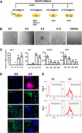
The next set of experiment was intended to show that their newly reprogrammed were of a cardiac nature. First, the cells clearly expressed heart-specific genes. Flk-1 and Isl-1 are genes that earmark cardiac progenitor cells, and by the eighth day of induction, the vast majority of cells expressed both these genes.


Generation of protein-induced cardiac progenitor cells by modified transcript proteins. (A): Strategy of protein-induced cardiac progenitor cell (piCPC) generation. (B): Cell colonies were initially observed around days 4–8 and could be passaged to many small colonies around day 12. Representative phase contrast images are shown. The control was untreated human dermal fibroblasts in vehicle medium after 8 days. Scale bars = 100 μm. (C): quantitative polymerase chain reaction analysis of cardiac progenitor genes Flk-1 and Isl-1 in piCPCs. Fibroblast markers Col1a2 and FSP1 were also detected (∗, p < .05; ∗∗, p < .01 vs. day 0 control; error bars indicate SD; n = 3). (D): Representative fluorescent images are shown with typical cardiac progenitor markers Flk-1 (red) and Isl-1 (green) and fibroblast markers ColI (green) and FSP-1 (S100A4) (green) before and after reprogramming at day 8. DAPI staining was performed to visualize nuclei (blue) and all images were merged. Scale bars, 100 μm. (E): Flow cytometry analysis demonstrated Flk-1 and Isl-1 expressions were increased from d0 to d8 separately. Abbreviations: bFGF, basic fibroblast growth factor; BMP4, bone morphogenetic protein 4; ColI, collagen I; d, day; DAPI, 4′,6-diamidino-2-phenylindole; FSP1, fibroblast-specific protein 1; mGHMT, modified Gata4/Hand2/Mef2c/Tbx5.

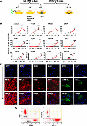
Second, cardiac cells can differentiate into three different cell types: heart muscle cells, blood vessels cells, and smooth muscle cells that surround the blood vessels. In mesoderm progenitors made from embryonic stem cells, inhibition of the Wnt signaling pathway can drive such cells to become heart muscle cells (see Chen, et al Nat Chem Biol 5:100–107; Willems E, et al Circ Res 109:360–364; Hudson J, et al Stem Cells Dev 21:1513–1523). However, Wang, Yu and company showed that treating the cells with a small molecule called IWR-1 that inhibits Wnt signaling drove their cells to differentiate into, not only heart muscle cells, but also endothelial (blood vessel) cells and smooth muscle cells when the cells were grown on gelatin coated dishes. When left to differentiate in culture, the cells beat synchronously and released calcium in a wave-like fashion that spread from one cell to another, suggesting that some cells were acting as pacemakers and setting the beat.


Protein-induced cardiac progenitor cells (piCPCs) differentiated into three cardiac lineages: cardiomyocytes, endothelial cells, and smooth muscle cells. (A): Schematic representation of the strategy to differentiate piCPCs in differentiation medium with IWR1 factor. (B): Quantitative data of mRNA expression of cardiac lineage marker genes (∗, p < .05; ∗∗, p < .01; and ∗∗∗, p < .001 vs. day 0 control; error bars indicate SD; n = 3). (C): Immunofluorescent staining for MHC, MYL2, CD31, CD34, smMHC, and αSMA. The combination of the four factors, GHMT, induces abundant MHC and Myl2, and some expression of CD31 and smMHC 28 days after transduction. Nuclei were counter stained with DAPI. Scale bars = 100 μm. (D): Flow cytometry analysis for cTnI, CD31, and smMHC. mGHMT plus IWR1 significantly enhances cTnI expression, and, to a lesser extent, CD31 and smMHC expression. Abbreviations: αSMA, α-smooth muscle actin; BMP4, bone morphogenetic protein 4; cTnI, cardiac troponin I; cTnT, cardiac troponin T; d, day; DAPI, 4′,6-diamidino-2-phenylindole; GHMT, Gata4/Hand2/Mef2c/Tbx5; mGHMT, modified GHMT; MHC, myosin heavy chain; MYL2, myosin light chain 2; smMHC, smooth muscle myosin heavy chain.

Then these cells were transplanted into the heart of mice that had suffered heart attacks. When compared to control hearts that received fluid, but no cells, the hearts of the animals that received protein-induced CPCs showed decreased scarring by 4 weeks after the transplantations. They also showed the growth of new heart muscle. A variety of staining experiments established that the engrafted protein-induced CPCs positive for heart muscle- and endothelial-specific cell markers. These experiments showed that transplantation of cardiac progenitor cells can not only help attenuate remodeling of the left ventricular after a heart attack, but that the protein-induced CPCs (piCPCs) can develop into cells of the cardiac lineage.

In vivo delivery of protein-induced cardiac progenitor cells improves cardiac function after myocardial infarction. (A): EF, FS, LVDd, and LVDs were analyzed by echocardiography (∗, p < .05; ∗∗, p < .01; ∗∗∗, p < .001 vs. relevant 1 week; all data are presented as mean ± SD, n = 8). (B): Transplanted cells were detected by magnetic resonance imaging 4 weeks after myocardial infarction (MI). Red arrow points to the signal loss due to SPIO-labeled cells. (C): Masson trichrome staining on heart sections 4 weeks after MI injection in sham, vehicle, and piCPC groups. Scale bar = 0.5 cm. (D): Immunofluorescent staining for cTnI (red), CD31 (red), and anti-dextran (SPIO, green) of heart sections after piCPCs were transplanted 4 weeks after MI. White arrows point to transplanted cells or colocalization of cTnI or CD31 with SPIO. Scale bars = 100 μm. Abbreviations: cTnI, cardiac troponin I; DAPI, 4′,6-diamidino-2-phenylindole; EF, ejection fraction; FS, fractional shortening; LVDd, left ventricular internal diameter at end-diastole; LVDs, left ventricular internal diameter at end-systole; piCPCs, protein-induced cardiac progenitor cell; SPIO, superparamagnetic iron oxide; W, week.

These are exciting results. It shows that direct reprogramming can occur without introducing genes into cells by means that can complicate the safety of the implanted cells. Also, because the cells are differentiated into progenitor cells, they still have the ability to proliferate and expand their numbers, which is essential for proper regeneration of a damaged tissue.

After a heart attack, the ventricle wall scars over and can become thin. However, piCPCs that have been directly reprogrammed from mature, adult cells can be used to replace dead heart muscle in a living animal.

Despite these exciting advances, further questions remain. For example, are the physiological properties of cells made from piCPCs similar enough to match the functional parameters of the heart into which they are inserting themselves? More work is necessary to answer that question. Functional equivalence is important, since a heart that does not function similarly from one end to the other can become arrhythmic, which is clinically dangerous. Further work is also required to precisely determine how well cells derived from piCPCs mature and coupling with neighboring cells. Therefore, larger animal studies and further studies in culture dishes will be necessary before this technique can come to the clinic. Nevertheless, this is a tremendous start to what will hopefully be a powerful and fruitful technique for healing damaged hearts.

Small Molecule Supercharges Human Cardiac Stem Cells


HO-1 or heme oxygenase is an enzyme that degrades heme groups to biliverdin, iron, and carbon monoxide. It is induced in cells in response to oxidative stress. Overexpression of HO-1 can make cells more resistant to oxidative stress. The highest levels of HO-1 are found in the spleen, where old red blood cells are sequestrated and destroyed.

Mesenchymal stem cells (MSCs) from bone marrow have been genetically engineered to overexpress HO-1 survive much better when implanted into the hearts of animals that have recently suffered a heart attack (Zeng B, et Al, Biomed Sci. 2010 Oct 7;17:80; Yang JJ et al Tohoku J Exp Med. 2012;226(3):231-41). Such cells also increase the density of blood vessels in infarcted tissue, and HO-1 has been postulated to increase blood vessel production (Jang YB et al Chin Med J (Engl). 2011 Feb;124(3):401-7).

These previous experiments show that HO-1 can increase the survival and therapeutic abilities of MSCs. Can increasing the levels of HO-1 do the same for other types of stem cells?

Stuart Atkinson at the Stem Cell Portal web site has highlighted a new paper that was published in the journal Stem Cells that has examined increasing the levels of HO-1 in Cardiac Stem Cells (CSCs).

CSCs are a resident stem cell in the heart that can be isolated from heart patients during heart surgeries. Animal studies and clinical trials have shown that implantation of CSCs soon after a heart attack can produce significant increases in heart function (Bearzi C, et al. Proc Natl Acad Sci U S A 2007;104:14068-14073; Bolli R, et al Lancet. 2011 Nov 26;378(9806):1847-57). Unfortunately, the success of this clinical has been called into questioned by some problems with the data reported in this paper. However, animal studies suggest that the effectiveness of CSCs is compromised by their limited ability to survive in the heart after a heart attack (Hong KU, et al. PLoS One 2014;9:e96725). Therefore, increasing the survival of CSCs might increase their therapeutic efficacy.

Atkinson notes that the compound cobalt protoporphyrin (CoPP) can induce the expression of higher levels of HO-1 and thereby increase the resistance of the cells to oxidative stress and augment cell survival. Therefore, Robert Bolli from the University of Louisville, Kentucky and his colleagues, in collaboration with researchers from the Albany Medical College have treated CSCs with CoPP and these tested their ability to heal the heart after a heart attack.

Bolli and others isolated human CSCs from patients undergoing CABG (cardiac artery bypass graft) surgery, and grew them in culture to beef up the numbers of cells. After a short time in culture, the CSCs were incubated with CoPP for 12 hours. Then Bolli and his team transplanted these human CSCs that were also labeled with green fluorescent protein (GFP) into the hearts of mice that had suffered rather massive heart attacks and had undergone 35 days of reperfusion. The GFP allowed Bolli and others to detect the presence of the implanted CSCs in the rodent heart tissue.

When these hearts of these mice were examined one and five weeks after CSC transplantation, the CoPP-treated CSCs showed substantially higher levels of survival in the mouse hearts. The other two groups of mice included those transplanted with non-pretreated CSCs, and mice treated with the culture medium used to grow the CSCs, and the pretreated CSCs survival significantly better than the non-pretreated CSCs.

CoPP pretreatment seems to augment cell survival, but do the surviving cells increase heart function? Bolli and others used echocardiogram to measure heart function, and echocardiographic assessment 5 weeks after CSC transplantation showed that the CoPP-preconditioned CSCs elicited greater improvement in remodeling of the left ventricle. Additionally, the hearts of the animals that received CoPP-pretreated CSCs showed improved movement of the walls of the heart during its pumping cycle, and better overall performance of the heart in general. Both pretreated and the non-pretreated CSCs, but not CSC culture growth medium shrank the amount of scar tissue in the heart and grew new heart tissue. However, The CoPP-pretreated CSCs were obviously superior to the non-pretreated CSCs at increasing the mass of heart muscle (see here for pictures).

These experiments might very well unravel a burning controversy surrounding CSCs. Bolli’s experiment show that can definitely grow new heart muscle. However, the bulk of the experiments with CSCs strongly suggest that these cells improve heart function by secreting pro-healing molecules without directly contributing to the regrowth of heart muscle. These papers probably observed the effects of CSCs that were transplanted into the heart, but did not survive very long. Bolli and his colleagues, on the other hand, were able to implant CSCs and survived for a much longer time in the hearts. Incidentally, Bolli and his team showed that the implanted CSCs expressed heart muscle-specific genes, which corroborated that these cells were differentiating into heart muscle cells, even though the proportion of cells that formed new heart muscle was relatively small.

In summary, CoPP pretreatment of cell seems to be feasible, safe, and effective as a means to improve CSC-based therapy. Even though It is likely that paracrine mechanisms are essential for CSC-based healing, the ability of CSCs to differentiate into heart muscle cells also seems to be an essential part of the means by which CSCs heal the heart after a heart attack. Thus more work is certainly warranted, but this is a fine start to what might be a simple, but effective way to increase the effectiveness of our own CSCs.