Microparticles and Local Control of Stem Cells


Using stem cells to grow three-dimensional structures, such as organs or damaged body parts, requires that scientists have the ability to control the growth and behavior of those cells. Also, adapting such a technology to an off-the-shelf kind of process so that it does not cost an arm and a leg is also important.

A research project by scientists from Atlanta, Georgia has used gelatin-based microparticles to deliver growth factors to specific areas of aggregates of stem cells that are differentiating. This localized delivery of growth factors provides spatial control of cell differentiation, which enables the creation of complex, three-dimensional tissues. The local delivery of growth factors also decreases the amount of growth factor used and, consequently, the cost of the procedure.

This particular microparticle technique was used on mouse embryonic stem cells and it proved to provide better control over the kinetics of cell differentiation since it delivered that promote cell differentiation or inhibit it.

Todd McDevitt, associate professor in the Wallace H. Coulter Department of Biomedical Engineering at Georgia Tech and Emory University, said, “By trapping these growth factors within microparticle materials first, we are concentrating the signal they provide to the stem cells. We can then put the microparticle materials physically inside the multicellular aggregate system that we use for differentiation for the stem cells. We have good evidence that this technique can work, and that we can use it to provide advantages in several areas.”

The differentiation of stem cells is largely controlled by external cues, including protein growth factors that direct cell proliferation, and differentiation that are available in the three-dimensional environment in which the cells live. In most experiments, stem cells are grown in liquid culture and growth factor is equally accessible to the growth factors. This makes the cultures quite homogeneous. But delivering the growth factors via microparticles gives better control of the spatial and temporal presentation of these growth factors to the stem cells. This gives scientists the means to make heterogeneous structures from stem cell cultures.

When embryonic stem cells grow in culture, they tend to clump together. When the growth medium is withdrawn or if growth factors that induce differentiation are added, the cells form an “embryoid body” that is stuff with cells differentiating into all kinds of cell types. When McDevitt and his co-workers added microparticles with the growth factors BMP4 (bone morphogen protein 4) or Noggin (which inhibits BMP4 signaling), they centrifuged the cells and found that the microparticles found their way into the interior of the embryoid bodies.

When they examined the embryoid bodies, with confocal microscopy they found that BMP4 directed the cells to make mesodermal and endodermal derived cell types. However, because the microparticles were in direct contact with the cells, they needed 12 times less growth factor than was required by solution-based techniques.

“One of the major , in a practical sense, is that we are using much less growth factor,” said McDevitt. “From a bioprocessing standpoint, a lot of the cost involved in making stem cell products is related to the cost of the molecules that must be added to make the stem cells differentiate.”

Beyond more focuses signaling, the microparticles also provided localized control that was not available through other techniques. It allowed researchers to create spatial differences in the aggregates and this is an important possible first step toward forming more complex structures with different tissue types such as vascularization and stromal cells.

“To build tissues, we need to be able to take stem cells and use them to make many cell types which are grouped together in particular spatial patterns,” explained Andres M. Bratt-Leal, the paper’s first author and a former graduate student in McDevitt’s lab. “This spatial patterning is what gives the ability to perform higher order functions.”

Once the stem cell aggregates were made and treated with growth factor-endowed microparticles, McDevitt and his colleagues saw spheres of cells with differentiating cells.

“We can see the microparticles had effects on one population that were different from the population that didn’t have the particles,” said McDevitt. “This may allow us to emulate aspects of how development occurs. We can ask questions about how tissues are naturally patterned. With this material incorporation we have the ability to better control the environment in which these cells develop.”

The microparticles could provide better control over the kinetics of cell differentiation; slowing it down with molecules that antagonize differentiation or speed up with other molecules that promote stem cell differentiation.

Despite the fact that McDevitt and his colleagues used mouse embryonic stem cells in this paper, he and his co-workers are already testing this technology on human embryonic stem cells, and the results have been comparable.

“Our findings will provide a significant new tool for tissue engineering, bioprocessing of stem cells and for better studying early development processes such as axis formation in embryos,” said Bratt-Leal. “During development, particular tissues are formed by gradients of signaling molecules. We can now better mimic these signal gradients using our system.”
Mouse Model of Huntington’s Disease Shows Replacement of Lost Neurons by Endogenous Stem Cell Populations


Huntington’s disease (HD) is a debilitating and invariably fatal disease that results from mutations in the IT15 gene. IT151 stands for “interesting transcript 15,” but it is more commonly referred to as the “huntingtin” gene. Mutations in the front of the gene (exon 1 for those who are interested) expand a run of CAG codons, and these mutations are probably the result of DNA polymerase slippage. Because CAG codons encode the amino acid glutamine, the mutant proteins contain long polyglutamine repeats and these repeats tend to clump inside neurons.

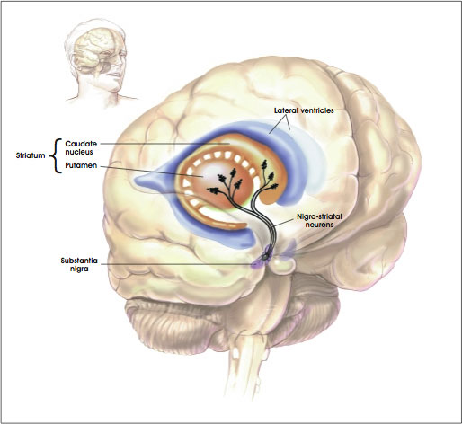
These protein aggregates form in neurons of the “striatum.” The striatum is a region of the brain that is also called the striate nucleus of the striate body. The striatum receives its name from the fact that it is organized in striped layers of gray and white matter. The striate nucleus is part of the cerebrum or forebrain.

Striatum

Mutant Huntington (Htt) protein has a toxic that causes cell death by means of unknown mechanisms. Clinically, the most obvious symptoms of HD involve involuntary movements of the arms, legs, and face. But the severe cognitive and personality changes are the most devastating to HD patients and most troubling for their caregivers.

Researchers are using animal models of HD to study the disease pathogenesis, to elucidate areas of the brain involved in structural and functional decline, and to evaluate potential therapeutic interventions. These animal models include injecting toxins into the brain to kill off those populations of neurons that typically die in HD patients, and transgenic models in which animals are bred with either extra mutant copies of the Htt gene or a pair of copies of the mutant Htt gene that have replaced the original, normal copies. All of these model systems have limitations, but they are all useful in some way for assessing the pathology of HD.

This long introduction leads us to new data from the laboratory of Steve Goldman, the co-director of the University of Rochester Medical Center’s Center for Translational Medicine. Goldman and his colleagues triggered the production of new neurons in mice that had a rodent form of HD. These new neurons successfully integrated into the brain’s existing neural networks and dramatically extended the survival of the mice.

“This study demonstrates the feasibility of a completely new concept to treat Huntington’s disease, by recruiting the brain’s endogenous neural stem cells to regenerate cell lost the disease,” said Goldman.

One of the types of neurons most commonly affected in HD patients is the medium spinal neuron, which is critical to motor control. Goldman banked on findings from previous studies in his laboratory on canaries. Songbirds such as canaries have the ability to lay down new neurons in the adult brain when mating season comes. The male birds, in response to a flush of male sex hormones,, grow a gaggle of new neurons in the vocal control centers of the brain, and this provides the bird the means to sing specific songs in order to attract mates. This event is known as adult neurogenesis, and Goldman and Fernando Nottebohm of the Rockefeller University discovered this phenomenon in the early 1980s.

“Our work with canaries essentially provided us with the information we needed to understand how to add new neurons to adult brain tissue,” said Goldman. Once we mastered how this happened in birds, we set about how to replicate the process in the adult mammalian brain.”

Humans possess the ability to make new neurons, but Goldman’s lab demonstrated in the 1990s that a font of neuronal precursor cells exist in the lining of the ventricles (these are structures at the very center of the brain and spinal cord that are filled with cerebrospinal fluid). In early development, these cells are actively producing neurons.

Shortly after birth, the neural stem cells stop generating neurons and produce support cells called glia. Some parts of the human brain continue to produce neurons into adulthood, the most prominent example is the hippocampus, where memories are formed and stored. However, the striatum, new neuron production is switched off in adulthood.

Goldman sought to switch neuron production back on in the striatum. He tested a cadre of growth factors that would switch the neural stem cells of the striatum (a region that is ravaged by HD) from producing new glia to producing new neurons. Goldman, however, had some help from his recent work in canaries. Namely that once mating season was upon the birds, targeted expression of brain-derived neurotrophic factor (BDNF) flared up in the vocal centers of the brain, where many new neurons were being produced.

Goldman used genetically engineered viruses to express BDNF and another protein called “Noggin” in the striatum. Goldman and others found that a single intraventricular injection of the adenoviruses expressing BDNF and Noggin triggered the sustained recruitment of new neurons in both normal of R6/2 (HD) mice. These treated mice also showed that the newly formed neurons were recruited to form new medium spiny neurons; the ones destroyed in HD. These new neurons also matured and achieved circuit integration.

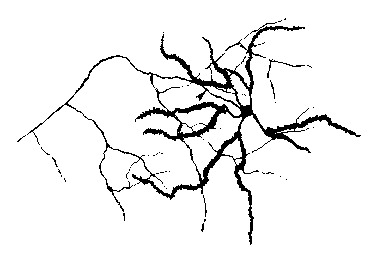
Medium Spinal Neuron

Also the treated mice showed delayed deterioration of motor function and substantially increased survival.

When the same experiments were conducted in squirrel monkeys, there was a similar addition of new striatal neurons.

Thus, induced neuronal addition may therefore represent a promising avenue for decreasing the ravages of HD and increasing cognitive ability.