Mesenchymal Stem Cells Secreting Brain-Derived Neurotrophic Factor Delay The Effects of Huntington’s Disease in Mice


Jan Nolta and her colleagues at the Stem Cell Program and Institute for Regenerative Cures at UC Davis have published a remarkable paper in the journal Molecular Therapy regarding Huntington’s disease and a potential stem cell-based strategy to delay the ravages of this disease.

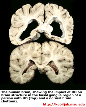
Huntington’s disease (HD) is an inherited neurodegenerative disease. It is inherited as an autosomal dominant disease, which means that someone need only inherit one copy of the disease-causing allele of the HTT gene to have this disease. HD is characterized by progressive cell death in the brain, particularly in a portion of the brain known as the striatum and by widespread brain atrophy.

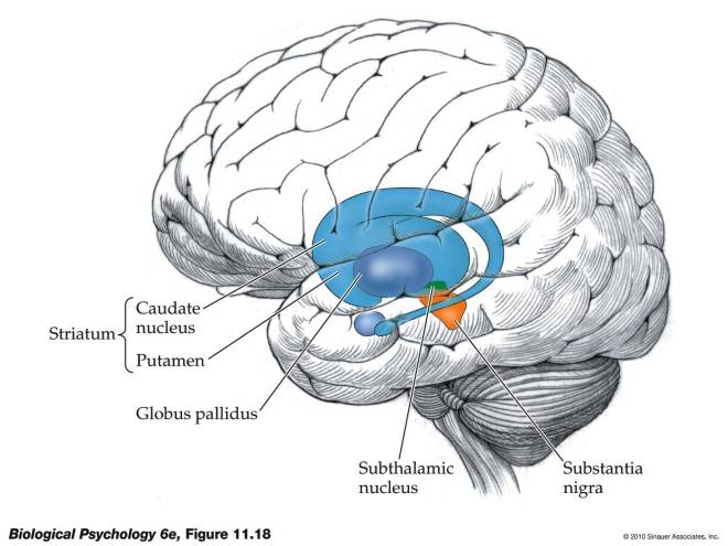
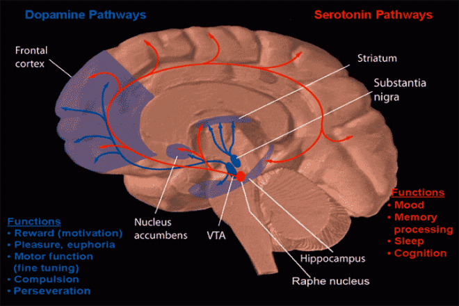
The portion of the brain known as the striatum lies underneath the surface of the forebrain (subcortical) and it receives neural inputs from the cerebral cortex and is the primary source of neural inputs to the basal ganglia system. The basal ganglia system (BGS) is located underneath the surface of the brain but even deeper within the cerebral hemispheres. The BGS is part of the corpus striatum, it consists of the subthalamic nucleus and the substantia nigra. The BGS help with voluntary motor control, procedural learning relating to routine behaviors. otherwise known as “habits,” eye movements, and cognitive, and emotional functions. The ventral striatum is very important in addiction because it is the reward center on consists of the nucleus accumbens, olfactory tubercle, and islands of Calleja.
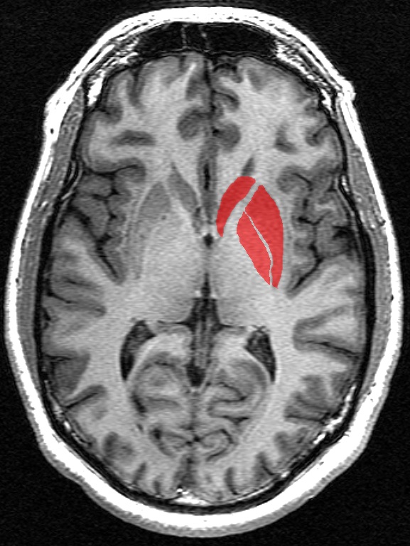
This is a transverse section of the striatum from a structural MR image. The striatum, in red, includes the caudate nucleus (top), the putamen (right), and, when including the term 'corpus' striatum, the globus pallidus (lower left).
This is a transverse section of the striatum from a structural MR image. The striatum, in red, includes the caudate nucleus (top), the putamen (right), and, when including the term ‘corpus’ striatum, the globus pallidus (lower left).

HD takes its largest toll on the striatum, which affects voluntary movement, routine behaviors, and personality. Disturbances of both involuntary and voluntary movements occur in individuals with HD. Chorea, an involuntary movement disorder consisting of nonrepetitive, non-periodic jerking of limbs, face, or trunk, is the major sign of the disease. Chorea is present in more than 90% of individuals, increasing during the first ten years. The choreic movements are continuously present during waking hours, cannot be suppressed voluntarily, and are worsened by stress. HD patients show impaired voluntary motor function early on and show a clumsiness in common daily activities.

With advancing disease duration, other involuntary movements such as slowness of movement (bradykinesia), rigidity, and involuntary muscle contractions that cause repetitive or twisting movements (dystonia) occur.  Eye movement becomes progressively worse. So-called “gaze fixation” is observed in ~75% of symptomatic individuals.  Unclear speech occurs early and Swallowing difficulties occur later.

Animal models of HD used in the past have injected molecules into the brain that kill off striatal cells and mimic at least some of the characteristics of HD in laboratory animals. Unfortunately, such a model system is fat too clean, since implanted cells tend to survive perfectly well. However the brains of HD patients are like unto a toxic waste dumps and implanted cells are quickly killed off. Therefore, a better animal model system was required, and it came in the form of R6/2 and YAC128 mice. R6/2 mice have a part of the human HTT gene that has 150 CAG triplets, and show the characteristic cell death in the striatum and behavioral deficits. The only problem with this mouse strain is that the neurodegenerative decline is very rapid rather than slow and progressive. YAC128 mice have a full-length copy of the HTT gene and show a slower, more progressive neurological decline that more closely approximates the human clinical condition.

In this paper from the Nolta laboratory, they used a growth factor that is known to decrease precipitously in HD brains; a growth factor called Brain-Derived Neurotrophic Factor (BDNF). BDNF is known to mediate the survival and function of striatal neurons and the reduction of BDNF in the brains of HD patients correlates with the onset of symptoms and the greater the reduction in BDNF, the greater the severity of the disease (see Her LS & Goldstein LS, J Neurosci 2008; 28, 13662-13676).

However injecting BDNF into the brain is problematic, since the protein has a very short half-life. Delivering the growth factor by means of genetically engineered viruses shows promise, but most of the viral vectors used in such experiments are recognized by the immune system as foreign invaders. Therefore, Nolta and her colleagues decided to genetically engineer mesenchymal stem cells (MSCs) to overexpress BDNF and implant these cells into the brains of R6/2 and YAC128 mice.

MSCs have an added advantage over viral vectors: these cells migrate to damaged areas where they can exert their healing properties (see Olson SD et al., Mol Neurobiol 45; 2012: 87-98).

Nolta and her coworkers actually tested human MSCs in HD model mice. After completing all the necessary control experiments to ensure that their isolated and engineered MSCs were secreting BDNF, Nolta and others implanted them into the brains of R6/2 and YAC128 mice.

HD mice show greater anxiety, which is manifested in a so-called “open field assay” by not remaining the center of the field. The control HD mice did not stay long in the center of the open field, but the normal mice did. The MSC-BDNF-implanted mice spend far more time in the center of the field. Mind you, not as much as wild-type mice, but significantly more than their HD counterparts.

Next the volume of the striata of these mice were determined and compared to the normal mice. While all the HD mice showed shrinking of the striatum, the MSC-NDNF-implanted YAC128 mice show significantly less shrinking of the striatum. Then the degree of neurogenesis (formation of new neurons) was measured in normal, HD, HD + implanted MSCs, and HS + MSC-BDNF mice. This experiment measures the degree of healing that is occurring in the brain. The brain from HD + MSC and HD + MSC-BDNF mice showed significantly more new brain cell growth. This is probably the reason for the delayed onset of symptoms and the delayed shrinking of the striatum.

Finally, Nolta and others measured the lifespans of the R6/2 mice and compared them with R6/2 mice that had been implanted with MSCs-BDNF. Animals transplanted with the MSCs that made the most BDNF lived 15% longer than the nontreated R6/2 mice.

MSCs have been shown in several experiments to promote neuronal growth, decrease cell death and decrease inflammation through the secretion of trophic factors. MSCs can modify the toxic environment that is part of the brain of an HD patient and help damaged tissue out by inducing neural regeneration and protection (see Crigler L, et al., Experimental neurology, 198; 2–6, 54-64; Kassis I, et al., Archives of Neurology 65; 2008: 753-761).

The downside of using MSCs that they will only survive in the brain for a few months. However, several studies have shown that the benefits of modified MSC implantation persist after the MSCs are gone, since the neural reconstruction wrought by the secreted BDNF stay after the MSCs have died off (see Arregui L, et al., Cell Mol Neurobiol 31; 2011: 1229-1243 and many others).

At best this treatment would delay the ravages of HD, but delaying this disease might very well be the first step towards a cure. Hopefully, clinical trials will not be fat behind.
Society for Neuroscience Meeting


I am in Washington DC at the Society for Neuroscience 2014 meeting. There is some incredible science here. Let me just share a few of the things I saw today:

The Thompson laboratory from UC Irvine (my alma mater – go anteaters!) made a model system for the blood-brain barrier from induced pluripotent stem cells. These scientists made iPSCs that had similar genetic defects to those observed in patients with Huntington’s disease. These iPSCs were then differentiated into blood-brain barrier cells and showed that these cells showed defects similar to those seen in patients with Huntington’s disease. The barrier leaked, which makes this a good model to study blood brain barrier defects in patients with neurological diseases.

Another poster described the use of vesicles from human fat-based stem cells to treat laboratory animals with a type of Huntington’s disease. These vesicles attenuated Huntington’s disease pathology and delayed its onset.

There were several other brilliant posters, and tomorrow, there will be even more. I will blog about those as time permits.

Nerve Growth Factor-Secreting Mesenchymal Stem Cells To Treat Huntington’s Disease


Vicki Wheelock at the UC Davis Medical Center has registered clinical trial number NCT01937923, which is otherwise known as “PRE-CELL.” This clinical trial will use various imaging techniques, laboratory tests, and clinical evaluations of Huntington’s disease (HD) patients to map the disease progression over 12-18 months. This trial will then hopefully identify candidates for a new trial in which these patients will be implanted with mesenchymal stem cells that secrete nerve growth factors. This represents one of the first clinical trials to examine the use of mesenchymal stem cells in the treatment of HD

The rationale for this study comes from a 2012 study in mice. Ofer Sadan, Eldad Melamed, and Daniel Offen from the Rabin Medical Center in Tel Aviv University, Israel, used R6/2 mice to test the efficacy of nerve growth factor-secreting mesenchymal stem cells isolated from bone marrow . In this paper, Sadan and others isolated mesenchymal stem cells from the bone marrow of healthy human volunteers and mice and then cultured them in special growth media that induces these cells to secrete special nerve growth factors. These so-called NTF+ cells were then transplanted into the striatum of R6/2 mice.

R6/2 mice express part of the human HTT gene; specifically the part that causes HD. Since HD is an inherited disease, there is a specific gene responsible for the vast majority of HD cases, and that gene is the human HTT gene, which encodes the Huntington protein. The function of the Huntington protein is uncertain, but it is found at high levels in neurons, even though it is found in other tissues as well, and dysfunctional Huntington protein affects neuron health.

Huntingtin Function

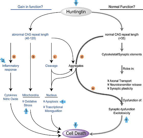
The HTT gene in HD patients contains the insertion of extra copies of the CAG triplet. The more CAG triplets are inserted into the HTT gene, the more severe the HD caused by the mutation. The hitch is that normal copies of the HTT gene has multiple copies of this CAG repeat. CAG encodes the amino acid glutamine, and Huntington contains a stretch of glutamine residues that seem to allow the protein to interact with other proteins found in neurons. When this glutamine stretch becomes too long, the protein is toxic and it begins to kill the cells. How long is too long? Research has pretty clearly shown that people whose HTT genes contain less than 28 CAG virtually never develop HD. People with between 28–35 CAG repeats, are usually unaffected, but their children are at increased risk of developing HD. People whose HTT genes contain 36–40 CAG repeats may or may not show HD symptoms, and those who have over 40 copies almost always are afflicted with HD.

hunt_gene_big

Now, back to R6/2 mice. These animals contain a part of the human HTT gene that has 150 CAG triplets. These mice show the characteristic cell death in the striatum and have behavioral deficits. In short R6/2 mice are pretty good model systems to study HD.

Sadan and others implanted MSCs that had been conditioned in culture to express high levels of nerve growth factors. Then these cells were transplanted into the striatum of R6/2 mice. R6/2 mice were also injected with buffer as a control.

The results showed that injections of NTF+ MSCs before the onset of symptoms did little good. The mice still showed cell death in the brains and behavioral deficits. However, NTF+ MSCs injected later (6.5 weeks), resulted in temporary improvement in the ability of the R6/2 mice to move and these cells also extended their life span. These results were published in the journal PLoS Currents (2012 Jul 10;4:e4f7f6dc013d4e).

Other work, also by Sadan and others, showed that injected MSCs tended to migrate to the damaged areas. When the injected cells were labeled with iron particles, they could be robustly observed with MRIs, and MRIs clearly showed that the injected cells migrated to the damaged areas in the brain (Stem Cells 2008; 26(10):2542-51). Another paper by Sadan and others also demonstrated that the striatum of NTF+ MSC-injected mice show less cell death than control mice (Sadan, et al. Exp Neurol. 2012; 234(2): 417-27). Other workers have also shown that implanted MSCs can provide improve symptoms in R6/2 mice and that they primary means by which they do this is by the secretion of nerve growth factors (Lee ST, et al. Ann Neurol 2009; 66(5): 671-81).

Thus, there is ample reason to suspect the PRECELL trial may lead to a stem cell-based clinical trial that will yield valuable clinical information. The animal data shows definite value in using preconditioned MSCs as a treatment for HD, and if the proper patients are identified by the PRE-CELL trials, then hopefully it will lead to a “CELL” trial in which HD patients are treated with NTF+ MSCs.

Mind you, this treatment will only delay HD at best and buy them time. Such treatments will not cure them. The NTF+ MSCs survive for a finite period of time in the hostile environment of the striatum of the HD patient, and the relief they will provide will be temporary. MSCs do not differentiate into neurons in this case, and they do not replace dead neurons, but they only help spare living neurons from suffering the same fate.

Huntington disease striatum

There is an MSC cell line that does make neurons, and if this cell line were used in combination with NTF+ MSCs, then perhaps neural replacement could be a possibility.  Also neural precursor cells could be used in combination with NTF+ MSCs to increase their survival.  Even then, as long as diseased neurons are producing toxic products, until gene therapy is perfected to the point that the actual genetic lesion in the striatal neurons is fixed, the deterioration of the striatum is inevitable. However, treatments like this could, potentially, delay this deterioration. This clinical trial should give us more information on exactly that question.

Two more points are worth mentioning.  When fetal striatal grafts were implanted into the brains of HD patients, the grafts underwent disease-like degeneration, and actually made the patients worse (see Cicchetti et al. PNAS 2009; 106(30): 12483-8 and Cicchetti F, et al. Brain 2011; 134(pt 3): 641-52).  Straight fetal implants do not seem to work.  Please let’s put the kibosh on these gruesome experiments.  Secondly, when neuronal precursor cells differentiated from human embryonic stem cells were implanted into HD rodents, the implanted cells formed some neurons and improved behavior to some extent, but non-neuronal differentiation remained a problem (Song J, et al., Neurosci Lett 2007; 423(1): 58-61).  Having non-brain cells in your brain is a significant safety problem.  Thus, embryonic stem cell-derived neuronal precursor cells do not seem to be the best bet to date either.  So, this present clinical trial seems to be making the most of what is presently safely available.

Transplantation of Neurons Made from Bioreactor-Grown Human Neural Precursor Cells Restores Brains of Rats With Huntington’s Disease


Huntington’s disease (HD) is an inherited disorder of the central nervous system characterized by progressive dementia, involuntary movements, and emotional deterioration.  The brain is affected in patients with HD and the part of the brain that takes the biggest beating is the “neostriatum.”

The term “neostriatum” is almost certainly not a word that you hear terribly often in conversation.  Therefore I will try to explain what it is.  The outer layers of the brain are known as the cerebral cortex and they are composed of so-called “grey matter.”  The cerebral cortex consists of grey matter because it is loaded with cells known as neurons.  Beneath the cortical layer of the brain, lies a whole host of extensions of these neurons that reside in the cerebral cortex.  These extensions are called “axons,” and beneath the cerebral cortex lies white matter, which consists, largely, of bundles of axons.  Think of the neurons are plugs and the axons as extension cords.  The neurons are plugged into each other by means of extension cords that extend from the cerebral cortex.

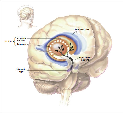
Now the cerebral cortex is not the only game in town.  There are also clusters of neurons that lie beneath the cerebral cortex called “nuclei.”  One of these nuclei beneath the cerebral cortex plays an extremely important role in voluntary motion, and this structure is called the “basal ganglia.”  Here’s a picture to make things a little clearer:

Basal ganglia

As you can see in the figure, the striatum consists of two structures: the putamen and the caudate nucleus.  The striatum or striate nucleus receives neural inputs from the cerebral cortex and inputs this neural information to the basal ganglia.

From a functional perspective, the striatum helps coordinate motivation with body movement.  It facilitates and balances motivation with both higher-level and lower-level functions – for example, inhibiting one’s behavior in a complex social interaction and fine-motor functions involved in inhibiting small voluntary movement.

Some of the neural outputs from the striatum are excitatory – they stimulate other neurons.  Other signals are inhibitory – they prevent the neurons to which they are connected from becoming stimulated.  Inhibitory neurons release a chemical called “GABA.”  These GABA-using neurons are very important for the work of the striatum, and it is exactly these neurons that die off at the greatest rate in patients with HD.  Therefore, treatments for patients with HD have focused on replacing or protecting these GABA-using neurons.

Experimentally, you can induce an HD-like disease in rodents if you inject a chemical into their brains called quinolinic acid.  Quinolinic acid causes many of the GABA-using neurons in the striatum to pack up and die, and for this reason, this chemical is heavily used in the laboratories of scientists who study HD and HD treatments.

In a paper by Marcus McLeod and others who did their work in the laboratory of Ivar Mendez, who was at Dalhousie University in Nova Scotia, Canada, but has since moved to the University of Saskatchewan, GABA-using neurons were made from cultured human neural precursor cells (hNPCs) and then implanted into the brains of rats that had been injected with quinolinic acid.  The results were spectacularly successful.  This work was published in the journal Cell Transplantation.

A definite twist with this particular paper is the way the GABA-using neurons were grown in culture; they were grown in bioreactors.  Bioreactors are devices that support biological cells, processes, or organisms.  They keep the environment of the cells constant, and provide a far superior way to grow cells or tissues in the laboratory.  McLeod and his colleagues used human neural progenitor cells and grew them to large numbers in bioreactors.  These expanded hNPCs were then differentiated them into GABA-using neurons and then injected into the brains of rats who has been treated with quinolinic acid.

The rat model allows the scientist to inject only one side of the brain with quinolinic acid.  This leaves the intact side of the brain as a control tissue that can be compared with the injected one.  The injected rats showed the characteristic death of the GABA-using neurons and the behavioral features that result from the death of these neurons.  Such animals do not walk normally when they are led through a cylinder, and they have trouble finding their way through a maze.  The animals that received the transplantations of the GABA-using neurons, however, performed almost as well in these tests as normal rats; not quite as well, but almost as well.  The rats treated with quinolinic acid did quite poorly, as expected.

Upon post-mortem examination, the rats transplanted with GABA-using neurons shows a host of new GABA-using neurons in their striatums.  These cells also underwent further maturation after transplantation, and they also made connections with other neurons.

Now this paper shows that the injected cells not only survived the transplantations, but they also matured, made connections and promoted recovery of many of the behavioral symptoms of HD.  This procedure certainly has promise.

Having said all that, there are two caveats to these experiments.  The rodent model is a good model as far as it goes, but it seems clear that the actual human disease turns the environment of the brain into a very inhospitable place.  Transplanted cells in the case of human HD patients do not usually survive terribly well.  It seems to me that treatments like this must be coupled with other treatments that seek to improve the actual cerebral environment.  The second caveat to this experiment is that the neural progenitor cells were taken 10-week-old from aborted fetuses.  While these scientists did not perform the abortions that ended the lives of these babies, it is more than little troubling that this research was done using the corpses of those babies whose lives were prematurely ended.

Nevertheless, despite these caveats, this paper represents a definite advance in the regenerative strategies available to treat HD patients.

Mouse Model of Huntington’s Disease Shows Replacement of Lost Neurons by Endogenous Stem Cell Populations


Huntington’s disease (HD) is a debilitating and invariably fatal disease that results from mutations in the IT15 gene. IT151 stands for “interesting transcript 15,” but it is more commonly referred to as the “huntingtin” gene. Mutations in the front of the gene (exon 1 for those who are interested) expand a run of CAG codons, and these mutations are probably the result of DNA polymerase slippage. Because CAG codons encode the amino acid glutamine, the mutant proteins contain long polyglutamine repeats and these repeats tend to clump inside neurons.

These protein aggregates form in neurons of the “striatum.” The striatum is a region of the brain that is also called the striate nucleus of the striate body. The striatum receives its name from the fact that it is organized in striped layers of gray and white matter. The striate nucleus is part of the cerebrum or forebrain.

Striatum

Mutant Huntington (Htt) protein has a toxic that causes cell death by means of unknown mechanisms. Clinically, the most obvious symptoms of HD involve involuntary movements of the arms, legs, and face. But the severe cognitive and personality changes are the most devastating to HD patients and most troubling for their caregivers.

Researchers are using animal models of HD to study the disease pathogenesis, to elucidate areas of the brain involved in structural and functional decline, and to evaluate potential therapeutic interventions. These animal models include injecting toxins into the brain to kill off those populations of neurons that typically die in HD patients, and transgenic models in which animals are bred with either extra mutant copies of the Htt gene or a pair of copies of the mutant Htt gene that have replaced the original, normal copies. All of these model systems have limitations, but they are all useful in some way for assessing the pathology of HD.

This long introduction leads us to new data from the laboratory of Steve Goldman, the co-director of the University of Rochester Medical Center’s Center for Translational Medicine. Goldman and his colleagues triggered the production of new neurons in mice that had a rodent form of HD. These new neurons successfully integrated into the brain’s existing neural networks and dramatically extended the survival of the mice.

“This study demonstrates the feasibility of a completely new concept to treat Huntington’s disease, by recruiting the brain’s endogenous neural stem cells to regenerate cell lost the disease,” said Goldman.

One of the types of neurons most commonly affected in HD patients is the medium spinal neuron, which is critical to motor control. Goldman banked on findings from previous studies in his laboratory on canaries. Songbirds such as canaries have the ability to lay down new neurons in the adult brain when mating season comes. The male birds, in response to a flush of male sex hormones,, grow a gaggle of new neurons in the vocal control centers of the brain, and this provides the bird the means to sing specific songs in order to attract mates. This event is known as adult neurogenesis, and Goldman and Fernando Nottebohm of the Rockefeller University discovered this phenomenon in the early 1980s.

“Our work with canaries essentially provided us with the information we needed to understand how to add new neurons to adult brain tissue,” said Goldman. Once we mastered how this happened in birds, we set about how to replicate the process in the adult mammalian brain.”

Humans possess the ability to make new neurons, but Goldman’s lab demonstrated in the 1990s that a font of neuronal precursor cells exist in the lining of the ventricles (these are structures at the very center of the brain and spinal cord that are filled with cerebrospinal fluid). In early development, these cells are actively producing neurons.

Shortly after birth, the neural stem cells stop generating neurons and produce support cells called glia. Some parts of the human brain continue to produce neurons into adulthood, the most prominent example is the hippocampus, where memories are formed and stored. However, the striatum, new neuron production is switched off in adulthood.

Goldman sought to switch neuron production back on in the striatum. He tested a cadre of growth factors that would switch the neural stem cells of the striatum (a region that is ravaged by HD) from producing new glia to producing new neurons. Goldman, however, had some help from his recent work in canaries. Namely that once mating season was upon the birds, targeted expression of brain-derived neurotrophic factor (BDNF) flared up in the vocal centers of the brain, where many new neurons were being produced.

Goldman used genetically engineered viruses to express BDNF and another protein called “Noggin” in the striatum. Goldman and others found that a single intraventricular injection of the adenoviruses expressing BDNF and Noggin triggered the sustained recruitment of new neurons in both normal of R6/2 (HD) mice. These treated mice also showed that the newly formed neurons were recruited to form new medium spiny neurons; the ones destroyed in HD. These new neurons also matured and achieved circuit integration.

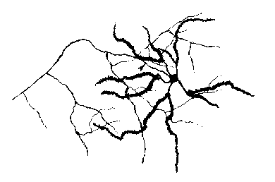
Medium Spinal Neuron

Also the treated mice showed delayed deterioration of motor function and substantially increased survival.

When the same experiments were conducted in squirrel monkeys, there was a similar addition of new striatal neurons.

Thus, induced neuronal addition may therefore represent a promising avenue for decreasing the ravages of HD and increasing cognitive ability.

Huntington Disease therapy fails


Huntington Disease in a fatal, inherited disease that causes degeneration of the central nervous system. It clinically manifests itself as severe movement and cognitive problems, and the patient gradually loses control of their body in a slow, painful slide to death that is difficult to watch.

A treatment for Huntington Disease that generated a far amount of hope in the 1990s was to transplant healthy neural tissue from fetuses. In particular, the striatum — the brain region most severely affected in Huntington disease was replaced by fetal neural tissue. Unfortunately, this tissue came from babies who were killed by selective abortion. Unfortunately, clinical follow-up of this approach has shown that technique does not work (Cicchetti, F. et al. Proc. Natl Acad. Sci. USA advance online publication doi:10.1073/pnas.0904239106 (2009).

Source: http://www.glaucoma.org/uploads/eye-anatomy-2012_650.gif

University of South Florida neurosurgeon Thomas Freeman and his colleagues have conducted a post-mortem analysis of the brains of three people with Huntington’s disease who received fetal striatal-tissue transplants a decade before they died. The results were rather clear – instead of slowing or stopping the progression of the disease, the grafts degenerated even more severely than the patients’ own tissue.

Early results for this procedure generated some hope.  Animal experiment in rats (Kendall, A. L. et al. Nature Med. 4, 727-729 (1998) and non-human primates (Isacson, O. et al. Nature Med. 1, 1189-1194 (1995) showed that transplanted tissue could replace lost striatal neurons and improve behavioural symptoms.  Also, early clinical results tended to support the efficacy of this technique.  Patients who had received these striatial grafts showed modest improvements and autopsies showed that the grafts of fetal neural tissue had survived and integrated into the brain (see Hauser, R. A. et al. Neurology 58, 687-695 (2002), and Bachoud-Lévi, A.-C. et al. Lancet Neurol. 5, 303-309).

Unfortunately, this more recent examination shows that the animal models were deceptive.  The animals were treated with chemicals that destroyed the striatum but left the rest of the brain intact.  In the case of human patients, the entire brain is diseased, and dying neurons release extensive amounts of neurotransmitters that kill neurons by overdosing them on these neurotransmitters.  The transplanted tissue is killed by neurotransmitter overdose.

This has implications for stem cell treatments of Huntington Disease.  Transplanted stem cells or neural progenitor cells will be subjected to this same cocktail of death.  Therefore another strategy is needed.

Fortunately, some cells can surround transplanted cells and protect them from death by neurotransmitter overdose.  For example, co-transplantation of testicular Sertoli cells with neural grafts not only produce an area of localized immuno-suppression (due to local secretion of GDNF by the Sertoli cells) but they can push stem cells into dopaminergic neurons, which are killed in Parkinson Disease (see Halberstadt C, Emerich DF, Gores P. Expert Opin Biol Ther. 4, (2004): 813-25).  Therefore, some new thinking on this front might provide a new treatment scheme.