New Autoimmune Treatment Removes Rogue Immune Cells Without Suppressing the Immune System


New preclinical experiments by scientists at the University of Pennsylvania have established that genetically engineered T-cells can drive severe autoimmune diseases into remission without suppressing the patient’s immune system. If the principles applied in this study also prove to be true in human patients, they can potentially revolutionize the treatment of autoimmune diseases.

Autoimmune diseases result when your immune system recognizes your own cells and tissues as foreign and mounts and immune response against them. Autoimmune diseases like systemic lupus erythematosus (also known as “lupus”), rheumatoid arthritis, scleroderma, multiple sclerosis, celiac disease, Sjögren’s syndrome, polymyalgia rheumatic, or ankylosing spondylitis can deeply affect the health of an individual and can also cause large amounts of tissue damage.

Treatment of autoimmune diseases usually requires high doses of drugs that suppress the immune system, such as corticosteroids, or various types of biological agents that also cause a host of undesirable side effects.

This new study, however, by scientists from the Perelman School of Medicine at the University of Pennsylvania have adapted an already-existing technology to remove the subset of antibody-making cells that cause the autoimmune disease. This strategy removes the rogue immune cells without harming the rest of the immune system.

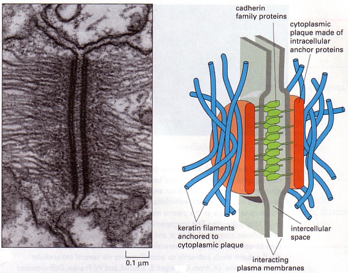
In these experiments, the University of Pennsylvania team examined an autoimmune disease called pemphigus vulgaris or PV. PV results when the immune system recognizes a protein called desmoglein-3 (Dsg3) as foreign and attacks it. Dsg3 helps form attachment sites called “desmosomes” that normally adhere skin cells together to form tight, tough sheets. Desmosomes are also found between epithelial cells, myocardial cells, and other cell types.

desmosomes

Current therapies for autoimmune diseases like PV use drugs like prednisone and rituximab, which suppress large parts of the immune system. Consequently, prednisone and rituximab can leave patients vulnerable to potentially fatal opportunistic infections and cancers.

To treat PV, University of Pennsylvania researcher Aimee Payne and her colleagues used a mouse version of PV that is fatal in mice. Their experimental treatment, however, successfully treated this otherwise fatal autoimmune disease without causing any unintended side effects, which might harm healthy tissue. The results from these experiments were published in the journal Science.

“This is a powerful strategy for targeting just autoimmune cells and sparing the good immune cells that protect us from infection,” said Dr Payne, who serves as the Albert M. Kligman Associate Professor of Dermatology at the Perelman School of Medicine.

In collaboration with Dr. Michael Milone, assistant professor of Pathology and Laboratory Medicine, Payne and her colleagues adapted the Chimeric Antigen Receptor T-Cell (CART-Cell) technology that is being successfully used to experimentally treat malignant cells in certain leukemias and lymphomas. “Our study effectively opens up the application of this anti-cancer technology to the treatment of a much wider range of diseases, including autoimmunity and transplant rejection,” Milone said.

Aimee Payne, Michael Milone, Christoph Ellebrecht, left to right

CART-Cells are T-lymphocytes that have been extracted from the peripheral blood of cancer patients and then genetically engineered to express a receptor that specifically recognizes a protein on the surface of tumor cells. These chimeric antigen receptor (CAR)-expressing cytotoxic T-lymphocytes have the ability to recognize and destroy tumor cells, which shrinks the tumor and potentially cures the patient.

CAAR technology

The core concepts behind CAR T-cells were first described in the late 1980s. Unfortunately, technical challenges prevented the development of this technology until later. However, since 2011, experimental CAR T cell treatments for B cell leukemias and lymphomas have been successful in some patients for whom all standard therapies had failed.

Antibody-producing B-lymphocytes or B-cells can also cause autoimmunity. A few years ago, a postdoctoral researcher in Payne’s laboratory named Dr. Christoph T. Ellebrecht came upon CAR T cell technology as a potential strategy for deleting rogue B-cells that make antibodies against a patient’s own tissues. Soon Payne and her team had teamed up with Milone’s, which studies CAR T cell technology. Their goal was to find a new way to treat autoimmune diseases.

“We thought we could adapt this technology that’s really good at killing all B cells in the body to target specifically the B cells that make antibodies that cause autoimmune disease,” said Milone.

“Targeting just the cells that cause autoimmunity has been the ultimate goal for therapy in this field,” noted Payne.

Because an excellent mouse model existed for PV, Payne and Milone decided to examine pemphigus vulgaris. Since PV consists of a patient’s antibodies attacking those molecules that normally keep skin cells together, it can cause extensive skin blistering and is almost always fatal. PV is treatable with broadly immunosuppressive drugs such as prednisone, mycophenolate mofetil, and rituximab.

However, to treat PV without causing broad immunosuppression, the Penn team designed an artificial CAR-type receptor that would home the patient’s own genetically engineered T-cells exclusively to those B-cells that produce harmful anti-Dsg3 antibodies.

Payne and Milone and their colleagues developed a “chimeric autoantibody receptor,” or CAAR, that displays fragments of the Dsg3 on their cell surfaces. Since the Dsg3 protein is the target of the PV-causing B-cells, the CAAR acts as a lure for the rogue B cells that target Dsg3. The CAAR effectively brings the cells into fatal contact with the therapeutic T cells.

After testing a battery of different cultured, genetically engineered T-cells, these teams eventually found a CAAR that worked well in cell culture and enabled host T cells to efficiently destroy anti-desmoglein-producing B-cells. These cultured cells worked so well that they even killed B-cells isolated from PV patients. The engineered CAAR T cells also performed successfully in a mouse model of PV. The CAAR T-cell effectively killed desmoglein-specific B cells, prevented blistering, and other manifestations of autoimmunity in the animals. “We were able to show that the treatment killed all the Dsg3-specific B cells, a proof of concept that this approach works,” Payne said.

Not only were these treatments devoid of undesirable side effects in the laboratory mice they studied, but they maintained their potency despite the presence of high levels of anti-Dsg3 antibodies that might have swamped out their CAARs.

Next, Payne plans to test her treatment in dogs, which can also develop PV and often die from it. “If we can use this technology to cure PV safely in dogs, it would be a breakthrough for veterinary medicine, and would hopefully pave the way for trials of this therapy in human pemphigus patients,” Payne said.

Penn scientists would also like to develop applications of CAAR T cell technology for other types of autoimmunity. Organ transplant rejection, which is also related to autoimmunity, complicates organ transplants, and normally requires long-term immunosuppressive drug therapy, may also be treatable with CAAR T cell technology.

“If you can identify a specific marker of a B cell that you want to target, then in principle this strategy can work,” Payne said.

SanBio, Inc Moves Forward With Clinical Stem Cell Trial for Traumatic Brain Injury in Japan


Traumatic brain injuries can result from a variety of causes, ranging from car accidents, falls, occupational hazards, and sports injuries. The cause of traumatic brain injury (TBI) differs from that of ischemic stroke, but many of the clinical manifestations are somewhat similar (motor deficits). Such injuries can cause lifelong motor deficits, and there are currently no approved medicines for the treatment of persistent disability from traumatic brain injury.

SanBio, Inc., has completed the regulatory requirements to conduct a clinical trial using their proprietary SB623 regenerative cell therapy to treat patients who suffer from TBI. The obligatory 30-day review period of clinical trial notification by the Japanese Pharmaceuticals and Medical Devices Agency (PMDA) was completed on March 7, 2016. No safety concerns were voiced, and the trial can proceed.

SanBio’s clinical trial is entitled “Stem cell therapy for traumatic brain injury” or STEMTRA, and it will study the safety and efficacy of SB623 cell therapy in treating patients who suffer from chronic motor impairments following a TBI.

Enrollment in this clinical trial started in the United States in October, 2015. The trial will include clinical sites and patients in Japan and will enroll ~52 patients. The enrollment of Japanese patients is expected to accelerate the overall enrollment of human subjects.

SanBio spokesperson, Damien Bates, the Chief Medical Officer and Head of Medical Research at SanBio, said: “SanBio’s regenerative cell medicine, SB623, has improved outcomes in patients with persistent motor deficits due to ischemic stroke, and our preclinical data suggest that it may also help TBI patients.  This is the first global Phase 2 clinical trial for TBI allogeneic stem cells, and the approval to conduct the trial in Japan, as well as in the United States, brings us one step closer to determining SB623’s efficacy for treatment whose who suffer from the effects of traumatic brain injury.”

SB623 are modified mesenchymal stem cells that transiently express a modified human Notch1 gene that only contains the intracellular domain of the Notch1 protein. This activated gene drives mesenchymal stem cells to form a cell type that habitually supports neural cells and promotes their health, survival, and healing.  When administered into damaged neural tissue, SB623 reverses neural damage. Since SB623 cells are allogeneic (from a donor), a single donor’s cells can be used to treat many patients. In cell culture and animal models, SB623 cells restore function to damaged neurons associated with stroke, traumatic brain injury, retinal diseases, and Parkinson’s disease. SB623 cells function by promoting the body’s natural regenerative process.

SanBio recently completed a US-based Phase 1/2a clinical trial for SB623 in patients with chronic motor impairments six months to five years following an ischemic stroke. The results of this trial demonstrated that SB623 can improve motor function following a stroke. On the strength of these results, SanBio initiated a Phase 2b randomized, double-blind, clinical trial of 156 subjects began enrollment in December 2015.  This trial is entitled ACTIsSIMA (“Allogeneic Cell Therapy for Ischemic Stroke to Improve Motor Abilities”).

Since the therapeutic mechanism of action of SB623 cells and the proposed route of administration are similar in the two trials (the stroke and TBI trials), the results of the TBI trial should be similar to those of the stroke trial.

The Japanese regulatory agencies grant marketing approval for regenerative medicines earlier countries as a result of an amendment to the Pharmaceutical Affairs Law in 2014. This particular amendment defined regenerative medicine products as a new category in addition to conventional drugs and medical devices, and the conditional and term-limited accelerated approval system for regenerative medicine products has started.

Two regenerative medicine products have already gained marketing approval under this new system, and the government-led industrialization of regenerative medicine products has gradually been realized.

SanBio has begun the preparation of clinical trial facilities in Japan and expects the launch of the clinical trial in 2016. the company hopes to market the medicine in Japan by taking advantage of the accelerated approval system.

New Gene Therapy Treatment Stops Deadly Brain Cancer in its Tracks


Brain cancers called Diffuse Intrinsic Pontine Gliomas (DIPGs) are often a death sentence. These aggressive, fast-growing, drug-resistant tumors are deadly and they originate from glial cells in the brain.

However a recent report published in the journal Cancer Cell details an experimental gene therapy that stops DIPGs in their tracks. This study included researchers from several different institutions, but was led by scientists at Cincinnati Children’s Hospital Medical Center. The study examined human cancer cells and a mouse model of DIPG.

DIPGs seem to require a gene called Olig2 (which encodes a transcription factor) to grow and survive. The majority of gliomas express the protein encoded by the Olig2 gene and removing this gene halts tumor growth and liquidating Olig2-producing cells inhibits tumor formation. This collaborative team designed a technique scientists found a way to use a gene therapy to shut down Olig2 expression.

“We find that elimination of dividing Olig2-expressing cells blocks initiation and progression of glioma in animal models and further show that Olig2 is the molecular arbiter of genetic adaptability that makes high-grade gliomas aggressive and treatment resistant,” said Qing Richard Lu, PhD, lead investigator and scientific director of the Brain Tumor Center at Cincinnati Children’s. “By finding a way to inhibit Olig2 in tumor forming cells, we were able to change the tumor cells’ makeup and sensitize them to targeted molecular treatment. This suggests a proof of principle for stratified therapy in distinct subtypes of malignant gliomas.”

DIPGs originate from supporting brain cells called oligodendrocytes. Oligodendrocytes make the insulation that surrounds the axons of various nerves in the central nervous system. Olig2 expression appears at the early stages of brain cell development, and is also present in the early-stage dividing and replicating cells in tumors. Olig2 also participates in the transformation of normal oligodendrocyte progenitor cells (OPCs) into cancer cells that divide uncontrollably. Olig2 also facilitates the adaptability of gliomas that helps them evade chemotherapeutic regimens. Indeed, clinically speaking, DIPGs may initially respond to chemotherapeutic agents, but they tend to quickly adapt to these drugs and develop high-levels of resistance to them.

Lu and his colleagues and collaborators eliminated Olig2-positive dividing cells from DIPG tumors that were still in the early stages of tumor formation. Lu and his colleagues used an ingenious technique to remove Oligo2 expression: by genetically engineering a herpes simplex virus-based vector, they delivered a suicide gene (Thymidine kinase) into replicating Olig2-positive cancer cells. Since herpes simplex viruses (HSVs) have the ability to grow in neurons that do not divide a great deal, the HSV-vectors are well suited to this purpose. After infecting the early DIPG cells with the HSV vectors, they administered an anti-herpes drug already in clinical use, ganciclovir (GCV), which kills any cells that have the thymidine kinase gene. The Olig2-deleted tumors were not able to grow.

In follow-up work, Lu and his colleagues observed a fascinating fate for the Olig2- tumors. These cells differentiated into astrocyte-like cells that continued to form tumors, but expressed the epidermal growth factor receptor (EGFR) gene at high levels. EGFR is an effective target for several chemotherapy drugs. In repeated tests in mouse models, Olig2 inhibition consistently transformed the glioma-forming cells into EGFR-expressing astrocyte-like cells. Then these tumors were treated with an EGFR-targeted chemotherapy drug called gefitinib. These treatments stopped the growth of new tumor cells and tumor expansion.

According to Dr. Lu, with additional testing, verification, and, of course, refinement, this experimental therapy that he and his colleagues have designed, could help prevent the recurrence of brain cancer in patients who have undergone initial rounds of successful treatment. Lu also added that these new treatments would probably be used in combination with other existing therapies like radiation, surgery, other chemotherapies and targeted molecular treatments.

Lu and his team will continue their research with other human cell lines and “humanized” mouse models of high-grade glioma. Such mouse models use genetically engineered mice that can grow brain tumors derived from the tumor cells of specific human patients. These tumor cells come from the tumors of patients whose families have donated biopsied tumor samples for research. This allows researchers to test different targeted drugs in their therapeutic protocol that may best match the genetic makeup of tumors from specific individuals.

The entire research team cautions the experimental therapeutic approach they describe will require extensive additional research. Therefore, this type of treatment is years away from possible clinical testing. Having said that, Lu said the data are a significant research breakthrough, since this study identifies a definite weakness in these stubborn cancers that almost always relapse and kill the patients who get them.

First Patient Randomized for ACTIsSIMA Trial for Chronic Stroke


SanBio, a regenerative medicine company in Mountain View, California, has announced the randomization of the first enrolled patient in the ACTIsSIMA Phase 2B clinical trial. This trial will examine the efficacy of SanBio’s proprietary SB623 product in patients who suffer from chronic motor deficits as a result of strokes. SB623 consists of modified adult bone-marrow-derived stem cells. A secondary purpose of this trial is to evaluate the safety of SB623 in these patients.

Ischemic strokes account for about 87 percent of all strokes in the United States. Ischemic strokes occur when there is an obstruction in one or more of the blood vessels that provide blood and oxygen to the brain. On the order of 800,000 cases of ischemic stroke occur in the United States every year, and it is the leading cause of acquired disability in the United States. Present drug treatments for stroke either try to prevent strokes or address patients who have recently suffered a stroke. Unfortunately, there are no medical treatments currently available for people who live with the effects of stroke, months or even years after suffering a stroke.

SB623 cells are derived from bone marrow mesenchymal stem cells extracted from healthy donors. These cells are designed to promote recovery from injury by triggering the brain’s natural regenerative ability. SB623 cells have been genetically engineered to express a modified version of the Notch gene (NICD) that conveys upon the cells the ability to promote the formation of new blood vessels and the survival of endothelial cells that form these new blood vessels (see J Transl Med. 2013, 11:81. doi: 10.1186/1479-5876-11-81).

SB623 was tested in a Phase 1/2A clinical trial in which SB623 was implanted into stroke patients and produced some improved motor function.

This follow-up trial, ACTIsSIMA, will treat stroke patients with SB623 cells in order to examine the safety and efficacy of SB623 cells. All patients in this trial have suffered from a stroke anywhere from six months to five years. Also, all patients must exhibit chronic motor impairments.

Damien Bates, M.D., Chief Medical Officer & Head of Research at SanBio, said, “Our previous trial suggested there was potential for SB623 to improve outcomes for patients with lasting motor deficits following an ischemic stroke. Randomization of the first subject marks an exciting step toward further evaluating this treatment as a promising new option for patients.”

For this trial, SanBio is collaborating with Sunovion Pharmaceuticals, Inc. Sunovion is a wholly owned subsidiary of Sumitomo Dainippon Pharma Co., Ltd., and SanBio and Sumitomo Dainippon Pharma have entered into a joint development and license agreement for exclusive marketing rights in North America for SB623 for chronic stroke.

The ACTIsSIMA trial will include approximately 60 clinical trial sites throughout the United States, and total enrollment is expected to reach 156 patients.

VM202 is a Safe, Beneficial Treatment for Limb Ischemia


The Korean biotechnology company ViroMed Co., Ltd. has announced the publication of a Phase 2 study that evaluated their VM202 product in patients with critical limb ischemia. This study involved 52 patients in the United States and showed that VM202 is not only safe, but also produced significant clinical benefits.

VM202 is a plasmid (small circle of DNA) that encodes the human hepatic growth factor (HGF) gene. When injected into muscles, VM202 is readily taken up by nearby cells that then quickly synthesize the two isoforms of HGF. Heightened HGF concentrations can treat ischemic cardiovascular diseases by inducing the formation of new blood vessels (angiogenesis). These new collateral vessels increase blood flow and tissue perfusion in the sick tissue, which effectively treats any tissue ischemia.

VM202

Severe obstruction of the arteries that feed the extremities (hands, feet and legs) is the cause of critical limb ischemia (CLI). The term “ischemia” refers to the starvation of a tissue for oxygen. The lack of sufficient blood flow to an organ or tissue can cause severe pain and even skin ulcers, sores, or gangrene. CLI-induced pain can awaken the patient during the night, and, therefore, is called “rest pain.” Rest pains often occur in the leg and is usually temporarily relieved by dangling the leg over the bed or getting up and walking.

CLI does not improve on its own. It is a severe condition that requires immediate by a vascular surgeon or vascular specialist.

Look at the right side of these angiograms and you will see that a vessel is obstructed and blood is not flowing through it. This is an example of Critical Limb Ischemia.

In this Phase 2 study, patients were divided into three groups, one of which received a placebo treatment, the second of which received a low-dose treatment VM202, and a third group that received a high-dose of VM202.

Both patient groups that received VM202 showed improvement compared to the placebo group, but patients in the higher-dose group showed significantly better ulcer healing and higher tissue oxygen levels than the placebo group. For example, 62 percent of the ulcers healed in patients treated with high-dose VM202 compared to only 11 percent of ulcers in patients who were treated with the placebo. Also, 71 percent of patients who received the high-dose VM202 showed improved oxygen concentrations in their tissues, compared to only 33 percent of patients who were treated with the placebo.

Emerson C. Perin, Director of the Stem Cell Center at the Texas Heart Institute and the principal investigator of this Phase 2 study, said: “These positive results are exciting, and VM202 shows great promise for treating patients with this debilitating disease who often have limited therapeutic options. We are looking forward to conducting a phase III trial to better understand the potential of this novel approach, especially in treating non-healing ulcers, which is a serious symptom that often leads to amputation because of the lack of medical therapies available.”

ViroMed has already been granted an IND or Investigational New Drug approval by the USFDA to initiate a Phase 3 study in diabetic patients who suffer from non-chronic ischemic foot ulcers. This study will enroll 300 subjects who will be divided into a VM202 group and a placebo group. The treatment regiment will mimic that of this smaller Phase 2 study and will only follow patients for seven months. This time, ViroMed is interested in determining if VM202 helps wound closure, which will constitute the primary efficacy endpoint on this new study.

Godspeed ViroMed!!

Noninvasive, Targeted, and Non-Viral Ultrasound-Mediated GDNF-Plasmid Delivery for Treatment of Parkinson’s Disease


A growth factor called Glial cell line-derived neurotrophic factor (GDNF) has the remarkable ability to supports the growth and survival of dopamine-using neurons. Dopamine-using neurons are the cells that die off in Parkinson’s disease (PD). Providing GDNF to dopamine-using neurons can help them survive , but getting GDNF genes into the central nervous system relies on invasive intracerebral injections in order to pass through the blood-brain barrier.

Typically, genes are placed into the central nervous system by means of genetically engineered viruses. Viruses, however, are often recognized by the immune system and are destroyed before they can deliver their genetic payload. Therefore, non-viral gene delivery that can pass through the blood-brain barrier is an attractive alternative, since it is non-invasive. Unfortunately, such a high-yield technique is not yet available.

A new study by workers in the laboratories of Hao-Li Liu from Chang Gung University and Chih-Kuang Yeh from National Tsing Hua University, Taiwan has utilized a novel, non-viral gene delivery system to deliver genes into the central nervous system.

In this study, Lui and Yeh and their research teams used tiny bubbles made from positively-charged molecules to carry genes across the blood brain barrier. These bubbles formed stable complexes with GDNF genes, and when the skulls of laboratory animals were exposed to focused ultrasound, the bubble-gene complexes permeated the blood brain barrier and induced local GDNF expression.

(A) Schematic of GDNFp-cMBs and mechanism for controlled gene transfection of GDNFp-cMBs into brain triggered by FUS. (B) Left: Microscope bright-field images; middle: TEM images; right: PI staining image of cMBs and GDNFp-cMBs. (C) Size distributions of cMBs, GDNFp-cMBs and nMBs. (D) Zeta potential of nMB and cMB before and after adding GDNFp. (E) DNA loading efficiency of GDNFp onto nMB and cMB. The left axis was the amount of GDNFp bound onto MBs (solid line). The right axis was the GDNFp loaded efficiency onto MBs (dotted line). Single asterisk, p < 0.05, versus nMBs. Data were analyzes by Student’s paired t-test presented as mean ± SEM (n = 6 per group).

In fact, this technique outperformed intracerebral injection in terms of targeted GDNF delivery. The amount of GDNF expressed in these laboratory animals that received the GDNF gene/microbubbles + ultrasound protocol was significantly higher than those animals that had genes directly injected into their brains. Furthermore, these higher levels of GDNF genes increased the levels of neuroprotection from PD. Animals that had a form of PD and had received nonviral GDNF gene therapy showed reduced disease progression and restored behavioral function.

This interesting study explores the potential of using ultrasound-induced passage through the blood brain barrier to bring genes into the central nervous system. This noninvasive technique successfully delivered genes into the brain to delay the effects of, and possibly treat, a neurodegenerative disease.

This study was published in Scientific Reports 6, Article number: 19579 (2016), doi:10.1038/srep19579.


Accelerated Reprogramming and Gene Editing Protocol Can Make Fixed Cells Much Faster


Sara Howden and her colleagues at the Morgridge Institute for Research and the Murdoch Children’s Research Institute in Australia have devised a protocol that can significantly decrease the time involved in reprogramming mature adult cells while genetically repairing them at the same time. Such an advance is essential for making future therapies possible.

Howden and others demonstrated that genetically repaired cells can be derived from patient skin cells in as little as two weeks. This is much shorter than the multistep approaches that take more than three months.

How were they able to shorten the time necessary to do this? They combined two integral steps in the procedure. Adult cells were reprogrammed to an embryonic stem cell-like state in order to be differentiated into the cells that we want. Secondly, the cells must undergo gene editing in order to correct the disease-causing mutation.

By in this new protocol developed by Howden and her colleagues, they combined the reprogramming and gene editing steps.

To test their new protocol, Howden and her team used cells isolated from a patient with an inherited retinal degeneration disorder, and an infant with severe immunodeficiency. In both cases, the team not only derived induced pluripotent stem cell lines from the adult cells of these patients, but they were also able to repair the genetic lesion that causes the genetic disease.

This protocol might advance transplant medicine by making gene-correction therapies available to patients in a much timelier fashion and at lower cost.

Presently, making induced pluripotent stem cell lines from a patient’s cells, genetically repairing those cells, expanding them, differentiating them, and then isolating the right cells from transplantation, while checking the cells all along the way and properly characterizing them for safety reasons would take too long and cost too much.

With this new approach, however, Howden and others used the CRISPR/Cas9 technology to edit the damaged genes while reprogramming the cells, greatly reducing the time required to make the cells for transplantation.

Faster reprogramming also decreases the amount of time the cells remain in culture, which minimizes the risks of gene instability or epigenetic changes that can sometimes occur when culturing cells outside the human body.

Howden’s next goal is to adapt her protocol to work with blood cells so that blood samples rather than skin biopsies can be used to secure the cells for reprogramming/gene editing procedure. Blood cells also do not require the expansion that skin cells require, which would even further shorten the time needed to make the desired cell types.

The accelerated pace of the reprogramming procedure could make a genuine difference in those cases where medical interventions are required in as little time as possible. For example, children born with severe combined immunodeficiency usually die within the first few years of life from massive infections.

Howden cautioned, however, that she and her team must first derive a long-term source of blood cells from pluripotent stem cells before such treatments are viable and demonstrate the safety of such treatments as well.

See Stem Cell Reports, 2015: DOI: 10.1016/j.stemcr.2015.10.009.

Researchers Grow Retinal Ganglion Cells in the Laboratory


Researchers from laboratory of Donald Zack at The Johns Hopkins University in Baltimore, Maryland have used genome editing methods to efficiently differentiate human pluripotent stem cells into retinal ganglion cells. Retinal ganglion cells are found in the retina that and helps transmit visual signals from the eye to the brain. Abnormalities or death of ganglion cells can cause vision loss, and conditions such as glaucoma and multiple sclerosis can wreak havoc on ganglion cells.

“Our work could lead not only to a better understanding of the biology of the optic nerve, but also to a cell-based human model that could be used to discover drugs that stop or treat blinding conditions,” said Zack, who is the Guerrieri Family Professor of Ophthalmology at the Johns Hopkins University School of Medicine. “And, eventually it could lead to the development of cell transplant therapies that restore vision in patients with glaucoma and MS.”

Published in the journal Scientific Reports, Zack and his team genetically modified a line of human embryonic stem cells so that they would fluoresce once they differentiated into retinal ganglion cells. Then they used these cells to develop new differentiation methods and characterize the resulting cells.

To genetically modify their cells, Zack and others used the CRISPR-Cas9 system. CRISPR stands for “clustered regularly interspaced short palindromic repeats” and these are short segments DNA, which are found in bacteria, contain short repeated sequences. Following each repeated sequence is a short spacer that usually comes from previous exposures to a bacterial virus or plasmid. Bacteria use the CRISPR/Cas system as a kind of immune system that prevents cells from being invaded by foreign DNA. CRISPRs are found in approximately 40% of sequenced bacterial genomes and 90% of sequenced archaeal genomes.

When bacteria are invaded by a virus, the particular Cas nucleases capture the viral DNA, cut it and insert it into the CRISPR array. When the bacterial cell is infected by a virus, an RNA is transcribed from the CRISPR array called the crRNA. This crRNA then hybridizes with the invading DNA or RNA and the double-stranded RNA or DNA/RNA hybrid is degraded by Cas proteins.

The CRISPR/Cas system is a useful laboratory tool for gene editing or adding, disrupting or changing the sequences of particular genes. If Cas9 and the appropriate crRNA are delivered into cells, you can cut a genome almost anywhere. CRISPR has a huge number of potential applications.

Zack and his group used the CRISPR/Cas system to insert a fluorescent protein gene into the DNA of their stem cells line. This red fluorescent protein would be expressed if a gene called BRN3B (POU4F2) was also expressed. BRN3B is expressed by mature retinal ganglion cells. Therefore, once these cells differentiated into retinal ganglion cells, they would glow red when viewed with a fluorescence microscope.

After differentiating their cells, Zack and his coworkers used a technique called fluorescence-activated cell sorting to isolate fully differentiated cells from other cells. The pure cell culture contained cells that displayed the biological and physical properties observed in retinal ganglion cells produced naturally, according to Zack.

As an added bonus, Valentin Sluch, a former graduate student in Zack’s laboratory, and her colleagues discovered that soaking the pluripotent stem cells in a chemical called “forskolin” at the commencement of the differentiation protocol significantly improved the efficiency of differentiation. Forskolin is a labdane diterpene found in the roots of the Indian Coleus plant (Coleus forskohlii), which belongs to the mint family.  It is used by some people as a weight loss supplement by some people.

“By the 30th day of culture, there were obvious clumps of fluorescent cells visible under the microscope,” said Sluch, who is now a postdoctoral scholar working at Novartis. Sluch continued, “I was very excited when it first worked. I just jumped up from the microscope and ran [to get a colleague]. It seems we can now isolate the cells and study them in a pure culture, which is something that wasn’t possible before.”

“We really see this as just the beginning,” adds Zack. In follow-up studies using CRISPR, his lab is looking to find other genes that are important for ganglion cell survival and function. “We hope that these cells can eventually lead to new treatments for glaucoma and other forms of optic nerve disease.”

To use these cells to develop new treatments for Multiple Sclerosis, Zack is collaborating with Dr. Peter Calabresi, professor of neurology and director of the Johns Hopkins Multiple Sclerosis Center.

Gene Therapy for Stroke Applied with Eye Drops


Administering growth factors to the brains of patients with neurodegenerative diseases can prevent neurons from dying and maintain the structure of their brains. For example, a recently published clinical trial by Nagahara and others from the Department of Neuroscience and the University of California, San Diego examined 10 Alzheimer’s disease (AD) patients and showed that these patients responded to Nerve Growth Factor gene therapy. When they compared treated and nontreated sides of the brain in 3 patients who underwent gene transfer, expansion of cholinergic neurons was observed on the NGF-treated side. Both neurons exhibiting the typical pathology of AD and neurons free of such pathology expressed NGF, which indicates that degenerating cells can be infected with therapeutic genes. No adverse pathological effects related to NGF were observed. In the words of this study, “These findings indicate that neurons of the degenerating brain retain the ability to respond to growth factors with axonal sprouting, cell hypertrophy, and activation of functional markers. [Neuronal s]prouting induced by NGF persists for 10 years after gene transfer. Growth factor therapy appears safe over extended periods and merits continued testing as a means of treating neurodegenerative disorders.” See JAMA Neurol. 2015 Oct 1;72(10):1139-47.

Another study that also shows that the brains of AD patients can respond to growth factors comes from a paper by Ferreira and others from the Journal of Alzheimers Disease. These authors hail from the Karolinska Institutet, Stockholm, Sweden, and they implanted encapsulated NGF-delivery systems into the brains of AD patients. Six AD patients received the treatment during twelve months. These patients were classified as responders and non-responders according to their twelve-month change in the Mini-Mental State Examination (MMSE), which is a standard. In order to set a proper standard of MMSE decline and brain atrophy in AD patients, Ferreira and other examined 131 AD patients for longitudinal changes in MMSE and brain atrophy. When these results provided a baseline, the NGF-treated were then compared with these baseline data. Those patients who did not respond to the implanted NGF showed more brain atrophy, and neuronal degeneration as evidenced by higher CSF levels of T-tau and neurofilaments than responding patients. The responders showed better clinical status and less pathological levels of cerebrospinal fluid (CSF) Aβ1-42, and less brain shrinkage and better progression in the clinical variables and CSF biomarkers. In particular, two responders showed less brain shrinkage than what was normally experienced in the baseline data. From these experiments, Ferreira and others concluded that encapsulated biodelivery of NGF might have the potential to become a new treatment strategy for AD.

Now new, even simpler treatment strategy has been developed by a research team funded by the National Institute of Biomedical Imaging and Bioengineering for delivering gene therapy to the brains of AD patients. This team invented an eye drop cocktail that can deliver the gene for a growth factor called granulocyte colony stimulating factor (G-CSF) to the brain. They have tested these eye drops on mice with stroke-like injuries.

When treated with these eye drops, the mice experienced a significant reduction in shrinkage of the brain, neurological defects, and death. Ingeniously, this research group also devised a way to use Magnetic Imaging Systems to monitor how well the gene delivery worked. This one-two punch of an inexpensive and noninvasive delivery system combined with a monitoring technique that is equally noninvasive might have the ability to improve gene therapy studies in laboratory animals. Such a strategy might also be transferable to human patients. Imagine that acute brain injury might be treatable in the near future by emergency medical workers by means of eye drops that carry a therapeutic gene.

The growth factor G-CSF (granulocyte-colony stimulating factor) has more than proven itself in several animal studies. In model systems for stroke, AD, and Parkinson’s disease, G-CSF promotes neuronal survival and decreases inflammation (See McCollum M, et al., Mol Neurobiol. 2010 Jun;41(2-3):410-9; Frank T, et al., Brain. 2012 Jun;135(Pt 6):1914-25; Prakash A, Medhi B, Chopra K. Pharmacol Biochem Behav. 2013 Sep;110:46-57; Theoret JK, et al., Eur J Neurosci. 2015 Oct 16. doi: 10.1111/ejn.13105). Unfortunately, when G-CSF was when tested in a human trial in more than 400 stroke patients, it failed to improve neurological outcomes in stroke patients. Therefore, it is fair to say that the excitement this growth factor once generated is not what is used to be. A caveat with this clinical trial, however, is that G-CSF expression in the brains of these patients might have been rather poor in comparison to the expression achieved in mice. To properly establish the efficacy or lack of efficacy of gene therapies in human patients, scientists MUST convincingly determine that the gene is expressed in the target tissue of test subjects. This has been a perennial problem that has dogged many gene therapy trials.

Philip K. Liu, Ph.D., of the Martinos Center for Biomedical Imaging at Harvard Medical School, and his collaborators, H. Prentice and J. Wu of Florida Atlantic University, developed the novel MRI-based techniques for monitoring G-CSF treatment and the eye drop-based delivery system as well. MRI can efficiently confirm successful administration and expression of G-CSF in the brain after gene therapy delivery. This work was published in the July issue of the journal Gene Therapy.

“This new, rapid, non-invasive administration and evaluation of gene therapy has the potential to be successfully translated to humans,” says Richard Conroy, Ph.D., Director of the NIBIB Division of Applied Science and Technology. “The use of MRI to specifically image and verify gene expression, now gives us a clearer picture of how effective the gene therapy is. The dramatic reduction in brain atrophy in mice, if verified in humans, could lead to highly effective emergency treatments for stroke and other diseases that often cause brain damage such as heart attack.”

Liu’s motivation for this project was to develop a gene delivery method that was simple, and could rapidly and effectively deliver the genes to the brain. A simple gene delivery technique would obviate the need for highly trained staff and expensive, sophisticated equipment. They also sought to successfully demonstrate the efficacy of their technology in laboratory animals so that it could be translated to humans.

To test their system, they deprived mice of blood flow to their brains, and then administered a genetically-engineered adenovirus that had the G-CSF gene inserted into its genome. This particular adenovirus is known to be quite safe in humans and can also efficiently infect brain cells. The adenovirus was also safely and effectively administered through eye drops. The simplicity of the eye drops means that it is easy to give multiple gene therapy treatments. By delivering the G-CSF gene at multiple time points after the induced blockage, Liu and others found that the treated mice showed significant reductions in deaths, brain atrophy, and neurological deficits as measured by behavioral testing of these mice.

MRI examinations also confirmed that G-CSF was expressed in treated mouse brains. Liu and his group used an MRI contrast agent tethered to a segment of DNA that targets the G-CSF gene. This inventive strategy enabled MRI imaging of G-CSF gene expression in mouse brains. The brains of mice treated with the recombinant adenovirus showed significant expression of the G-CSF gene. Control mice treated with the same adenovirus carrying the contrast agent bound to a different piece of DNA produced no MRI signal in the brain.

Control mice that did not receive G-CSF in eye drops, MRI scan identified areas of the brain with reduced metabolic activity and shrinkage as a result of the stroke. Mice treated with the G-CSF gene therapy, however, kept their usual levels of metabolic activity and did not have any evidence of brain atrophy. On average, after a stroke, mouse brain striatum size decreased more than 3-fold, from 15 square millimeters in normal mice to less than 5 square millimeters. But in contrast, G-CSF-treated mice retained an average striatum volume of more than 13 square millimeters, which is close to normal brain volume.

“We are very excited about the potential of this system for eventual use in the clinic,” says Liu, “The eye drop administration allows us to do additional treatments with ease when necessary. The MRI allows us to track gene expression and treatment success over time. The fact that both methods are non-invasive increases the ability to develop, and successfully test gene therapy treatments in humans.”

Liu and his collaborators are now jumping through the multitudes of hoops to take this work to a clinical trial. They are trying to secure FDA approval for the use of the G-CSF gene therapy in human patients. Finally, they also need to invite collaborating with physicians to develop their clinical trial protocol.

New Gene Therapy Effectively Treats All Muscles in Dogs With Muscular Dystrophy


The X-linked genetic disease, muscular dystrophy, affects the structure and function of skeletal muscles. Muscular dystrophy patients harbor mutations in a gene that encodes a protein known as dystrophin. Dystrophin attaches the internal skeleton of skeletal muscle cells to the cell membrane. In turn, proteins in the skeletal muscle membrane attach to the intracellular matrix that acts as the foundational material upon which muscle cells (and other cells) sit. Therefore, the dystrophin protein serves to attach skeletal muscle cells to the extracellular matrix. The loss of dystrophin causes muscles to separate from the cell matrix and detach from each other. The lack of attachment of muscles to each other causes them to degenerate and die.

Dystrophin

The death of skeletal muscles in muscular dystrophy patients leads to the replacement of what was once skeletal muscle with scar tissue, fatty tissue, or even bone. Because muscular dystrophy is caused by mutations in an X-linked gene, the majority of muscular dystrophy patients are boys. The losses of muscle structure, function, and mass cause patients to lose their ability to walk and eventually breath (since the diaphragm is a skeletal muscle) as they age. Thus muscular dystrophy tends to put patients in wheelchairs and condemn them to respirators.

The most common form of muscular dystrophy is called Duchenne Muscular Dystrophy or DMD. Close to 250,000 people in the United States suffer from muscular dystrophy. Treatment options are very limited and usually palliative. However, a research team from the University of Missouri has successfully treated dogs that suffer from DMD. They are optimistic that human clinical trials can be planned in the next few years.

This is a remarkable finding, especially, when you consider that the dystrophin gene is extremely large. In fact, the dystrophin gene is the largest gene in the human genome. This makes gene therapy treatments for DMD problematic.

Dongsheng Duan, who serves as the lead scientist in this study, and is the Margaret Proctor Mulligan Professor in Medical Research at the MU School of Medicine “This is the most common muscle disease in boys, and there is currently no effective therapy. This discovery took our research team more than 10 years, but we believe we are on the cusp of having a treatment for the disease.

Duan continued: “Due to its size, it is impossible to deliver the entire gene with a gene therapy vector, which is the vehicle that carries the therapeutic gene to the correct site in the body,” Duan said. “Through previous research, we were able to develop a miniature version of this gene called a microgene. This minimized dystrophin protected all muscles in the body of diseased mice.”

Duan and his colleagues worked for almost ten years to develop a viable strategy that can safely transfer the micro-dystrophin gene to every muscle in a the body of dogs that have a canine form of DMD. Dogs are an excellent model system for human medicine, since dogs are about the same size as a human boy. Successful treatment of DMA dogs can provide the foundation for human clinical trials.

In this new study, Duan and his team demonstrated that by using a common virus to deliver the micro-dystrophin gene to all the muscles in the body of a diseased dog. Duan and others injected DMA dogs with this genetically engineered virus when they were two-three months old. For dogs, this is about the time when they begin to show some of the DMD-associated signs and symptoms. Now, these dogs are six-seven months old and they are experiencing normal development and muscular activity.

“The virus we are using is one of the most common viruses; it is also a virus that produces no symptoms in the human body, making this a safe way to spread the dystrophin gene throughout the body,” Duan said. “These dogs develop DMD naturally in a similar manner as humans. It’s important to treat DMD early before the disease does a lot of damage as this therapy has the greatest impact at the early stages in life.”

New Gene Therapy for Retinitis Pigmentosa Treats Early and Late Stages of the Disease in Dogs


Collaboration between scientists from the University of Pennsylvania and the University of Florida, Gainesville has hit pay dirt when it comes to treating an inherited eye disease. This study used gene therapy to treat the disease and the results of this research project make a definitive contribution to the development of gene therapies for people with the blinding eye disorders for which there is presently no cure.

The disease in question is called retinitis pigmentosa, which is a group of rare, genetic disorders characterized by the degradation and subsequent loss of photoreceptors in the retina. People who suffer from retinitis pigmentosa have difficulty seeing at night and experience a loss of peripheral vision.

As mentioned, retinitis pigmentosa is an inherited disorder that results from mutations in any one of more than 50 different genes. These genes encode proteins that are required for retinal photoreceptors, and mutations in these genes compromises photoreceptor survival and function.

In human patients, retinitis pigmentosa is the most common inherited disease that results in degeneration of the photoreceptors of the retina. Approximately 1 in 4,000 people are affected with retinitis pigmentosa and 10 to 20 percent have a particularly severe form called X-linked retinitis pigmentosa. This disease predominately affects males, who experience night blindness by age 10 and progressive loss of the visual field by age 45. 70 percent of people with the X-linked retinitis pigmentosa harbor loss-of-function mutations in the retinitis pigmentosa GTPase Regulator (RPGR) gene. RPGR encodes a protein that maintains the health and survival of retinal photoreceptors. There are two types of photoreceptors; rods that give us the ability to see in dim light, and cones that allow us to see fine detail and color in bright light. Loss of the RPGR protein damages both types of photoreceptors.

Because there are no treatments for retinitis pigmentosa, gene therapy might be the best option to treat this disease. Fortunately, some varieties of dogs have a naturally occurring, late-stage retinitis pigmentosa that closely resembles the human disease. In previous experiments, gene therapies were used in diseased dogs, but such studies showed that benefits from gene therapy were only observed when it was used in the earliest stages of the disease.

“The study shows that a corrective gene can stop the loss of photoreceptors in the retina, and provides good proof of concept for gene therapy at the intermediate stage of the disease, thus widening the therapeutic window,” said Neeraj Agarwal, Ph.D., a program director at National Eye Institute, a part of the National Institutes of Health, who funded this research.

The dogs used in this study all suffered from a naturally occurring canine form of RPGR X-linked retinitis pigmentosa that is observed in some mixed breeds. These animals provided an excellent model system for their gene therapy tests, since affected dogs with early to late stages of the disease could be treated with the experimental therapy in one eye while the other untreated eye could be evaluated in parallel as a control.

To treat these blind dogs, the team utilized adeno-associated virus (AAV). They engineered AAV particles that possessed the entire RPGR gene. Then they devised a way to deliver these viruses to the retinal cells so that the viruses could infect the retinal cells and produce normal copies of the RPGR protein.

When the eyes treated with the AAV vectors were subjected to detailed imaging, it was clear that the gene therapy protocol arrested the thinning of the retinal layer. This shows that the treatment halted the degeneration of the photoreceptors in the affected dogs. When the treated eyes were compared with the untreated eye, the structure of the rod and cone photoreceptors was obviously improved and better preserved in the treated eye in comparison to the untreated eye. When the neural physiology of the retinas from the treated and untreated eyes was compared, once again, the retinas from the eyes treated with the gene therapy were more normal than the untreated eyes. In fact, the gene therapy halted the photoreceptor cell death associated with retinitis pigmentosa for two and a half years, which was the length of the study.

The team also treated dogs who suffered from later-stage disease in the hope that the gene therapy could not only improve the condition of dogs in the early stages of the disease, but also those with later stages of the disease. Interestingly, the gene therapy also froze the loss of retinal thickness and preserved the structure of surviving photoreceptors, but the retinas in the untreated eyes continued to thin and their photoreceptor function deteriorated as well. When the dogs were sent through an obstacle course and a maze under dim light, the animals did significantly better when they used their eye that had been treated with the gene therapy compared with their performance when they used the untreated eye. This shows that this gene therapy also works in dogs suffering from the late-stages of retinitis pigmentosa.

Can such a therapy be used in people in human clinical trials? Not yet. More safety testing must be done in order to properly determine if it is safe over long periods of time, according to this study’s co-leaders, Gustavo Aguirre, V.M.D., Ph.D., and William Beltran, D.V.M., Ph.D., of the University of Pennsylvania. Other collaborators, University of Pennsylvania scientists Artur Cideciyan, Ph.D., and Samuel Jacobson, M.D., Ph.D. are presently screening potential patients who have RPGR mutations as a prolegomena for a future clinical trial.

Their results are published in Proceedings of the National Academy of Sciences.

U of Penn Group Releases Hopeful Results of CAR T-Cells Trial


Chimeric Antigen Receptor T-Cells (CART-cells) are a type of genetically engineered type of immune cell that represents one of the most promising avenues of cancer therapy. Such treatments can induce sustained remissions in patients with stubborn disease.

Studies with CART-cells have been tested in patients with relapsed and stubborn chronic lymphocytic leukemia (CLL). Now a new publication by Porter and others reports the results of a clinical trial that examined CART-cells as a treatment for blood-based cancers. This study reports that infused CART-cells were functional up to 4 years after treatment. Patients also achieved completely remission, and no patient who achieved complete remission relapsed, and no minimal residual disease was detected, suggesting that in a subset of patients, CAR T cells may drive disease eradication.

Patients enrolled in this study suffered from CLL and had a poor prognosis. The CART-cells employed in this study targeted the molecule CD19. Porter and others report the mature results of the treatment of 14 patients with relapsed and refractory CLL.

The patient’s own T-Cells were extracted from circulating blood, and genetically engineered to express a CD19-directed receptor. Patients received doses of 0.14 × 10[8] to 11 × 10[8] CTL019 cells. Patients were monitored for toxicity, response, expansion, and persistence of circulating CTL019 T cells.

The overall response rate in these heavily pretreated CLL patients was 8 of 14 (57%), and there were 4 complete remissions (CR) and 4 partial remissions (PR). The expansion of the CAR T-cells in culture correlated with clinical responses; the better the engineered T-cells grew in culture the better they performed in the Patient’s bodies. Furthermore, the CAR T-cells persisted and remained functional beyond 4 years in the first two patients achieving Complete Remission. None of the patients who experienced Complete Remission have relapsed.

All the patients who responded to the treatment developed “B cell aplastic” (abnormally low B-cell levels) and experienced cytokine release syndrome, which was part and partial of T cell proliferation.

Minimal residual disease was not detectable in patients who achieved Complete Remission, suggesting that disease eradication may be possible in some patients with advanced CLL.

Gene Therapy Sprouts New Neurons in Alzheimer’s Patients’ Brains


The journal JAMA Neurology has published a new study that describes an experimental gene therapy that reduces the rate at which nerve cells in the brains of Alzheimer’s patients degenerate and die. See Tuszynski, M. H., et al. (2015). Nerve Growth Factor Gene Therapy: Activation of Neuronal Responses in Alzheimer Disease. JAMA Neurology, published online August 24, 2015. DOI: 10.1001/jamaneurol.2015.1807.

In this study, targeted injections of a growth factor called “nerve growth factor” or NGF into the brain of patients rescued dying cells around the injection site, enhanced the growth of these cells and induced them to sprout new nerve fibers. Surprisingly, in some cases, these beneficial effects persisted for 10 years after the therapy was first delivered.

Alzheimer’s disease (AD) is the world’s leading form of dementia. It affects approximately 47 million people worldwide, and this number is expected to almost double every 20 years. Despite the huge amounts of time, effort, and money devoted to developing an effective cure, the vast majority of new drugs for AD have failed in clinical trials.

While these new results are preliminary findings, they come from the first human trials designed to test the potential benefits of NGF gene therapy for AD patients.

NGF was discovered in the 1940s by Rita Levi-Montalcini, who demonstrated, quite convincingly, that a small protein that she had isolated and purified promoted the survival of certain sub-types of sensory neurons during development of the nervous system. Since that time, others have shown that it also promotes the survival of neurons that produce acetylcholine in the basal forebrain; these cells die off at an alarming rate in AD patients.

In 2001, Mark Tuszynski and his coworkers at the University of California, San Diego School of Medicine initiated a clinical trial based on these laboratory findings. This trial was the first of its kind, and it was designed to investigate the ability of NGF gene therapy to slow or prevent the neuronal degeneration and cell death characteristic of AD.

In phase I of this trial, eight patients with mild AD received so-called “ex vivo” therapy to deliver the NGF gene directly into the brain. This trial extracted skin fibroblasts from the skin on the patient’s backs, and then genetically engineered those cells to express the NGF genes. These NGF-expressing cells were then implanted into the patients’ basal forebrain. Since NGF is too large to cross the blood-brain barrier, it had to be administered directly into the brain. Also, outside the brain, exogenous NGF can stimulate other nerve cells can cause unwanted side-effects such as pain and weight loss.

One of these patients died just 5 weeks after receiving the therapy. Tuszynski’s team secured permission to perform an autopsy of this patient, and in 2005 they reported that the treatment led to robust growth responses, and did not cause any adverse effects (Tuszynski, M. H., et al. (2005). A phase 1 clinical trial of nerve growth factor gene therapy for Alzheimer disease. Nature Medicine, 11: 551 – 555).

The latest results come from postmortem examination of these patients’ brains, all of whom had also been recruited in a safety trial between March 2001 and October 2012. Additionally, two other were included who had received in vivo therapy that included injecting a modified virus that carried the NGF gene into the basal forebrain.

Some of the participants died about one year after undergoing therapy, and others survived for 10 years after the treatment. These autopsies showed that all of them had responded to the treatment.

Essentially, all the brain tissue samples taken from around the implantation sites contained diseased neurons, as expected, but the cells were overgrown, and had sprouted axonal fibers that had grown towards the region into which NGF had been delivered. In contrast, samples taken from the untreated side of the brain exhibited no such response.

This trial was conducted to test the safety of the treatment and it did confirm that none of the patients experienced long-term adverse effects from the treatment, even after long periods of time. These results also suggest that NGF is successfully taken up by nerve cells following targeted delivery. Also the cells synthesize NGF protein so that its concentration dramatically increases in and around the delivery site. Probably the most exciting part of these findings is that the responses to NGF can persist for many years after the gene has been delivered into the brain.

Cholinergic Neuronal Hypertrophy and Sprouting Shown is labeling for p75, a neurotrophin receptor expressed on cholinergic neurons of the nucleus basalis of Meynert. Images were obtained 3 years after adeno-associated viral vectors (serotype 2)–nerve growth factor (AAV2-NGF) delivery (A-C) and 7 years after ex vivo gene transfer (D-F). A-C, Cholinergic neurons are labeled for p75 within the zone of transduction (A), 3 mm from the zone of transduction (B), and in a control Alzheimer disease (AD) brain of the same Braak stage (C). Cells near the NGF transduction region appear larger. The inset shows higher-magnification views of a typical neuron from each region. D, Shown is a graft of fibroblasts transduced to secrete NGF (yellow arrowhead) adjacent to the nucleus basalis of Meynert (red arrowheads). E, The graft (G) at higher magnification is densely penetrated by p75-labeled axons arising from the nucleus basalis of Meynert. These axons are sprouting toward the graft, a classic trophic response. F, Shown are p75-labeled axons from the nucleus basalis of Meynert sprouting toward the graft. Individual axons coursing toward the graft are evident (arrowheads). The bar represents 125 µm in A through C, 500 µm in D, and 100 µm in E and F.
Cholinergic Neuronal Hypertrophy and Sprouting
Shown is labeling for p75, a neurotrophin receptor expressed on cholinergic neurons of the nucleus basalis of Meynert. Images were obtained 3 years after adeno-associated viral vectors (serotype 2)–nerve growth factor (AAV2-NGF) delivery (A-C) and 7 years after ex vivo gene transfer (D-F). A-C, Cholinergic neurons are labeled for p75 within the zone of transduction (A), 3 mm from the zone of transduction (B), and in a control Alzheimer disease (AD) brain of the same Braak stage (C). Cells near the NGF transduction region appear larger. The inset shows higher-magnification views of a typical neuron from each region. D, Shown is a graft of fibroblasts transduced to secrete NGF (yellow arrowhead) adjacent to the nucleus basalis of Meynert (red arrowheads). E, The graft (G) at higher magnification is densely penetrated by p75-labeled axons arising from the nucleus basalis of Meynert. These axons are sprouting toward the graft, a classic trophic response. F, Shown are p75-labeled axons from the nucleus basalis of Meynert sprouting toward the graft. Individual axons coursing toward the graft are evident (arrowheads). The bar represents 125 µm in A through C, 500 µm in D, and 100 µm in E and F.

Now, does the observed cellular response to NGF alleviate disease symptoms? Although phase II trials testing the efficacy of the treatment are ongoing, preliminary findings from the initial study suggest that the therapy did indeed slow the rate at which mental function declined in one of the patients involved. These new results indicate that gene therapy is a viable strategy for treating Alzheimer’s and other neurodegenerative diseases, and warrants further research and development.

So What About Three-Person Embryos?


In 2013, Deiter Egli’s group at Harvard University successfully transferred chromosomes that were in the process of dividing and segregating (known as an incompletely assembled spindle-chromosome complex) from one human egg into another egg whose nucleus had been removed (Nature 493, 632–637 (31 January 2013) doi:10.1038/nature11800). They prevented the eggs from prematurely re-entering meiosis by cooling the chromosome/spindle complex to room temperature. This allowed normal polar body extrusion, efficient development to the blastocyst stage, and, eventually, the derivation of normal stem cells.

3-person-ivf

Egli’s technique allows the genome of one egg to initiate development in the cytoplasm of another egg. Why is this significant? Because within out cells is a bean-shaped vesicle called a mitochondrion. Mitochondria make the energy for our cells. To do this, mitochondria use a variety of proteins encoded on genes found in the nuclear genome. However, mitochondria also have their own genome that encodes some crucial mitochondrial proteins and RNAs. The human mitochondrial genome is a small, circular DNA molecule that encodes 37 different genes.

C:TypesetSBG36-32013-1302013-130.vp

Mutations in genes encoded by the mitochondrial genome tend to have rather catastrophic consequences for the fertility of women. When the egg undergoes fertilization, the vast majority of the mitochondria of the sperm are degraded and their mitochondrial DNA is eliminated (Katsumi Kasashima, Yasumitsu Nagao, and Hitoshi Endo. Reprod Med Biol. 2014; 13(1): 11–20). Research has shown that the father’s mitochondrial genome can make some very small contribution to the embryo, a phenomenon known as “paternal leakage,” but it is usually pretty small (Kuijper B1, Lane N, Pomiankowski A. J Evol Biol. 2015 Feb;28(2):468-80). Therefore, if the mother carries a deleterious mutation in her mitochondrial DNA, her eggs will usually not be able to progress through fertilization successfully and support the growth and development of the embryo. Consequently, the mother will be infertile.

This new technique by Egli, however, allows mothers who are infertile because of mutations in their mitochondria DNA, to have children who are genetically related to them. All that is needed are eggs from a healthy donor, and a laboratory that has the know-how and will to do this procedure. The mother’s eggs are harvested by standard IVF technologies, fertilized by the father’s spermatozoa, and after fertilization has ended, the chromosome-spindle complex is lifted from the young embryos and transferred into enucleated donor eggs that contain mitochondria with normal genomes. Development will then ensue without a hitch. Right?

Well not so fast. As it turns out, this procedure has been carried out in several different animal species, and the results are decidedly mixed (see Reinhardt and others, Science 2013;341:1345).

If we begin with insects, we can move new mitochondrial genomes into embryos by standard genetic techniques. If we do so in the fruit fly, Drosophila melanogaster, such mitochondrial transfer produces fly embryos that develop normally, but the animals show altered juvenile viability, adult male animals show accelerated aging and reduced fertility. Genetically, it is clear that transferring new mitochondria into an egg messes up the expression of nuclear genes. Identical experiments in the seed beetle causes altered development and metabolic rates, reduced fertility in males and reduced survival in females. Similar studies in copepods (Tigriopus californicus) causes reduced juvenile viability, and reduced mitochondrial function and energy production in adults.

If mice are subjected to these same experiments, the animals develop normally and survive to adulthood, but these adult mice show reduced growth and exercise ability and reduced learning ability in males.

The above-mentioned experiments used standard genetic breeding techniques to generate animal strains that had a mismatch between the nuclear and mitochondrial genome.  Such techniques are demonstrably non-invasive.  However, the technology applied in Egli’s laboratory were invasive, and included removing chromosome/spindle complexes and transferring them to donor eggs that had been enucleated. Therefore, the effects of these invasive procedures had to be tested as well. If such invasive procedures were tested in cultured mouse cells, the hybrid cells showed altered cellular respiration and growth. In short, their mitochondria worked poorly inside their new homes.

If Egli’s technique was used in non-human primates, macaques in particular, the animals developed to the juvenile stage and appeared normal.

On the strength (or weakness) of these experiments, some reproductive specialists in countries where such techniques can be performed without fear of prosecution have used mitochondrial transfer in human embryos. Again the results are quite mixed. Healthy children have been born by this procedure, but several others have not. Helen Pearson reported in Nature News on the 14th of October, 2005 about two Chinese babies that were made with mitochondrial transfer that died in utero at 24 and 29 weeks. Other outcomes include a miscarriage, an abortion of a fetus that had Turner Syndrome, at least two children with mixed mitochondria that studies linked with cognitive dysfunction and obesity, and a child born with a severe developmental disorder. I do not call these hopeful results.

Another experiment that gives me pause was published in the journal Cell Reports in June of 2014 by Joerg Patrick Burgstaller and others. This paper showed that even small amounts of diseased mitochondrial DNA in an embryo would spread throughout the organism. The amount of spread is wide and varied, but even small amounts of variant mitochondrial DNA did spread. This significance of this is stark for this debate. You see, Egli’s original paper in Nature showed that very small amounts of the original mitochondrial DNA are transferred to the donor egg. Granted it below 1% of the total mitochondrial DNA in the embryo, but it is still detectable. Burgstaller and others have shown that even with this small amount of mitochondrial DNA, it will still spread throughout the developing baby and given them a body with some cells that have most the diseased mitochondrial DNA, and others that have the normal mitochondrial DNA, and other cells that have a mixture of the two. Therefore, Egli’s technique is NOT a cure for conditions linked to mitochondrial DNA mutations. Let me repeat this for every one – Egli’s technique is NOT a cure for conditions linked to mitochondrial DNA mutations.

No vertebrates have yet been studied who have gone through mitochondrial replacement and survived to reproductive age. Given the decidedly mixed record of this technology in a variety of animal models and the paucity of data so far, this technology is clearly not ready for use in humans.

However, that has not stopped scientists and politicians in the United Kingdom from pushing this technology forward as a fertility treatment for infertile women who harbor mitochondrial DNA mutations.  Some in the scientific community warned about the potential dangers of this technology.  Their concerns were largely ignored and in many cases severely criticized.  Even worse, some thought that three-person embryos could grease the slippery slope in which this technology or similar ones like cloning would be applied as generalized treatments for infertility.  That concern was labeled ridiculous. No longer.

Science magazine reported that cloning magnate Shoukhrat Mitalipov has formed a partnership with disgraced fraudster Woo Suk Hwang.  The two are teaming up to form a joint commercial venture to use Mitalipov’s cloning techniques as a way to treat infertility and perhaps other diseases.  Mitalipov’s commercial venture Mitogenome Therapuetics and Hwang along with the company BoyaLife, which will reportedly put up more than $90 million into the effort.  Mitalipov has also generated news reports by asking FDA approval to use so-called 3-person IVF “mitochondrial transfer” technology, which shares some technical elements with cloning, to treat infertility. This surprised some in the UK, including members of Parliament who were hoodwinked into voting to approve the three-person embryo procedure by being told that this technology would only be used to treat mitochondrial diseases.

The slippery slope is real and unless citizens rise up and make noise, we are going to be dragged where angels fear to tread by over-zealous scientists who are willing to sacrifice young children for the sake of their own fame and success.  This technology is not ready for use in humans.  The approval of this technology in the UK is a very bad idea.  It will also spread to the use of cloning in general as a treatment for diseases, and we will then move to fetus farming.  May God give us the strength to say enough is enough.

The United States FDA’s Cellular, Tissue and Gene Therapies Advisory Committee will be holding a public hearing to “discuss considerations for the design of early-phase clinical trials of cellular and gene therapy products” including the three-parent IVF method. The public has until October 15 to send in written comments. If you are interested in making your views known, go here.

Gene Therapy for Blind Mice Might Lead to Human Trials


When a mouse sees an owl, it scurries for cover as fast as it can to escape the back-breaking talons of the swooping owl. If the mouse has defective vision and cannot properly see the owl, then the mouse is simply doomed. So ingrained is this escape response into the psyche of mice, that simply showing laboratory mice a video of a swooping owl will send them off into various directions for safety. Consequently, vision scientists can use this behavior to test treatments of blindness.

Rob Lucas of the University of Manchester, UK and his colleagues have played videos of swooping owls to normal and blind laboratory mice, and to blind mice that were treated with an experimental treatment for blindness. Lucas and others showed the owl video to mice that were so blind that they did not respond to the video. Then after giving these blind mice a treatment for blindness, those same mice were shown the same owl video, and they reacted as though they could see the swooping owl just fine. As Lucas explained: “You could say they were trying to escape, but we don’t know for sure. What we can say is that they react to the owl in the same way as sighted mice, whereas the untreated mice didn’t do anything.”

This best evidence acquired by Lucas and others to date that injecting the gene for a pigment that detects light into the eyes of blind mice can help them see again.

The gene therapy used by Lucas and others is meant to treat those types of blindness that are caused by damaged or missing photoreceptor, which are the cells in the neural retina that detect light. There are two types of photoreceptors in the retina: rods and cones. Rods and cones contain a pigment known as an “opsin,” which allows them to respond to light.  Opsin genes encode proteins that contain a vitamin A-based cofactor that helps it respond to light. The amino acid sequence of each opsin gene allows it to specifically respond to a range of frequencies of light. Different types of cones express specific opsins that allow them to specialize in the colors they can detect. Mutations in the opsin genes can cause blindness, and Lucas and his colleagues are interested in replacing the defective opsin genes in the retinas of laboratory mice. The majority of gene therapy experiments to treat blindness to date have concentrated on replacing faulty genes in rarer, specific forms of inherited blindness, such as

When a mouse sees an owl, it scurries for cover as fast as it can to escape the back-breaking talons of the swooping owl. If the mouse has defective vision and cannot properly see the owl, then the mouse is simply doomed. So ingrained is this escape response into the psyche of mice, that simply showing laboratory mice a video of a swooping owl will send them off into various directions for safety. Consequently, vision scientists can use this behavior to test treatments of blindness.

Rob Lucas of the University of Manchester, UK and his colleagues have played videos of swooping owls to normal and blind laboratory mice, and to blind mice that were treated with an experimental treatment for blindness. Lucas and others showed the owl video to mice that were so blind that they did not respond to the video. Then after giving these blind mice a treatment for blindness, those same mice were shown the same owl video, and they reacted as though they could see the swooping owl just fine. As Lucas explained: “You could say they were trying to escape, but we don’t know for sure. What we can say is that they react to the owl in the same way as sighted mice, whereas the untreated mice didn’t do anything.”

This best evidence acquired by Lucas and others to date that injecting the gene for a pigment that detects light into the eyes of blind mice can help them see again.

The gene therapy used by Lucas and others is meant to treat those types of blindness that are caused by damaged or missing photoreceptor, which are the cells in the neural retina that detect light. There are two types of photoreceptors in the retina: rods and cones. Rods and cones contain a pigment known as an “opsin,” which allows them to respond to light. Opsin genes encode proteins that contain a vitamin A-based cofactor that helps it respond to light. The amino acid sequence of each opsin gene allows it to specifically respond to a range of frequencies of light. Different types of cones express specific opsins that allow them to specialize in the colors they can detect. Mutations in the opsin genes can cause blindness, and Lucas and his colleagues are interested in replacing the defective opsin genes in the retinas of laboratory mice. The majority of gene therapy experiments to treat blindness to date have concentrated on replacing faulty genes in rarer, specific forms of inherited blindness, such as Leber congenital amaurosis.

Structures of opsins and of the chromophore retinal. (a) A model of the secondary structure of bovine rhodopsin. Amino-acid residues that are highly conserved in the whole opsin family are shown with a gray background. The retinal-binding site (K296) and the counterion position (E113) are marked with bold circles, as is E181, the counterion in opsins other than the vertebrate visual and non-visual ones. C110 and C187 form a disulfide bond. (b) The chemical structures of the 11-cis and all-trans forms of retinal. (c) The crystal structure of bovine rhodopsin (Protein DataBank ID: 1U19 [PDB:1U19]). The chromophore 11-cis-retinal, K296 and E113 are shown in stick representation in the ringed area. (d) The structure of the Schiff base linkage formed by retinal within the bovine opsin, together with the counterion that stabilizes it.
Structures of opsins and of the chromophore retinal. (a) A model of the secondary structure of bovine rhodopsin. Amino-acid residues that are highly conserved in the whole opsin family are shown with a gray background. The retinal-binding site (K296) and the counter ion position (E113) are marked with bold circles, as is E181, the counter ion in opsins other than the vertebrate visual and non-visual ones. C110 and C187 form a disulfide bond. (b) The chemical structures of the 11-cis and all-trans forms of retinal. (c) The crystal structure of bovine rhodopsin (Protein DataBank ID: 1U19 [PDB:1U19]). The chromophore 11-cis-retinal, K296 and E113 are shown in stick representation in the ringed area. (d) The structure of the Schiff base linkage formed by retinal within the bovine opsin, together with the counter ion that stabilizes it.  Taken from Terakita A, The opsins. Genome Biol 2005; 6(5):213.
The new treatment strategy employed by Lucas and others seek to enable other cells that lie just above the photoreceptors to capture light. Rod and cone cells normally detect light and convert it into an electrochemical signal that is sent to bipolar and then ganglion cells above them, which processing these signals and send them to the brain. By engineering bipolar or even ganglion cells to produce their own light-detecting pigment, they can to some extent compensate for the lost receptors, although the resolution of the vision is poor.

retina_schema

Lucas and others used the human gene for rhodopsin, the pigment used by rod cells to detect light and hooked this gene to a genetic “switch” that would only turn on the gene inside ganglion and bipolar cells. Then they inserted this DNA into a virus that infected the retinal cells of mice whose rods and cones had been destroyed.

After treatment, Lucas and his colleagues found that the mice could distinguish objects by their size quite well, but not as well as sighted mice. “The treated mice could discriminate black and white bars, but only ones that were 10 times thicker than what sighted mice could see,” says Lucas.

In earlier attempts, mice could only tell objects apart under extremely bright light. Therefore, this new finding is crucial. “Our mice could respond in ordinary light, the equivalent of looking at a computer monitor under ordinary office lighting,” says Lucas.

This is also the first time a human gene has been tested this way. The virus they used to deliver the gene therapy to mouse retinal cells has already been approved for use in humans, and Lucas says he hopes to begin trials of a human treatment in about five years.

“This is the most effective example yet of the use of genetic therapy to treat advanced retinal degeneration,” says Robin Ali, whose team at University College London has given gene therapy treatments of people with Leber congenital amaurosis.

But Robert Lanza, chief medical officer at Ocata Therapeutics in Marlborough, Massachusetts, warns that we don’t yet know how long the beneficial effects of the new treatment might last, since it seems that the sight in people with Leber congenital amaurosis who were treated with gene therapy between one and three years ago has begun to wane.

See Current Biology DOI: 10.1016/j.cub.2015.07.029.

First Clinical Trial for Genetically Engineered Stem Cell Treatment for Pulmonary Arterial Hypertension


A Canadian research team has published the results of the world’s first clinical trial of a genetically enhanced stem cell therapy for pulmonary arterial hypertension (PAH).

PAH is a rare and deadly disease that mainly affects young women, and is characterized by very high blood pressure in those arteries that supply blood to the lungs. Some cases of PAH are caused by mutations in the BMPR2 gene, but in many cases the cause remains unknown. Currently, PAH patients are treated with combination of various drug and oxygen. Drug treatments include blood vessel dilators, such as epoprosternol (Flolan or the inhaled form known as iloprost or Ventavis), endothelin receptor antagonists, such as bosentan (Tracleer) or ambrisentan (Letaris), sildenafil (Viagra) or tadalafil (Cialis), high doses of calcium channel blockers, anticoagulants, and diuretics. Such treatments can improve symptoms and exercise capacity (at best), but they cannot repair the blood vessel damage to the lungs or cure the disease.

This new study, entitled “Endothelial NO-Synthase Gene-Enhanced Progenitor Cell Therapy for Pulmonary Arterial Hypertension: the PHACeT Trial“ was published in the journal Circulation Research, and was coauthored lead investigator Duncan J. Stewart of the Ottawa Hospital Research Institute, and his collaborators.

The paper describes PAH as a progressive and eventually lethal disease that is characterized by eventual loss of functional lung microvasculature. This paper also argues that cell-based therapies offer the possibility of repairing and regenerating the lung microcirculation. The paper also reports that stem-cell therapy has shown promise in a pre-clinical evaluation that utilized experimental models of PAH.

This trial was a phase 1, dose-escalating clinical study whose goal was to test the tolerability, feasibility, and side-effects of a genetically-enhanced stem cell therapy to repair and regenerate lung blood vessels in PAH patients. Seven PAH patients who volunteered for this study underwent a blood cell selection process known as apheresis in order to harvest a certain population of white blood cells from their blood. These white blood cells were grown in the laboratory under special conditions that specifically selected for stem-like cells called endothelial progenitor cells (EPCs). These EPCs were genetically engineered to produce greater amounts of nitric oxide synthase, which makes the signaling molecule, nitric oxide (NO), a natural substance that widens blood vessels and is essential for efficient vascular repair and regeneration. These genetically enhanced cells were then injected directly into the lung circulation of the patient from whom there were originally harvested.

Of these seven patients, five were female and two were male, and all seven patients received treatment from December 2006 to March 2010. Continued observation and follow-up exams of these patients showed that the cell infusion procedure was well tolerated, and, on the whole, these patients showed a trend towards improvement in total pulmonary resistance (TPR) over the three-day delivery period. However, there was one serious adverse event (death) that occurred immediately after discharge in a patient who had severe, end-stage disease.

These investigators concluded that delivery of EPCs overexpressing eNOS was tolerated in PAH patients, and also produced evidence of short-term improvements, associated with long-term benefits in functional and quality-of-life assessments. However, they caution that future studies will be needed in order to further establish the efficacy of this therapy.

It must be noted that this study was not designed to rigorously assess the benefits the stem cell therapy versus a placebo. However, this research group observed improved blood flow in the lungs of patients during days following the therapy, and enhanced ability to exercise and better quality of life for up to six months after the therapy. Once again, I must provide the caveat that since this was not a double-blinded, placebo-controlled study, it is no possible to determine for sure if these observed effects were due to the cells or to psychological effects.

The therapy was generally well-tolerated, but one patient who had very severe and disease and signs of poor prognosis died one day after treatment. As unfortunate as this is, it is an expected outcome, given how sick the patient was and given their declining condition prior to treatment.

“Pulmonary arterial hypertension is a deadly and incurable disease that often strikes people in the prime of their life,” says the Circulation Research paper’s senior author Dr. Duncan Stewart, a practicing cardiologist and Executive Vice-President of Research at The Ottawa Hospital, and a professor of medicine at the University of Ottawa. “We desperately need new therapies for this disease, and regenerative medicine approaches have shown great promise in laboratory models and in clinical trials for other conditions.”

“This trial shows that genetically-enhanced stem cell therapy is a promising treatment approach for pulmonary arterial hypertension,” observes Dr. Stewart. “Although this is an important start, we will need to do larger studies to establish whether this therapy can produce important and durable benefits for people suffering from this challenging disease.”

Dr. Stewart is also the lead researcher of the first clinical trial in the world of a genetically-enhanced stem cell therapy for heart attack.

Genetically Engineered Stem Cells to Treat Osteoporosis in Mice


Osteoporosis is a nasty condition characterized by weak and brittle bones often leading to devastating bone fractures and other injuries. Unfortunately, millions of people worldwide have been diagnosed with osteoporosis.

Osteoporosis

Contrary to popular belief, out bones are dynamic organs that undergo constant remodeling consisting of bone resorption and renewal. However, once bone resorption rates outpace bone renewal, bone densities decrease, which puts bones at risk of fractures. Medical researchers are would like to find new ways to not only discourage bone resorption, but generate new bone material to replace demineralized bone. Ideally, therapies would rejuvenate bone growth so that it the bone reverts back to its original density levels.

Now a promising strategy to accomplish this goal is relies on stem cell therapy. A collaborative study by Xiao-Bing Zhang and his colleagues from Loma Linda University and Jerry L. Pettis from the Memorial VA Medical Center has built on their prior work with genetically modified hematopoietic stem cells (HSCs) that identified a growth factor that caused a 45% increase in bone strength in mouse models. This work was published in the journal Proceedings of the National Academy of Sciences, USA.

Zhang and his coworkers wanted to find a gene therapy that promotes bone growth while minimizing side effects. To that end, Zhang’s group focused on a growth factor called PGDFB or “platelet-derived growth factor, subunit B.” The properties of this growth factor make it a promising candidate, since it is already FDA approved for treating bone defects in the jaw and mouth.

platelet-derived growth factor, subunit B

First, Zhang and others isolated HSCs from the bone marrow of donor mice. HSCs were chosen because they can be given intravenously, after which they will home in to one of the major sites of bone loss (the endosteal bone surface). The isolated HSCs were then genetically engineered to overexpress the growth factor PGDFB. Experimental mice were then irradiated to wipe out their own HSCs, and then these same mice were transplanted with the modified HSCs.

After four weeks, the upper leg bones of the mice (femur) were tested. Zhang and his colleagues found that PGDFB promoted new trabecular bone formation, but because the PGDFB was expressed at high levels, it negatively affected bone mineral density. Zhang and others then used weaker promoters to optimize the dosage of PGDFB expression in the HSCs. They discovered that the phosphoglycerate kinase promoter (PGK) worked well to mitigate the amount of PGDFB that is expressed in cells. When these HSCs were transplanted into irradiated mice, they observed increases in trabecular bone volume, thickness, and number as well as increases in connectivity density. Additionally, cortical bone volume increased by 20-30% while cortical porosity was reduced by 40%. Importantly, the lower dosage of PGDFB resulted in no observed decreases in bone mineral density due to osteomalacia or hyperparathyroidism.

These treated femurs and a control sample underwent three-point mechanical testing to test the integrity of the new bone. The PGK-PGDFB-treated femur displayed a 45% increase in maximum load-to-failure in the midshaft of the femur and a 46% increase in stiffness, indicating quality bone formation. Thus the new bone that is deposited it also of high quality.

The next step in this work would like to determine why this combination of a PGK promotor and PDGFB worked so well. Zhang and others have discovered that PDGFB promotes bone marrow mesenchymal stem cell formation and angiogenesis, which are two important factors in bone growth. They also found that optimizing the dosage of PDGFB is quite important for promoting osteoblast (bone-forming) cell formation.

Finally Zhang’s group investigated how osteoclastogenesis, or the creation of cells that reabsorb bone (osteoclasts) is affected by PDGFB with a PGK promotor. The treated femurs also had an increase in biomarkers for osteoclasts. This increase in both osteoblasts and osteoclasts indicates that the treated bones undergo the normal bone rebuilding and remodeling cycle.

Overall, this research provides a compelling investigational pathway for future cell therapies to treat osteoporosis. Mouse models show a fast-acting technique that result in bone formation and increasing bone strength.

Gene Therapy Helps Deaf Mice Hear


New research published in the journal Science Translational Medicine suggests that gene therapy treatments for inherited types of deafness might one day become a reality. This new report shows that fixing faulty DNA sequence in deaf mice can improve their auditory responses. In separate experiments, the drug-maker Novartis is testing a different form of gene therapy in people who have lost their hearing through damage or disease.

Safety missteps in the late 1990s and early 2000s set gene therapy research back several years. During those darker days, gene therapy scientists re-tooled and re-examined their basic assumptions about gene therapy. Even though gene therapy experiments were relatively successful in laboratory mice, humans are not mice, and a whole new set of gene therapy strategies were needed. Fortunately, as a result of this intense research, gene therapy is enjoying a modern-day renaissance. Positive clinical results were observed in clinical trials in 2013 with patients with a blood cancer called acute lymphocytic leukemia, or ALL, and last year in patients an inherited form of blindness called choroideremia.

“We are somewhat late in the auditory field but I think we are getting there now,” said Tobias Moser of the University Medical Center Gottingen, Germany, who was not involved in the new research. “It’s an exciting time for gene therapy in hearing.”

Currently, there are no approved disease-modifying treatments for disabling hearing loss; a condition that affects 360 million people, or 5 percent of the world’s population, according to the World Health Organization. Hearing aids can amplify sounds, and cochlear implants convert sounds into electrical signals for the brain to decode, but such devices cannot fully replicate natural hearing.

The vast majority of hearing loss in older people (known as presbycusis) is noise-induced or age-related, but at least half of deafness that occurs before a baby learns to speak is caused by defects in one of more than 70 individual genes. These are the infants Swiss and U.S. researchers hope to help, after showing that replacing a mutated gene improved the function of hair cells of the inner ear and partially restored hearing in deaf mice.

Scientists from the Ecole Polytechnique Federale de Lausanne and the Boston Children’s Hospital tested hearing in newborn mutant mice by seeing how high they jumped when startled by a noise (startle response). Next, this team focused on a gene called Tmc1. Mutations in Tmc1 commonly cause human genetic deafness, and accounting for 4 to 8 percent of cases of inherited human deafness. But other forms of hereditary deafness can also potentially be “fixed” using the same strategy.

For those who are interested, the Tmc1 gene encodes a protein called Transmembrane channel-like protein 1 (TMC1), which is a membrane-embedded protein that is in the plasma membrane of Hair cells in the cochlea of the inner ear. TMC1 works with another membrane protein called TMC2 to interact with a protein complex called the “Tip link” proteins.

Tip Link Protein Complex

These Tip link proteins, protocadherin 15 and cadherin 23 help TMC1 and TMC2 to transmit signaling into the hair cell when it is deformed by sound waves in the cochlea. Without TMC1, the movements of the hair cells fail to generate any signal within it, and without internal signals, the hair cell will not send any signals to the auditory nerve.

Inner Hair Cells

Jeffery Holt and his colleagues used a small virus called adeno-associated virus (AAV) and genetically modified it so that it would carry the TMC1 gene. Next, they found that by using the promoter of the chicken β-actin gene, the TMC1 gene would be highly expressed in cochlear hair cells. When the inner ears of mice were infected with the TMC1-carrying AAVs, the deaf mice showed restored sensory transduction, auditory brainstem responses, and acoustic startle reflexes. This suggest that gene therapy with Tmc1 is well suited for further development as a treatment of auditory function in deaf patients who carry Tmc1 mutations.

Jeffrey Holt of Boston Children’s said their technique still needed work to perfect it, but he is very hopeful that clinical trials in human patients will start within five to 10 years.

Work at Novartis is more advanced, with the first patient treated last October in an early-stage clinical trial that will recruit 45 people in the United States, with results due in 2017.  The Swiss company’s product, acquired in a 2010 deal with GenVec worth up to $214 million, delivers a gene called Atoh1 that acts as a master switch for turning on the growth of inner ear hair cells that are central to hearing.

Cystic Fibrosis Gene Therapy Causes Modest Improvements


Patients with Cystic Fibrosis (CF) have a mutation in a gene that encodes a chloride pump. Without a functional chloride pump, the production of mucus by the ductal systems of the lungs and other organs produce a very thick, difficult to move mucus that tends to clog the lungs and cause suffocation. In order to live, CF patients have to take a battery of pills every day just to keep the symptoms at bay. Gene therapy could be a simpler and more effective treatment. Several clinical trials have examined the use of various gene therapy vectors to treat CF patients, but these trials have not been overly successful.

It seems that gene therapy engineers some cells with the normal copy of the CF gene, but these cells are soon sloughed off and do little good. Therefore, a new paradigm is to repeatedly administer the gene vector. This new strategy has stabilized and slightly improved lung function in a clinical trial that tested 136 cystic fibrosis patients. Patients who received the gene therapy showed no decline of lung function, but instead had a 3% improvement on average, after taking the gene therapy once a month for a year. Patients who received the placebo showed a decline of 3-4% on average over the same period. These results were published in Lancet Respiratory Medicine.

This is the first evidence worldwide which shows that if you give gene therapy to CF patients it has a protective effect.

Prof Eric Alton, of Imperial College London, who led the trial, warned: “The effect is modest and it is variable. It is not ready to go straight into the clinic yet.”

Prof Alton and his colleagues at the UK Cystic Fibrosis Gene Therapy consortium includes scientists at Edinburgh and Oxford Universities as well as Imperial College. They hope to have a further trial next year.

Cystic fibrosis leads to a buildup of thick, sticky mucus that causes debilitating infections in the nose, throat and lungs. Patients’ average life expectancy is 41.

Trial participant Kieran Kelly usually takes about 40 pills, injections and inhaled medicines throughout the day. Mr. Kelly told BBC News: “I did feel a lot healthier. It might have been psychological, but I did feel better in myself. You have to live every day that you have,” he added. “You have to be as positive as you can, just live your life and enjoy it.”

Mr. Kelly’s fiancé, Nadia Lloyd, said: “You have to be quite hopeful. When we first met [nine years ago], the average life expectancy was 28. So every time you see medical developments, it is always so encouraging”.
Unfortunately, the two of them know the new gene therapy probably will not be ready in time to help Mr. Kelly. “The chances are that it will have an effect on anyone taking part in the trials are quite slim,” he said. It would be great if it does.” However Miss Lloyd said that Mr. Kelly has already benefited from drugs developed as a result of other people taking part in previous trials. She added: “What Kieran is doing could help so many people in the future. I am very proud of him.”

Prof Stuart Elborn, of Queen’s University in Belfast, said the results were “encouraging” but the therapy had been no more effective than some of the drugs currently available. He called for more small-scale tests to see if a larger dose would be more effective. “If I was on the board of a pharmaceutical company, I would require further studies to determine the best dose and whether the current treatment could be combined with other drugs to increase the effect,” he said. “It is too soon to proceed with larger phase-three trials costing many millions.”

Cystic Fibrosis Trust chief executive Ed Owen said: “The advantage of gene therapy is that it attacks the basic defect of cystic fibrosis and that has the potential to reduce the daily routine of drugs that those with cystic fibrosis endure each day and (offers the possibility) of long-term improvement to transform their lives”.

Regulating Gene Expression Pushes Tumors into Remission


A study published online by the journal Science Translational Medicine discusses a new experimental treatment for a rare, deadly leukemia (blood cancer) that can send the disease into remission even in those patients for whom the standard therapy has failed. Such a treatment can buy patients more time to have a stem cell transplant that could save their lives. This study was only a small pilot study, but these findings are potentially revolutionary.

“It was unbelievable, really, seeing a patient who had already failed Campath [the drug typically used to treat the disease] literally going back into remission,” said Thomas P. Loughran Jr., MD, the director of the University of Virginia Cancer Center, who also served as one of the lead researchers of this study. “We were able to get every single patient back into remission.”

This new approach for battling T-cell prolymphocytic leukemia combines immunotherapy, which boosts the body’s immune system, with the manipulation of gene activity. Such a strategy might cast the mold for treatments for not only T-cell prolymphocytic leukemia, but other cancers as well. “There’s been a revolution in the last few years seeing success with immunotherapy, and people speculated that perhaps if you combined epigenetic and immunotherapy, that might be even more spectacular,” Loughran said. “This is proof of principle that this might be true.”

The pilot study, led by Loughran at UVA and Elliot Epner, MD, at Pennsylvania State University College of Medicine, looked at eight patients with T-cell prolymphocytic leukemia, which is an aggressive cancer that is extremely difficult to treat. T-cell prolymphocytic leukemia is also extremely rare and appears mostly in older men.

Cancer cells in patients with T-cell prolymphocytic leukemia have a cell-surface protein called CD52. By using an antibody to CD52, in the form of a drug called alemtuzumab (a monoclonal antibody to CD52), some patients can be driven into complete remission that lasts, for the most part, between 6-12 months. Alemtuzumab binds to CD52 and directs white blood cells to destroy them (see here).  However, if patients do not receive a bone marrow transplant, the cancer will come back and the cells will not be as sensitive to alemtuzumab. The reason for this resistance is that the cancer cells have down-regulated the expression of CD52. In this reports, patients were given drugs that prevent genes from being silenced (known as histone deacetylase inhibitors). The data from this study shows that the co-administration of epigenetic agents can overcome resistance to alemtuzumab, since the histone deacetylase inhibitors prevented the cancer cells from down-regulating their CD-52 genes.

Alemtuzumab

Although this experimental approach did not cure the patients, it did send them all into remission. Furthermore, it works as well as predicted; patients could be re-treated and receive the same benefit each time. These treatments gave patients vital time as they looked for a suitable bone marrow/stem cell donor. Patients with T-cell prolymphocytic leukemia must have disease that is in remission in order to first receive the transplant.

There are limitations to this approach. Mounting toxicity limits how many times the treatment can be administered. Secondly, the suppression of the immune system can lead to infections and other complications. But the treatment has made a significant difference for all those patients who participated in this study. One patient was expected to live only four months but survived 34. Three others were still alive at the time the researchers were compiling the trial results.

The drugs used in the treatment are already commercially available, meaning doctors could, in theory, administer the treatment without further testing. Loughran, however, believes there needs to be additional study, hopefully in larger trials, but the rarity of the disease makes recruiting subjects difficult. Loughran encourages patients with the disease to consider seeking treatment at UVA. “We’d be very glad to see them here, if they want to come see us,” he said.