Mouse Model of Huntington’s Disease Shows Replacement of Lost Neurons by Endogenous Stem Cell Populations


Huntington’s disease (HD) is a debilitating and invariably fatal disease that results from mutations in the IT15 gene. IT151 stands for “interesting transcript 15,” but it is more commonly referred to as the “huntingtin” gene. Mutations in the front of the gene (exon 1 for those who are interested) expand a run of CAG codons, and these mutations are probably the result of DNA polymerase slippage. Because CAG codons encode the amino acid glutamine, the mutant proteins contain long polyglutamine repeats and these repeats tend to clump inside neurons.

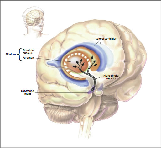
These protein aggregates form in neurons of the “striatum.” The striatum is a region of the brain that is also called the striate nucleus of the striate body. The striatum receives its name from the fact that it is organized in striped layers of gray and white matter. The striate nucleus is part of the cerebrum or forebrain.

Striatum

Mutant Huntington (Htt) protein has a toxic that causes cell death by means of unknown mechanisms. Clinically, the most obvious symptoms of HD involve involuntary movements of the arms, legs, and face. But the severe cognitive and personality changes are the most devastating to HD patients and most troubling for their caregivers.

Researchers are using animal models of HD to study the disease pathogenesis, to elucidate areas of the brain involved in structural and functional decline, and to evaluate potential therapeutic interventions. These animal models include injecting toxins into the brain to kill off those populations of neurons that typically die in HD patients, and transgenic models in which animals are bred with either extra mutant copies of the Htt gene or a pair of copies of the mutant Htt gene that have replaced the original, normal copies. All of these model systems have limitations, but they are all useful in some way for assessing the pathology of HD.

This long introduction leads us to new data from the laboratory of Steve Goldman, the co-director of the University of Rochester Medical Center’s Center for Translational Medicine. Goldman and his colleagues triggered the production of new neurons in mice that had a rodent form of HD. These new neurons successfully integrated into the brain’s existing neural networks and dramatically extended the survival of the mice.

“This study demonstrates the feasibility of a completely new concept to treat Huntington’s disease, by recruiting the brain’s endogenous neural stem cells to regenerate cell lost the disease,” said Goldman.

One of the types of neurons most commonly affected in HD patients is the medium spinal neuron, which is critical to motor control. Goldman banked on findings from previous studies in his laboratory on canaries. Songbirds such as canaries have the ability to lay down new neurons in the adult brain when mating season comes. The male birds, in response to a flush of male sex hormones,, grow a gaggle of new neurons in the vocal control centers of the brain, and this provides the bird the means to sing specific songs in order to attract mates. This event is known as adult neurogenesis, and Goldman and Fernando Nottebohm of the Rockefeller University discovered this phenomenon in the early 1980s.

“Our work with canaries essentially provided us with the information we needed to understand how to add new neurons to adult brain tissue,” said Goldman. Once we mastered how this happened in birds, we set about how to replicate the process in the adult mammalian brain.”

Humans possess the ability to make new neurons, but Goldman’s lab demonstrated in the 1990s that a font of neuronal precursor cells exist in the lining of the ventricles (these are structures at the very center of the brain and spinal cord that are filled with cerebrospinal fluid). In early development, these cells are actively producing neurons.

Shortly after birth, the neural stem cells stop generating neurons and produce support cells called glia. Some parts of the human brain continue to produce neurons into adulthood, the most prominent example is the hippocampus, where memories are formed and stored. However, the striatum, new neuron production is switched off in adulthood.

Goldman sought to switch neuron production back on in the striatum. He tested a cadre of growth factors that would switch the neural stem cells of the striatum (a region that is ravaged by HD) from producing new glia to producing new neurons. Goldman, however, had some help from his recent work in canaries. Namely that once mating season was upon the birds, targeted expression of brain-derived neurotrophic factor (BDNF) flared up in the vocal centers of the brain, where many new neurons were being produced.

Goldman used genetically engineered viruses to express BDNF and another protein called “Noggin” in the striatum. Goldman and others found that a single intraventricular injection of the adenoviruses expressing BDNF and Noggin triggered the sustained recruitment of new neurons in both normal of R6/2 (HD) mice. These treated mice also showed that the newly formed neurons were recruited to form new medium spiny neurons; the ones destroyed in HD. These new neurons also matured and achieved circuit integration.
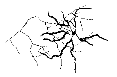
Medium Spinal Neuron

Also the treated mice showed delayed deterioration of motor function and substantially increased survival.

When the same experiments were conducted in squirrel monkeys, there was a similar addition of new striatal neurons.

Thus, induced neuronal addition may therefore represent a promising avenue for decreasing the ravages of HD and increasing cognitive ability.
Stem Cell Therapies for Myelin Disorders May Undergo Clinical Trials Soon


The highly-regarded journal Science has published a review article by University of Rochester Medical Center scientists Steve Goldman, M.D., Ph.D., Maiken Nedergaard, Ph.D., and Martha Windrem, Ph.D. that argues that stem cell researchers are very close to human application of stem cell therapies for a class of neurological diseases known as myelin disorders. Myelin disorders consist of a lengthy list of rather nasty diseases:

1) multiple sclerosis, which is a disease that affects the brain and spinal cord and damages the myelin sheath that surrounds and protects nerve cells. This damage slows down or blocks messages between the brain and body, which leads to the symptoms of MS, which include visual disturbances, muscle weakness, trouble with coordination and balance, sensations such as numbness, prickling, or “pins and needles,” and thinking and memory problems.

2) white matter stroke, a lack of blood flow to white matter, which is quite severe, since blood flow to the white matter far less than that of gray matter.

3) cerebral palsy, a group of disorders that involve the brain and nervous system functions, that include movement, learning, hearing, seeing, and thinking. There are several different types of cerebral palsy, including spastic, dyskinetic, ataxic, hypotonic, and mixed. Cerebral palsy is caused by injuries or abnormalities of the brain. Most of these problems occur as the baby grows in the womb, but they can happen at any time during the first 2 years of life, while the baby’s brain is still developing. In some people with cerebral palsy, parts of the brain are injured due to low levels of oxygen (hypoxia) in the area. It is not known why this occurs. Premature infants have a slightly higher risk of developing cerebral palsy. Cerebral palsy may also occur during early infancy as a result of several conditions, including: bleeding in the brain, brain infections, head injuries, infections in the mother during pregnancy, severe jaundice.

4) certain dementias, and

5) rare but fatal childhood disorders called pediatric leukodystrophies. Leukodystrophies are a varied group of diseases that primarily affect the white matter of the central nervous system (CNS). These diseases include both primary myelin disorders, axonal/neuronal degeneration and inflammatory disorders. There are two types of leukodystrophies: dysmyelinating diseases, which usually results from inherited defects in an enzyme pathway or organelle function that causes abnormal formation, destruction, or turnover of myelin, and demyelinating disorders that result in abnormal destruction of normal myelin and/or axons.

According to Goldman, “Stem cell biology has progressed in many ways over the last decade, and many potential opportunities for clinical translation have arisen. In particular, for diseases of the central nervous system, which have proven difficult to treat because of the brain’s great cellular complexity, we postulated that the simplest cell types might provide us the best opportunities for cell therapy.”

Myelin disorders share a common pathological factor and that is a cell that makes myelin, called an “oligodendrocyte.”  Oligodendrocytes arise from a cell found in the central nervous system called “glial progenitor cells (GPCs).  GPCs give rise to oligodendrocytes and “sister cells” called “astrocytes.”  Both cells serve rather critical functions in the central nervous system.

Astrocytes

Oligodendrocytes produce myelin, a fatty substance that insulates the fibrous connections between nerve cells that are responsible for transmitting signals throughout the body. When myelin-producing cells are lost or damaged in conditions such as multiple sclerosis and spinal cord injury, signals traveling between nerves are weakened or even lost.

Oligodendrocyte

Astrocytes are the unsung heroes of the central nervous system.  They were largely neglected for some time, but are now coming into their own as one of the main glial (support cells) in the brain.  Astrocytes secrete a cocktail of growth factors that keep neurons and oligodendrocytes healthy and help them properly signal to other cells.

Because they give rise to cells that are so central to the function of so many other brain cells, GPCs and their offspring represent a promising target for stem cell therapies.  An added bonus of using GPCs is that (unlike other cells in the central nervous system) they are rather homogeneous and are also don;t mind being manipulated and cultured.  Consequently, they are easy to transplant.  In fact, several animal studies have established that transplanted oligodendrocytes will disperse and repair or “remyelinate” damaged nerves.
“Glial cell dysfunction accounts for a broad spectrum of diseases, some of which – like the white matter degeneration of aging – are far more prevalent than we previously realized,” said Goldman. “Yet glial progenitor cells are relatively easy to work with, especially since we don’t have to worry about re-establishing precise point-to-point connections as we must with neurons. This gives us hope that we may begin to treat diseases of glia by direct transplantation of competent progenitor cells.”

Several key technological advances have made these recent advances in neural stem cell protocols possible.  First of all, superior imaging technologies with more advanced MRI scanners that provide sharper and more magnified images can provide more precise information about the specific damage in the central nervous system that result from myelin disorders.  Additionally, once cells are transplanted, these new scanning techniques allow scientists to more precisely trace their implanted cells and determine what those cells are doing.

Another important advance are the major obstacles that have recently been overcome in recent work.  First, there have been significant advances in the manipulation and handling of GPCs and their progeny.  Goldman’s lab  has pioneered the techniques for GPC manipulation.  He and his colleagues have determined the precise steps used in the induction of GPC differentiation into either oligodendrocytes or astrocytes.  Goldman’s lab has produced GPCs by reprogramming skin cells and he has also identified specific cell surface molecules that act as markers for GPCs.  The identification of markers is a huge advance because it allows him and his co-workers to isolate the differentiated cells away from those that might potentially cause tumors.

The Nedergaard lab has done a tremendous amount of work to understand the structure of the neural networks of the brain and their functional contributions to the brain as a whole.  Together, these two labs have developed models of normal human neural activity and brain disease that are based on laboratory animals that have been transplanted with human GPCs.  This enables the human neural cells to operated within a living brain instead of a culture dish.  Such experimental has already opened several new strategies for modeling and potentially treating human glial diseases.

The authors contend that these advances have accelerated research to the point where human clinical trials for myelin disorders will probably occur soon.  As an example, patients with multiple sclerosis would benefit from the invention of a new generation of stabilizing anti-inflammatory drugs, since multiple sclerosis results from thsensitizatione  of the immune system to the myelin sheath.  However, such drugs always have nasty side-effects.  If you do not believe me, just examine this short list of side effects for one of these drugs.  Instead MS patients would definitely benefit from a progenitor-based cell therapy that could repair the now permanent and untreatable damage to the central nervous system that results from this disease.  Also several childhood diseases of white matter are also excellent candidates for cell-based treatments.
“We have developed a tremendous amount of information about these cells and how to produce them,” said Goldman. “We understand the different cell populations, their genetic profiles, and how they behave in culture and in a variety of animal models. We also have better understanding of the disease target environments than ever before, and have the radiographic technologies to follow how patients do after transplantation. Moving into clinical trials for myelin disorders is really just a question of resources at this point.”