Minneapolis Heart Institute Foundation Tests Stem Cell Combination in Heart Attack Patients


The Minneapolis Heart Institute Foundation has announced a new clinical trial that will examine the ability of a stem cell combination to treat patients with ischemic heart failure.

In patients who have suffered from former heart attacks, clogged coronary blood vessels and heart muscle that hibernates can result in a heart that no longer works well enough to support the life of the patient. The lack of blood flow to vital parts of the heart and an increasing work load can result is so-called “Ischemic heart failure.” Such heart failure after a previous heart attack is one of the leading cause of death and morbidity in the world. According to the World Health Organization, ischemic heart disease affects more than 12% of the world’s population.

Stem cell therapy has been tested as a potential treatment for ischemic heart disease. Despite flashes of remarkable success, the overall efficacy of these treatments has been relatively modest. Most clinical trials have used the patient’s own bone marrow cells. In this case, the cell population is very mixed and it might not even be stem cell populations in the bone marrow that are eliciting recovery. Also, the quality of each patient’s bone marrow is probably quite varied, which makes standardizing such experiments remarkably difficult. Other clinical trials have used bone marrow derived mesenchymal cells [MSCs]. Several clinical trials with MSCs have seen some improvement in patients. MSCs seem to induce the formation of new blood vessels and also seem to induce endogenous stem cell populations in the heart to come to life and fix the heart. Other trials have used cardiac stem cells (CSCs) that were derived from biopsies of the heart. Even though fewer clinical trials have tested the efficacy of CSCs in human patients, the trials that have been conducted suggest that these cells can truly regenerate damaged heart tissue.

The Minneapolis Heart Institute Foundation® (MHIF) has announced a new clinical trial which will examine the combination of MSCs with CSCs to treatment patients with ischemic heart failure. This clinical trial, the CONCERT study, will be led by Principal Investigator Jay Traverse, MD. The CONCERT study will implant MSC’s and CSC’s in order to determine if the combination would be more successful than using either alone based on pre-clinical studies in swine demonstrating an enhanced synergistic effect of the combination.

CONCERT is sponsored by the National Institutes of Health and the Cardiovascular Cell Therapy Research Network (CCTRN), of which MHIF is a charter member. This will be a phase II clinical trial, which means that the focus of this leg of the study is to assess the relative safety of CSCs and MSCs, delivered either alone, or in combination, in comparison to placebo, and to measure the efficacy of the stem cell cocktail as well. To that end, researchers will measure and note any change or improvement in left ventricular (LV) function by cardiac MRI as well as changes in various clinical outcomes (survival, 6-minute walking, blood pressure, etc.), and quality of life.

This phase II study is a randomized, blinded, placebo-controlled study that will enroll 160 subjects at seven different CCTRN sites throughout the U.S. All recruited subjects will have ischemic cardiomyopathy and an ejection fraction 5%). This is significant, because some work in animals suggests that CSCs can make new heart muscle tissue that can shrink the heart scar. The first 16 patients were recently enrolled in a FDA-required safety run-in phase, but the remaining patients will be enrolled in the fall after a three-month safety analysis is performed. Incidentally, this is the first cardiac stem cell trial to perform MRIs on patients with defibrillators and pacemakers

“This combination of cells represents the most potent cell therapy product ever delivered to patients,” said Dr. Traverse. “Confirming that both types of stem cells together work better than either individual cell type could lead to improved patient outcomes and better quality of life for ischemic heart failure patients.”
Enrollment Completed in Phase 2 ALLSTAR Cardiac Clinical Trial


Capricor Therapeutics Inc. has announced the completion of patient enrollment in their Phase 2 ALLSTAR clinical trial.  ALLSTAR stands for ALLogeneic Heart STem Cells to Achieve Myocardial Regeneration, and this trial will test Capricor’s CAP-1002 product in patients suffering from cardiac dysfunction following a heart attack.

CAP-1002 cells are cardiosphere derived cells (CDCs) that were isolated from donors.  This investigational therapy is an off-the-shelf “ready to use” cardiac cell therapy that comes from donor heart tissue.  CAP-1002 cells are made to be directly infused into a patient’s coronary artery during a catheterization procedure.

These CDCs were tested in the CADUCEUS clinical trial, in which they were shown to decrease scar size and increase viable heart tissue when implanted into the hearts of heart attack patients.  One-year follow-up examinations of these confirmed the earlier results.

ALLSTAR will study a population similar to the one in the CADUCEUS study (patients who had experienced a heart attack 30-90 days earlier), except that ALLSTAR will treat patients 91-365 days after suffering a heart attack.  The extension of the patient pool was to see if the indication window for CAD-1002 could be extended.

The Capricor CEO Linda Marbàn said, “With the last patient in ALLSTAR having been dosed on September 30, we expect to report top-line 12-month primary efficacy outcome results in the fourth quarter of 2017.”

ALLSTAR is being sponsored by Capricor and is led by Drs. Timothy Henry and Rajendra Makkar of the Cedars-Sinai Heart Institute.  The trial is being conducted at approximately 25-40 sites across the U.S.

The Phase I portion of the trial was funded in part by the National Institutes of Health and completed enrollment in December 2013, and the Phase II portion of the trial is supported in large part by the California Institute for Regenerative Medicine (CIRM).

Stem Cell Therapy Might Improve Brain Function of Traumatic Brains Injury Patients


Accidents happen and sometimes really bad accidents happen; especially if they injure your head.  Traumatic brain injuries or TBIs can result from automobile accidents, explosions or other events that result from severe blows to the head.  TBIs  an adversely affect a patient and his/her family for long periods of time.  TBI patients can experience cognitive deficits that prevent them from thinking or speaking straight, and sensory deficits that prevent them from seeing, hearing or smelling properly.  Psychological problems can also result.  Essentially, TBIs represent a major challenge for modern medicine.

According to data from the Centers for Disease Control (CDC), 1.7 million Americans suffer from TBIs each year (of varying severity).  Of these, 275,000 are hospitalized for their injuries and approximately 52,000 of these patients die from their injuries.  In fact, TBIs contribute to one-third of all injury-related deaths in the United States each year.  More than 6.5 million patients are burdened by the deleterious effects of TBIs, and this leads to an economic burden of approximately $60 billion each year.

Currently, treatments for TBI are few and far between.  Neurosurgeons can use surgery to repair damaged blood vessels and tissues, and diminish swelling in the brain.  Beyond these rather invasive techniques, the options for clinicians are poor.

A new study by Charles S. Cox, professor of Pediatric Surgery and co-director of the Memorial Hermann Red Duke Trauma Institute, and his colleagues suggest that stem cell treatments might benefit TBI patients.  The results of this study were published in the journal Stem Cells.

This study enrolled 25 TBI patients.  Five of them received no treatment and served as controls, but the remaining 20 received gradually increasing dosages of their own bone marrow stem cells.  The harvesting, processing and infusion of the bone marrow cells occurred within 48 hours of injury.  Functional and cognitive results were measured with standard tests and brain imaging with magnetic resonance imaging and diffusion tensor imaging.

This work is an extension of extensive preclinical work done by Cox and his coworkers in laboratory animals and a phase I study that established that such stem cell transplantation are safe for human patients.  The implanted stem cells seem to quell brain inflammation and lessen the damage to the brain by the TBI.

Despite the fact that those TBI patients who received the stem cell treatments had greater degrees of brain damage, the treatment group showed better structural preservation of the brain and better functional outcomes than the control group.  Of particular interest was the decrease in indicators of inflammation as a result of the bone marrow cell-based infusions.

Cox said of this trial, “The data derived from this trial moves beyond just testing safety of this approach.”  He continued:  “We now have a hint of a treatment effect that mirrors our pre-clinical work, and were are now pursuing this approach in a phase IIb clinical trial sponsored by the Joint Warfighter Program within the US Army Medical Research Acquisition Activity, as well as our ongoing phase IIb pediatric severe TBI clinical trial; both using the same autonomous cell therapy.”

This an exciting study, but it is a small study.  While the safety of this procedure has been established, the precise dosage and long-term benefits will require further examination.  However it is a fine start to what may become the flowering of new strategies to treat TBI patients.

NurOwn, Modified Mesenchymal Stem Cells, Show Clinical Benefit in Phase 2 Trial in ALS Patients


BrainStorm Cell Therapeutics Inc. (BCLI) has developed a cell-based product they call “NurOwn.” NurOwn consists of mesenchymal stem cells that have been cultured to secrete a variety of neurotrophic factors (NTFs). These NTFs are a collection of different growth factors that promote the survival of neurons. NurOwn cells were originally developed in the laboratories of Professor Dani Offen and the late Professor Eldad Melamed, at Tel Aviv University. NurOwn cells have been studied extensively and they clearly have the capacity to migrate to damaged areas in the central nervous system (Sadan O, et al., Stem Cells. 2008 Oct;26(10):2542-51), decrease dopamine depletion in a Parkinson’s disease model system (Barhum Y, et al., J Mol Neurosci. 2010 May;41(1):129-37), can promote the survival of photoreceptors in the retina of animals who optic nerves were damaged (Levkovitch-Verbin H, et al., Invest Ophthalmol Vis Sci. 2010 Dec;51(12):6394-400), decrease quinolinic acid toxicity in an animal model of Huntington’s disease (Sadan O, et al., Exp Neurol. 2012 Apr;234(2):417-27), and improve motor function and survival in a genetic model of Huntington’s disease.

On the strength of these experiments, NurOwn cells have also been tested in clinical trials. Because NTF-secreting MSCs (or, MSC-NTF cells) are designed specifically to treat neurodegenerative diseases, most of the clinical trials, to date, have examined of safety and efficacy of MSC-NTFs in patients with neurological disorders. The safety of NurOwn cells was established in a small phase I/II trial with amyotrophic lateral sclerosis (ALS) patients. This was a small study (12 patients), but showed that, at least in this patients population, intrathecal (injected into the central nervous system) and intramuscular administration of MSC-NTF cells in ALS patients with is safe and patients even showed some indications of clinical benefits, but the study was too small to be definitive about the efficacy of these cells.

Now a recently completed randomized, double-blind, placebo-controlled phase 2 study of NurOwn in ALS patients has found that NurOwn is safe and well tolerated and may also confer clinical benefits upon ALS patients.

According to BrainStorm, this phase 2 study achieved its primary objective (safety and tolerability). No deaths were reported in the study and no patients discontinued participation because of an adverse event. All patients in both active treatment and placebo groups experienced at least one treatment-emergent adverse event that tended to be mild-to-moderate in intensity in both groups. Treatment-related adverse events, as determined by a blinded investigator, occurred slightly more frequently in active-treated patients than in placebo-treated patients (97.2 percent vs. 75.0 percent). The largest differences in frequencies were for the localized reactions of injection site pain and back pain, and fever, headache, and joint pain.

However, NurOwn also achieved multiple secondary efficacy endpoints in this trial. NurOwn showed clear evidence of a clinically significant benefit. Most significantly, the response rates were higher for NurOwn-treated subjects compared to placebo at all time points in the 24 weeks during which when the study was conducted.

This clinical trial conducted at three sites in the U.S: Massachusetts General Hospital, UMass Medical School and the Mayo Clinic. 48 patients were randomized to receive NurOwn cells administered via combined intramuscular and intrathecal injection (n= 36), or placebo (n=12). They were followed monthly for approximately three months before treatment and six months following treatment, and were assessed at 2, 4, 8, 12, 16 and 24 weeks.

The primary investigator in this trial, Robert H. Brown of the University of Massachusetts Medical Center and Medical School said, “These exciting findings clearly indicate that it is appropriate to conduct a longer study with repetitive dosing.”

Subjects treated with NurOwn in this trial showed slowing of progression of ALS and no safety concerns. NurOwn-treated patients also displayed increased levels of growth factors in the cerebrospinal fluid and decreased signs of inflammation after two weeks. These are good indicators that the MSC-NTF cells are orchestrating some kind of beneficial biological effect.

Based on these results, new trials are warranted that will examine repeat dosing at 8 to 12 weeks and employ a larger number of subjects.

Mesenchymal Stem Cells from Bone Marrow Improve Liver Function and Reduce Liver Scarring in Patients with Alcoholic Cirrhosis


Dr Soon Koo Baik from the Yonsei University Wonju College of Medicine, and Dr. Si Hyun Bae from The Catholic University of Korea and their colleagues have conducted an important phase 2 clinical trial that tests the ability of mesenchymal stem cells from bone marrow to treat cirrhosis of the liver. In this trial, seventy-two patients who had established cirrhosis of the liver participated in a multicenter, randomized, open-label, phase 2 trial (published in the journal Hepatology, DOI:10.1002/hep.28693).

The liver is a hugely important organ. Not only is it the largest internal organ in our bodies, but it serves as the main metabolic factory of the body because of the outsized role it plays in metabolism. The liver takes up and stores and processes nutrients from food. Once it processes fats, sugars, and amino acids, the liver delivers them to the rest of the body. The liver also makes new proteins, such as clotting and immune factors, produces bile, which helps the body absorb fats, cholesterol, and fat-soluble vitamins, and removes waste products the kidneys cannot remove, such as fats, cholesterol, toxins, and medications.

The condition known as cirrhosis is a condition in which the liver gradually deteriorates and becomes unable to function normally due to chronic, or long-lasting, injury. The accumulation of scar tissue in the liver is typically slow and gradual and as scar tissue replaces more healthy liver tissue, the liver begins to fail. Scar tissue also partially blocks the flow of blood through the liver. Chronic liver failure (also known as end-stage liver disease) culminates in the inability of the liver to perform important functions. Since the liver is an organ that have a good deal of regenerative ability, end-stage liver disease essentially becomes so damaged that it cannot effectively replace damaged cells.

Cirrhosis is most commonly called by chronic alcoholism, but so can chronic viral infections by viruses like hepatitis B virus and hepatitis C virus.  Additionally, particular genetic diseases can also cause cirrhosis in children or young adults.

Mesenchymal stem cells have the ability to secrete cocktails of pro-healing molecules that might be able to support the growth and survival of liver cells. A variety of experiments in animals have established that the administration of mesenchymal stem cells (MSCs) from bone marrow (Truong, NH, et al., Stem Cells Int. 2016;2016:5720413. doi: 10.1155/2016/5720413; Almeida-Porada G, et al., Exp Hematol. 2010;38:574–580; Berardis S, et al., World J Gastroenterol. 2015;21:742–758), and other sources (De Ugarte DA, et al., Cells Tissues Organs. 2003;174:101–109; in ‘t Anker PS, etr al., Haematologica. 2003;88:845–852; Lee OK, et al., Blood. 2004;103:1669–1675) can decrease inflammation within the liver, inhibit the death of liver cells and promote their survival, and promote the regeneration of residential liver cells.

In clinical trials, administration of MSCs to cirrhosis patients has established the safety of MSC-based treatments (Amin MA, et al., Clin Transplant. 2013;27:607–612; El-Ansary M, et al., Stem Cell Rev. 2012;8:972–981; Jang YO, et al., Liver Int. 2014;34:33–41; Kharaziha P, et al., Eur J Gastroenterol Hepatol. 2009;21:1199–1205; Mohamadnejad M, et al., Arch Iran Med. 2007;10:459–466). Unfortunately, the design of these trials involved the mixing of patients with alcohol-based cirrhosis, viral-based cirrhosis, and other types of cirrhosis. Therefore, it is impossible to draw any conclusions about the efficacy of MSC transplantations on the basis of these trials. However, one trial, by Jang, et al, examined the effect of MSCs from bone marrow in patients with alcoholic cirrhosis. After 11 patients received MSC implantations, improvements in liver tissue architecture were observed in 6/11 patients, and 10 patients showed recovery of liver function. These 10 patients had decreased expression of molecules that induce scarring in the liver (i.e. TGF-β1, collagen type I, and α-smooth muscle actin). Significantly, Jang and others observed these improvements in the absence of significant complications or side effects during the study period. On the strength of these results, a larger phase 2 study is certainly warranted (see F. Ezquer, et al., World J Gastroenterol. 2016 Jan 7; 22(1): 24–36).

In this Bak and Bae clinical trials, 72 patients were randomly assigned to three groups that consisted of a control group and two autologous bone marrow-based MSC groups that underwent either one-time or two-time hepatic arterial injections of 5 × 10[7] MSCs, 30 days after bone marrow aspiration. All patients also underwent a follow-up biopsy 6 months after enrollment and adverse events were monitored for 12 months.

The primary endpoint in this study was the improvement in the amount of scar tissue in biopsies (as assayed by Picrosirius-red staining). The secondary endpoints included liver function tests, a measure of the severity of cirrhosis called the Child-Pugh score, and another score called the Model for End-stage Liver Disease (MELD) score. The outcomes were analyzed by per-protocol analysis.

When it comes to the amount of scar tissue in the patient’s livers, patients that received one-time and two-time bone marrow-based MSC administrations, showed 25% (19.5±9.5% vs. 14.5±7.1%) and 37% (21.1±8.9% vs. 13.2±6.7%) reductions in the amount of liver scar after MSC administration, respectively (P0.05). The Child-Pugh scores of both BM-MSC groups (one-time: 7.6±1.0 vs. 6.3±1.3 and two-time: 7.8±1.2 vs. 6.8±1.6) were also significantly improved following BM-MSC transplantation (P<0.05) compared to the control group that did not receive MSCs. Most significantly, perhaps, is that the proportion of patients with adverse events did not differ among the three groups.

From this larger phase 2 study, it seems that transplantation of a patient’s own bone marrow-based MSCs can safely improve the degree of scarring in the liver of cirrhosis patients and also improve liver function in patients with alcoholic cirrhosis. This study seems to confirm what was observed in preclinical studies in laboratory animals and extends what was observed in the phase 1 studies. While more work is certainly required, these results are certainly hopeful.

Gene Therapy Sprouts New Neurons in Alzheimer’s Patients’ Brains


The journal JAMA Neurology has published a new study that describes an experimental gene therapy that reduces the rate at which nerve cells in the brains of Alzheimer’s patients degenerate and die. See Tuszynski, M. H., et al. (2015). Nerve Growth Factor Gene Therapy: Activation of Neuronal Responses in Alzheimer Disease. JAMA Neurology, published online August 24, 2015. DOI: 10.1001/jamaneurol.2015.1807.

In this study, targeted injections of a growth factor called “nerve growth factor” or NGF into the brain of patients rescued dying cells around the injection site, enhanced the growth of these cells and induced them to sprout new nerve fibers. Surprisingly, in some cases, these beneficial effects persisted for 10 years after the therapy was first delivered.

Alzheimer’s disease (AD) is the world’s leading form of dementia. It affects approximately 47 million people worldwide, and this number is expected to almost double every 20 years. Despite the huge amounts of time, effort, and money devoted to developing an effective cure, the vast majority of new drugs for AD have failed in clinical trials.

While these new results are preliminary findings, they come from the first human trials designed to test the potential benefits of NGF gene therapy for AD patients.

NGF was discovered in the 1940s by Rita Levi-Montalcini, who demonstrated, quite convincingly, that a small protein that she had isolated and purified promoted the survival of certain sub-types of sensory neurons during development of the nervous system. Since that time, others have shown that it also promotes the survival of neurons that produce acetylcholine in the basal forebrain; these cells die off at an alarming rate in AD patients.

In 2001, Mark Tuszynski and his coworkers at the University of California, San Diego School of Medicine initiated a clinical trial based on these laboratory findings. This trial was the first of its kind, and it was designed to investigate the ability of NGF gene therapy to slow or prevent the neuronal degeneration and cell death characteristic of AD.

In phase I of this trial, eight patients with mild AD received so-called “ex vivo” therapy to deliver the NGF gene directly into the brain. This trial extracted skin fibroblasts from the skin on the patient’s backs, and then genetically engineered those cells to express the NGF genes. These NGF-expressing cells were then implanted into the patients’ basal forebrain. Since NGF is too large to cross the blood-brain barrier, it had to be administered directly into the brain. Also, outside the brain, exogenous NGF can stimulate other nerve cells can cause unwanted side-effects such as pain and weight loss.

One of these patients died just 5 weeks after receiving the therapy. Tuszynski’s team secured permission to perform an autopsy of this patient, and in 2005 they reported that the treatment led to robust growth responses, and did not cause any adverse effects (Tuszynski, M. H., et al. (2005). A phase 1 clinical trial of nerve growth factor gene therapy for Alzheimer disease. Nature Medicine, 11: 551 – 555).

The latest results come from postmortem examination of these patients’ brains, all of whom had also been recruited in a safety trial between March 2001 and October 2012. Additionally, two other were included who had received in vivo therapy that included injecting a modified virus that carried the NGF gene into the basal forebrain.

Some of the participants died about one year after undergoing therapy, and others survived for 10 years after the treatment. These autopsies showed that all of them had responded to the treatment.

Essentially, all the brain tissue samples taken from around the implantation sites contained diseased neurons, as expected, but the cells were overgrown, and had sprouted axonal fibers that had grown towards the region into which NGF had been delivered. In contrast, samples taken from the untreated side of the brain exhibited no such response.

This trial was conducted to test the safety of the treatment and it did confirm that none of the patients experienced long-term adverse effects from the treatment, even after long periods of time. These results also suggest that NGF is successfully taken up by nerve cells following targeted delivery. Also the cells synthesize NGF protein so that its concentration dramatically increases in and around the delivery site. Probably the most exciting part of these findings is that the responses to NGF can persist for many years after the gene has been delivered into the brain.

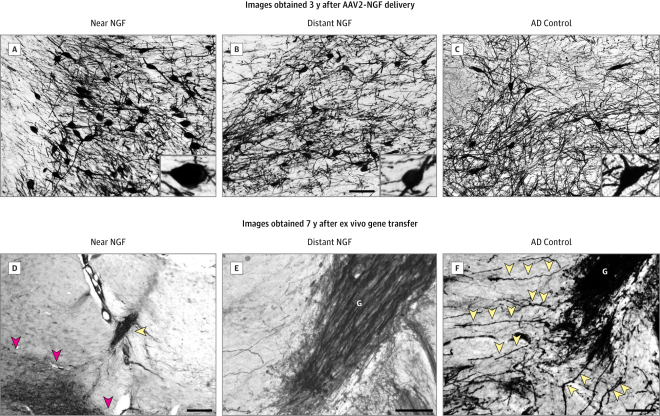
Cholinergic Neuronal Hypertrophy and Sprouting Shown is labeling for p75, a neurotrophin receptor expressed on cholinergic neurons of the nucleus basalis of Meynert. Images were obtained 3 years after adeno-associated viral vectors (serotype 2)–nerve growth factor (AAV2-NGF) delivery (A-C) and 7 years after ex vivo gene transfer (D-F). A-C, Cholinergic neurons are labeled for p75 within the zone of transduction (A), 3 mm from the zone of transduction (B), and in a control Alzheimer disease (AD) brain of the same Braak stage (C). Cells near the NGF transduction region appear larger. The inset shows higher-magnification views of a typical neuron from each region. D, Shown is a graft of fibroblasts transduced to secrete NGF (yellow arrowhead) adjacent to the nucleus basalis of Meynert (red arrowheads). E, The graft (G) at higher magnification is densely penetrated by p75-labeled axons arising from the nucleus basalis of Meynert. These axons are sprouting toward the graft, a classic trophic response. F, Shown are p75-labeled axons from the nucleus basalis of Meynert sprouting toward the graft. Individual axons coursing toward the graft are evident (arrowheads). The bar represents 125 µm in A through C, 500 µm in D, and 100 µm in E and F.
Cholinergic Neuronal Hypertrophy and Sprouting
Shown is labeling for p75, a neurotrophin receptor expressed on cholinergic neurons of the nucleus basalis of Meynert. Images were obtained 3 years after adeno-associated viral vectors (serotype 2)–nerve growth factor (AAV2-NGF) delivery (A-C) and 7 years after ex vivo gene transfer (D-F). A-C, Cholinergic neurons are labeled for p75 within the zone of transduction (A), 3 mm from the zone of transduction (B), and in a control Alzheimer disease (AD) brain of the same Braak stage (C). Cells near the NGF transduction region appear larger. The inset shows higher-magnification views of a typical neuron from each region. D, Shown is a graft of fibroblasts transduced to secrete NGF (yellow arrowhead) adjacent to the nucleus basalis of Meynert (red arrowheads). E, The graft (G) at higher magnification is densely penetrated by p75-labeled axons arising from the nucleus basalis of Meynert. These axons are sprouting toward the graft, a classic trophic response. F, Shown are p75-labeled axons from the nucleus basalis of Meynert sprouting toward the graft. Individual axons coursing toward the graft are evident (arrowheads). The bar represents 125 µm in A through C, 500 µm in D, and 100 µm in E and F.

Now, does the observed cellular response to NGF alleviate disease symptoms? Although phase II trials testing the efficacy of the treatment are ongoing, preliminary findings from the initial study suggest that the therapy did indeed slow the rate at which mental function declined in one of the patients involved. These new results indicate that gene therapy is a viable strategy for treating Alzheimer’s and other neurodegenerative diseases, and warrants further research and development.

Neuralstem Treats Final Patient in Phase 2 ALS Stem Cell Trial


NeuralStem, Inc. has announced that the final patient in its Phase 2 clinical trial that assessed the efficacy of its NSI-566 spinal cord-derived neural stem cell line in the treatment of amyotrophic lateral sclerosis (ALS), which is otherwise known as Lou Gehring’s disease.

ALS is a rapidly progressive, invariably fatal neurological disease that attacks the nerve cells responsible for controlling voluntary muscles; that is, muscle action we are able to control, such as those in the arms, legs, and face, etc.  ALS is a member of those disorders known as motor neuron diseases, all of which are characterized by the gradual degeneration and death of motor neurons.

Motor neurons are nerve cells located in the brain, brain stem, and spinal cord that serve as controlling units and vital communication links between the nervous system and the voluntary muscles of the body. Messages from motor neurons in the brain (so-called upper motor neurons) are transmitted to motor neurons in the spinal cord (so-called lower motor neurons) to particular muscles. In ALS, both the upper motor neurons and the lower motor neurons degenerate or die, and stop sending messages to muscles. Unable to function, the muscles gradually weaken, waste away (atrophy), and have very fine twitches (called fasciculations). Eventually, the ability of the brain to start and control voluntary movement is lost.

ALS causes weakness with a wide range of disabilities. Eventually, all muscles under voluntary control are affected, and individuals lose their strength and the ability to move their arms, legs, and body. When muscles in the diaphragm and chest wall fail, people lose the ability to breathe without ventilatory support. Most people with ALS die from respiratory failure, usually within 3 to 5 years from the onset of symptoms. However, about 10 percent of those with ALS survive for 10 or more years.

Although the disease usually does not impair a person’s mind or intelligence, several recent studies suggest that some persons with ALS may have depression or alterations in cognitive functions involving decision-making and memory.

ALS does not affect a person’s ability to see, smell, taste, hear, or recognize touch. Patients usually maintain control of eye muscles and bladder and bowel functions, although in the late stages of the disease most individuals will need help getting to and from the bathroom.

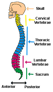
In this multicenter Phase 2 trial, 15 patients who still had the ability to walk were treated in five different dosing cohorts. The first 12 of these patients received injections only in the cervical regions of the spinal cord in increasing doses (5 injections of 200,000 cells per injection to injections of 4000,000 cells each . In the cervical region, these injected stem cells could potentially preserve the nerves that mediate breathing and this is precisely that this part of the trail aims to test.

spinal cord regions

In the final three patients injected in this trial, patients received a total of 40 injections of 400,000 cells each into both cervical and lumbar regions (a total of 16 million cells were injected. This is in contrast to the patients who participated in the Phase 1 study who received 15 injections of 100,000 cells each (total of 1.5 million cells). This trial will continue until six months past the final surgery, after which the data will be analyzed.

“By early next year, we will have six-month follow-up data on the last patients who received what we believe will be the maximum safe tolerated-dose for this therapy,” said Dr. Eva Feldman, principal investigator in this clinical trial, and a member of the ALS Clinic at the University of Michigan. Dr. Feldman also serves as an unpaid consultant to Neuralstem.

Patient’s Own Peripheral Blood Stem Cells Benefits Stroke Patients


A study conducted in Taiwan has examined the ability of a patient’s circulating peripheral blood stem cells to benefit stroke patients.

In this study, one of two groups of stroke patients received injections of their own peripheral blood stem cells (PBSCs) directly into the brain but the other group received standard care. Those patients who received the PBSCs experienced some improvement in stroke scales and functional capabilities. Patients who received their own PBSCs also were given injections of granulocyte-colony stimulating factor (G-CSF), which seems to protect the nervous system after trauma to it.

“In this phase 2 study, we provide the first evidence that intracerebral injection of autologous (self-donated) PBSCs can improve motor function in those who have suffered a ,” said stroke and have motor deficits as a result,” said Woei-Cheng Shyu of the China Medical University, who is the corresponding author of this study. “Our study demonstrates that this therapeutic strategy was feasible and safe in stroke patients who suffered a prior stroke, but within five years from the onset of symptoms.”

Strokes, also known as trans-ischemic attacks (TIAs) result from blockage in blood vessels that feed the brain. The lack of blood flow to the brain starves it of oxygen, and the cells of the brain begin to die off. Because neuronal death as a result of stroke limits functional recovery, stem cell therapy is advancing as a potentially effective regenerative treatment for stroke.

Also, in many types of stem cell trials, PBSCs are the stem cell of choice. The ease of isolating these cells without invasive procedures makes them the stem cell choice for many clinical trials. In order to utilize PBSCs, patients must amplify their supply of PBSCs, and injections of G-CSF seems to do just that.

In this study, all patients had suffered a prior stroke as long as five years prior to being treated.

At the end of the 12-month follow-up, the group of 15 patients with neurological deficits who received the PBSC injections into the brain experienced neurological and functional improvements, according to several different clinical measurements.

On the other hand, the 15-patient control group who had neurological deficits but did not receive the PBSC injections did not experience the same beneficial results.

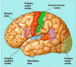
In the experimental group, nine of the 15 patients showed proper activation of the motor nerves after stimulating that part of the brain with a magnet. This procedure, called transcranial magnetic stimulation or TMS, places a magnet above skull, directly above the part of the brain you want to stimulate. The rapidly changing magnetic field generated by the magnet produces weak electrical currents in the brain, which stimulates nearby neurons. In this experiment, researchers targeted the precentral gyrus, which is the portion of the brain where the primary motor cortex. Because strokes sometimes kill off neurons in the primary motor cortex, stimulation of the primary motor cortex will not lead to stimulation of motor nerves, but in this experiment, 9 of 15 patients who received the PBSC injections who positive motor evoke potential or MEPs after TMS. Why this ratio was not 15 out of 15 remains unclear at this time.

primary motor cortex

One of the main conclusions of this work, is that “Despite this success, it should be noted that this was a preliminary study and, due to the small number of patients, are tentative. In the future we plan to conduct a multi-center, large-scale, double-blind, placebo-controlled randomized studies [sic] to better evaluate the effect of PBSC implantation in patients suffering from the effects of past stroke.”

Stem Cell Injections Reduce Lower Back Pain


W. Jeremy Beckworth and his co-workers at Emory Orthopaedics and Spine Center, in collaboration with several other orthopedic care groups, have participated in a clinical trial that demonstrated that a single injection of stem cells into degenerative intervertebral discs significantly reduced lower back pain for at least 12 months according. Beckworth’s clinical trial consisted of 100-patients and was a phase II, international clinical trial.

Beckworth, assistant professor of Orthopaedics and Rehab Medicine, gave patient injections of a subset of mesenchymal stem cells isolated from bone marrow stem cells called mesenchymal precursor cells (MPCs) in order to attenuate pain in patients with lower back pain. On average, Beckworth and his colleagues discovered that stem cell injections led to a reduction in pain levels greater than 50 percent at 12 months. Additionally, patients who received stem cell injections felt less of a need for pain medications, showed an improvement in function, and less need for further surgical and non-surgical spine interventions. These results were compiled from patients with moderate to severe disc-related lower back pain.

“These are very exciting findings,” explains Beckworth. “The results provide significant hope for a condition that has been very tough to treat. Discogenic low back pain, a painful degenerative disc, is the most common cause of chronic low back pain.”

This phase II clinical trial builds on a previously reported preclinical study showed that highly purified MPCs were able to repair and restore disc structure. All the data from this trial showed that there were statistically significant improvements in patients who received stem cell injections compared to those in control groups who received no such injections.

“Currently there is no adequate treatment for discogenic low back pain,” says Beckworth. “Both conservative and surgical treatments fall short. These positive results pave the way for a phase III study that may be starting later this year.”

New US Phase IIa Trial and Phase III Trial in Kazakhstan Examine CardioCell’s itMSC Therapy to Treat Heart Attack Patients


The regenerative medicine company CardioCell LLC has announced two new clinical trials in two different countries that utilize its allogeneic stem-cell therapy to treat subjects with acute myocardial infarction (AMI), which is a problem that faces more than 1.26 million Americans annually. The United States-based trial is a Phase IIa AMI clinical trial that is designed to evaluate the clinical safety and efficacy of the CardioCell Ischemia-Tolerant Mesenchymal Stem Cells or itMSCs. The second clinical trial in collaboration with the Ministry of Health in Kazakhstan is a Phase III AMI clinical trial on the intravenous administration of CardioCell’s itMSCs. This clinical trial is proceeding on the strength of the efficacy and safety of itMSCs showed in previous Phase II clinical trials.

CardioCell’s itMSCs are exclusively licensed from CardioCell’s parent company Stemedica Cell Technologies Inc. Normally, when mesenchymal stem cells from fat, bone marrow, or some other tissue source are grown in the laboratory, the cells are provided with normal concentrations of oxygen. However, CardioCell itMSCs are grown under low oxygen or hypoxic conditions. Such growth conditions more closely mimic the environment in which these stem cells normally live in the body. By growing these MSCs under these low-oxygen conditions, the cells become tolerant to low-oxygen conditions (ischemia-tolerant), and if transplanted into other low-oxygen environments, they will flourish rather than die.

Another advantage of itMSCs for regenerative treatments over other types of MSCs is that itMSCs secrete higher levels of growth factors that induce the formation of new blood vessels and promote tissue healing. These clinical trials have been designed to help determine if CardioCell’s itMSC-based therapies stimulate a regenerative response in acute heart attack patients.

“CardioCell’s new Phase IIa AMI study is built on the excellent safety data reported in previous Phase I clinical trials using our unique, hypoxically grown stem cells,” says Dr. Sergey Sikora, Ph.D., CardioCell’s president and CEO. “We are also pleased to report that the Ministry of Health in Kazakhstan is proceeding with a Phase III CardioCell-therapy study following its Phase II study that was highly promising in terms of efficacy and safety. Our studies target AMI patients who have depressed left ventricular ejection fraction (LVEF), which makes them prone to developing extensive scarring and therefore to the development of chronic heart failure. CardioCell hopes our itMSC therapies will inhibit the development of extensive scarring and, thus, the occurrence of chronic heart failure in these patients.”

The United States-based Phase IIa clinical trial will take place at Emory University, Sanford Health and Mercy Gilbert Medical Center. The CardioCell Phase IIa AMI trial is a double-blinded, multicenter, randomized study designed to assess the safety, tolerability and preliminary clinical efficacy of a single, intravenous dose of allogeneic mesenchymal bone-marrow cells infused into subjects with ST segment-elevation myocardial infarction (STEMI).

“While stem-cell therapy for cardiovascular disease is nothing new, CardioCell is bringing to the field a new, unique type of stem-cell technology that has the possibility of being more effective than other AMI treatments,” says MedStar Heart Institute’s Director of Translational and Vascular Biology Research and CardioCell’s Scientific Advisory Board Chair Dr. Stephen Epstein. “Evidence exists demonstrating that MSCs grown under hypoxic conditions express higher levels of molecules associated with angiogenesis and healing processes. There is also evidence indicating they migrate with greater avidity to various cytokines and growth factors and, most importantly, home more robustly to ischemic tissue. Studies like those underway using CardioCell’s technology are designed to determine if we can evoke a more potent healing response that will reduce the extent of myocardial cell death occurring during AMI and thereby decrease the amount of scar tissue resulting from the infarct. A therapy that could achieve this would have a major beneficial impact in reducing the occurrence of chronic heart failure.”

Kazakhstan’s National Scientific Medical Center is conducting a Phase III AMI clinical trial using CardioCell’s itMSCs, which are sponsored by local licensee Altaco. This clinical trial is entitled, “Intravenous Administration of itMSCs for AMI Patients,” and is proceeding based on a completed Phase II efficacy and safety study. However, the results of this previous Phase II study are preliminary because the sample group was so small. Despite these limitations, the findings demonstrated statistically significant elevation (more than 12 percent over the control group) in the ejection fraction of the left ventricle of the heart in patients who had received itMSCs. Also, a significant reduction in inflammation was also observed, as ascertained by lower CRP (C-reactive protein) levels in the blood of treated patients in comparison to control groups. Thus, Dr. Daniyar Jumaniyazov, M.D., Ph.D., principal investigator in Kazakhstan clinical trials states: “In our clinical Phase II trial for patients with AMI, treatment using itMSCs improved global and local myocardial function and normalized systolic and diastolic left ventricular filling, as compared to the control group. We are encouraged by these results and look forward to confirming them in a Phase III study.”

CardioCell’s treatment is the first to apply itMSC therapies for cardiovascular indications like AMI, chronic heart failure and peripheral artery disease. Manufactured by CardioCell’s parent company Stemedica and approved for use in clinical trials, itMSCs are manufactured under Stemedica’s patented, continuous-low-oxygen conditions and proprietary media, which provide itMSCs’ unique benefits: increased potency, safety and scalability. itMSCs differ from competing MSCs in two key areas. itMSCs demonstrate increased migratory ability towards the place of injury, and they show increased secretion of growth and transcription factors (e.g., VEGF, FGF and HIF-1), as demonstrated in a peer-reviewed publication (Vertelov et al., 2013). This can potentially lead to improved regenerative abilities of itMSCs. In addition, itMSCs have significantly fewer HLA-DR receptors on the cell surface than normal MSCs, which might reduce the propensity to cause immune responses. As another benefit, itMSCs are highly scalable. A single donor specimen can currently yield about 1 million patient treatments, and this number is expected to grow to 10 million once full robotization of Stemedica’s facility is complete.

Adult Stem Cells Used for Spinal Disc Repair


The Australian regenerative medicine company Mesoblast Limited announced the results of their 12-month clinical trial that examined the use of their “off-the-shelf” product to treat patients with disc-related low back pain.

This phase 2 clinical trial enrolled 100 patients with chronic moderate to severe “discogenic low back pain” and tested the ability of “mesenchymal precursor cells” to shore up degenerating intervertebral discs.

Intervertebral discs

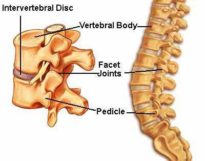
Intervertebral discs sit between each vertebra and act as shock absorbers. Each disc consist of an outer layer called the “annulus fibrosus.” The annulus fibrosus consists of several layers of fibrocartilage. The annulus fibrosus surrounds an inner layer called the nucleus pulposus, which contains loose fibers suspended in a mucoprotein gel with the consistency of jelly. This jelly-like center distributes pressure evenly across the disc. These discs absorb the impact of the body’s daily activities and keep the two vertebrae separated. The development of a prolapsed disc results when the jelly in the nucleus pulposus is forced out of the doughnut/disc, which may put pressure on the nerve located near the disc.

Intervertebral structure

More than six million people in the United States alone deal with chronic back pain that has persisted for at least three months, and 3.5 million people are affected by moderate or severe degenerative intervertebral disc disease.

In this clinical trial, Mesoblast Limited injected their mesenchymal precursor cells (MPCs) into the degenerating intervertebral discs of patients suffering from moderate to severe back pain. When compared with a control group, patients who received the MPC injections used less pain killers, went through fewer surgeries and non-surgical interventions, and had greater disc stability as ascertained by X-rays. MPC injections also were well tolerated and produced few side effects.

This phase 2 clinical trial extends earlier observations by Mesoblast Limited on laboratory animals. In preclinical trials, purified MPCs increased the quality of the jelly content of the nucleus pulposus and improved disc structure in sheep.

This present study enrolled 100 patients at 13 different sites across Australia and the United States with early disc degeneration and randomly assigned the subjects to one of four groups: 1) those who received saline injections; 2) those who received hyaluronic acid injections; 3) those who received low-dose MPCs in hyaluronic acid; and 4) those who received high-dose injections of MPCs in hyaluronic acid.

All patients received their injections in an outpatient procedure, and are being evaluated for safety and efficacy to evaluate long-term treatment effects.

At 12 months, the key findings were improvement in chronic low back pain, function, and disc stability. Also, no safety concerns emerged as a result of the treatment.

As this trial proceeds, more data should be forthcoming.

Phase 2 Clinical Trial that Tests Stem Cell Treatment for Heart Attack Patients to be Funded by California Institute for Regenerative Medicine


A new stem cell therapy that treats heart attack patients with cells from a donor has been approved to begin a Phase 2 clinical trial.

Capricor Therapeutics Inc. a regenerative medicine company, has developed this treatment, which extracts donor stem cells from the heart called “cardiosphere-derived cells,” and then infuses them into the heart of the heart attack patient by means of a heart catheter procedure, which is quite safe. These stem cells are introduced into the heart to reduce scarring in the heart and potentially replace dead heart muscle cells. One clinical trial called the CADUCEUS trial has already shown that cardiosphere-derived cells can reduce the size of the heart scar.

In a previous phase I study (phase I studies typically only ascertain the safety of a treatment), cardiosphere-derived cells were infused into the hearts of 14 heart attack patients. No major safety issues were observed with these treatments, and therefore, phase 2 studies were warranted.

Alan Trounson, Ph.D., president of the California Institute for Regenerative Medicine (CIRM), which is funding the trial, said this about the phase 2 trial approval: “This is really encouraging news and marks a potential milestone for the use of stem cells to treat heart disease. Funding this type of work is precisely what our Disease Team Awards were designed to do, to give promising treatments up to $20 million dollars to develop new treatments for some of the deadliest diseases in America.”

Capricor was given approval by the National Heart Lung and Blood Institute (NHLBI) Gene and Cell Therapy (GST) to move into the next phase of clinical trials after these regulatory bodies had thoroughly reviewed the safety data from the phase 1 study. After NHLBI and GST determined that the phase 1 study met all the required goals, CIRM also independently reviewed the safety data from the Phase 1 and other aspects of the Phase 2 clinical trial design and operations. Upon successful completion of the independent review, Capricor was given approval to move forward into the CIRM-funded Phase 2 component of the study

Capricor CEO Linda Marbán, Ph.D., said, “Meeting the safety endpoints in the Phase 1 portion of the trial is a giant leap forward for the field and for Capricor Therapeutics. By moving into the Phase 2 portion of this trial, we can now attempt to replicate the results in a larger population.”

For the next phase, an estimated 300 patients who have had heart attacks will be evaluated in a double-blind, randomized, placebo-controlled trial. One group of heart-attack patients will include people 30 to 90 days following the heart attack, and a second group will follow patients 91 days to one year after the incident. Other patients will receive placebos and neither the patients nor the treating physicians know who will receive what.  This clinical trial should definitely determine if an “off-the-shelf” stem cell product can improve the function of a heart attack patient’s heart.

The California Institute for Regenerative Medicine (CIRM) is funding this clinical trial, and for this CIRM should be lauded.  However, when CIRM was brought into existence through the passage of proposition 71, it sold itself as a state-funded entity that would deliver embryonic stem cell-based cures.  Now I know that director Alan Trounson has denied that, but Wesley Smith at the National Review “Human Exceptionalism” blog and the LA times blogger Michael Hiltzik have both documented that Trounson and others said exactly that.  Isn’t ironic that one of the promises intimated by means of embryo-destroying research is now being fulfilled by means of non-embryo-destroying procedures?  If taxpayer money is going to fund research like this, then I’m all for it, but CIRM has to first clean up its administrative act before they deserve a another penny of taxpayer money.