Stem Cell-Based Skin Graft for Severe Burns


Severe wounds are typically treated with full thickness skin grafts. Some new work by researchers from Michigan Tech and the First Affiliated Hospital of Sun Yat Sen University in Guangzhou, China might provide a way to use a patient’s own stem cells to make split thickness skin grafts (STSG). If this technique pans out, it would eliminate the needs for donors and could work well for large or complicated injury sites.

This work made new engineered tissues were able to capitalize on the body’s natural healing power. Dr. Feng Zhao at Michigan Tech and her Chinese colleagues used specially engineered skin that was “prevascularized, which is to say that Zhao and other designed it so that it could grow its own veins, capillaries and lymphatic channels.

This innovation is a very important one because on of the main reasons grafted tissues or implanted fabricated tissues fail to integrate into the recipient’s body is that the grafted tissue lacks proper vascular support. This leads to a condition called graft ischemia. Therefore, getting the skin to form its own vasculature is vital for the success of STSG.

STSG is a rather versatile procedure that can be used under unfavorable conditions, as in the case of patients who have a wound that has been infected, or in cases where the graft site possess less vasculature, where the chances of a full thickness skin graft successfully integrating would be rather low. Unfortunately, STSGs are more fragile than full thickness skin grafts and can contract significantly during the healing process.

In order to solve the problem of graft contraction and poor vascularization, Zhao and others grew sheets of human mesenchymal stem cells (MSCs) and mixed in with those MSCs, human umbilical cord vascular endothelial cells or HUVECs. HUVECs readily form blood vessels when induced, and growing mesenchymal stem cells tend to synthesize the right cocktail of factors to induce HUVECs to form blood vessels. Therefore this type of skin is truly poised to form its own vasculature and is rightly designated as “prevascularized” tissue.

Zhao and others tested their MSC/HUVEC sheets on the tails of mice that had lost some of their skin because of burns. The prevascularized MSC/HUVEC sheets significantly outperformed MSC-only sheets when it came to repairing the skin of these laboratory mice.

When implanted, the MSC/HUVEC sheets produced less contracted and puckered skin, lower amounts of inflammation, a thinner outer skin (epidermal) thickness along with more robust blood microcirculation in the skin tissue. And if that wasn’t enough, the MSC/HUVEC sheets also preserved skin-specific features like hair follicles and oil glands.

The success of the mixed MSC/HUVEC cell sheets was almost certainly due to the elevated levels of growth factors and small, signaling proteins called cytokines in the prevascularized stem cell sheets that stimulated significant healing in surrounding tissue. The greatest challenge regarding this method is that both STSG and the stem cell sheets are fragile and difficult to harvest.

An important next step in this research is to improve the mechanical properties of the cell sheets and devise new techniques to harvest these cells more easily.

According to Dr. Zhao: “The engineered stem cell sheet will overcome the limitation of current treatments for extensive and severe wounds, such as for acute burn injuries, and significantly improve the quality of life for patients suffering from burns.”

This paper can be found here: Lei Chen et al., “Pre-vascularization Enhances Therapeutic Effects of Human Mesenchymal Stem Cell Sheets in Full Thickness Skin Wound Re-pair,” Theranostics, October 2016 DOI: 10.7150/ thno.17031.
VM202 is a Safe, Beneficial Treatment for Limb Ischemia


The Korean biotechnology company ViroMed Co., Ltd. has announced the publication of a Phase 2 study that evaluated their VM202 product in patients with critical limb ischemia. This study involved 52 patients in the United States and showed that VM202 is not only safe, but also produced significant clinical benefits.

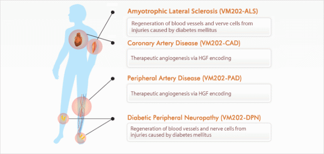
VM202 is a plasmid (small circle of DNA) that encodes the human hepatic growth factor (HGF) gene. When injected into muscles, VM202 is readily taken up by nearby cells that then quickly synthesize the two isoforms of HGF. Heightened HGF concentrations can treat ischemic cardiovascular diseases by inducing the formation of new blood vessels (angiogenesis). These new collateral vessels increase blood flow and tissue perfusion in the sick tissue, which effectively treats any tissue ischemia.

VM202

Severe obstruction of the arteries that feed the extremities (hands, feet and legs) is the cause of critical limb ischemia (CLI). The term “ischemia” refers to the starvation of a tissue for oxygen. The lack of sufficient blood flow to an organ or tissue can cause severe pain and even skin ulcers, sores, or gangrene. CLI-induced pain can awaken the patient during the night, and, therefore, is called “rest pain.” Rest pains often occur in the leg and is usually temporarily relieved by dangling the leg over the bed or getting up and walking.

CLI does not improve on its own. It is a severe condition that requires immediate by a vascular surgeon or vascular specialist.

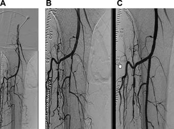
Look at the right side of these angiograms and you will see that a vessel is obstructed and blood is not flowing through it. This is an example of Critical Limb Ischemia.

In this Phase 2 study, patients were divided into three groups, one of which received a placebo treatment, the second of which received a low-dose treatment VM202, and a third group that received a high-dose of VM202.

Both patient groups that received VM202 showed improvement compared to the placebo group, but patients in the higher-dose group showed significantly better ulcer healing and higher tissue oxygen levels than the placebo group. For example, 62 percent of the ulcers healed in patients treated with high-dose VM202 compared to only 11 percent of ulcers in patients who were treated with the placebo. Also, 71 percent of patients who received the high-dose VM202 showed improved oxygen concentrations in their tissues, compared to only 33 percent of patients who were treated with the placebo.

Emerson C. Perin, Director of the Stem Cell Center at the Texas Heart Institute and the principal investigator of this Phase 2 study, said: “These positive results are exciting, and VM202 shows great promise for treating patients with this debilitating disease who often have limited therapeutic options. We are looking forward to conducting a phase III trial to better understand the potential of this novel approach, especially in treating non-healing ulcers, which is a serious symptom that often leads to amputation because of the lack of medical therapies available.”

ViroMed has already been granted an IND or Investigational New Drug approval by the USFDA to initiate a Phase 3 study in diabetic patients who suffer from non-chronic ischemic foot ulcers. This study will enroll 300 subjects who will be divided into a VM202 group and a placebo group. The treatment regiment will mimic that of this smaller Phase 2 study and will only follow patients for seven months. This time, ViroMed is interested in determining if VM202 helps wound closure, which will constitute the primary efficacy endpoint on this new study.

Godspeed ViroMed!!

First Clinical Trial for Genetically Engineered Stem Cell Treatment for Pulmonary Arterial Hypertension


A Canadian research team has published the results of the world’s first clinical trial of a genetically enhanced stem cell therapy for pulmonary arterial hypertension (PAH).

PAH is a rare and deadly disease that mainly affects young women, and is characterized by very high blood pressure in those arteries that supply blood to the lungs. Some cases of PAH are caused by mutations in the BMPR2 gene, but in many cases the cause remains unknown. Currently, PAH patients are treated with combination of various drug and oxygen. Drug treatments include blood vessel dilators, such as epoprosternol (Flolan or the inhaled form known as iloprost or Ventavis), endothelin receptor antagonists, such as bosentan (Tracleer) or ambrisentan (Letaris), sildenafil (Viagra) or tadalafil (Cialis), high doses of calcium channel blockers, anticoagulants, and diuretics. Such treatments can improve symptoms and exercise capacity (at best), but they cannot repair the blood vessel damage to the lungs or cure the disease.

This new study, entitled “Endothelial NO-Synthase Gene-Enhanced Progenitor Cell Therapy for Pulmonary Arterial Hypertension: the PHACeT Trial“ was published in the journal Circulation Research, and was coauthored lead investigator Duncan J. Stewart of the Ottawa Hospital Research Institute, and his collaborators.

The paper describes PAH as a progressive and eventually lethal disease that is characterized by eventual loss of functional lung microvasculature. This paper also argues that cell-based therapies offer the possibility of repairing and regenerating the lung microcirculation. The paper also reports that stem-cell therapy has shown promise in a pre-clinical evaluation that utilized experimental models of PAH.

This trial was a phase 1, dose-escalating clinical study whose goal was to test the tolerability, feasibility, and side-effects of a genetically-enhanced stem cell therapy to repair and regenerate lung blood vessels in PAH patients. Seven PAH patients who volunteered for this study underwent a blood cell selection process known as apheresis in order to harvest a certain population of white blood cells from their blood. These white blood cells were grown in the laboratory under special conditions that specifically selected for stem-like cells called endothelial progenitor cells (EPCs). These EPCs were genetically engineered to produce greater amounts of nitric oxide synthase, which makes the signaling molecule, nitric oxide (NO), a natural substance that widens blood vessels and is essential for efficient vascular repair and regeneration. These genetically enhanced cells were then injected directly into the lung circulation of the patient from whom there were originally harvested.

Of these seven patients, five were female and two were male, and all seven patients received treatment from December 2006 to March 2010. Continued observation and follow-up exams of these patients showed that the cell infusion procedure was well tolerated, and, on the whole, these patients showed a trend towards improvement in total pulmonary resistance (TPR) over the three-day delivery period. However, there was one serious adverse event (death) that occurred immediately after discharge in a patient who had severe, end-stage disease.

These investigators concluded that delivery of EPCs overexpressing eNOS was tolerated in PAH patients, and also produced evidence of short-term improvements, associated with long-term benefits in functional and quality-of-life assessments. However, they caution that future studies will be needed in order to further establish the efficacy of this therapy.

It must be noted that this study was not designed to rigorously assess the benefits the stem cell therapy versus a placebo. However, this research group observed improved blood flow in the lungs of patients during days following the therapy, and enhanced ability to exercise and better quality of life for up to six months after the therapy. Once again, I must provide the caveat that since this was not a double-blinded, placebo-controlled study, it is no possible to determine for sure if these observed effects were due to the cells or to psychological effects.

The therapy was generally well-tolerated, but one patient who had very severe and disease and signs of poor prognosis died one day after treatment. As unfortunate as this is, it is an expected outcome, given how sick the patient was and given their declining condition prior to treatment.

“Pulmonary arterial hypertension is a deadly and incurable disease that often strikes people in the prime of their life,” says the Circulation Research paper’s senior author Dr. Duncan Stewart, a practicing cardiologist and Executive Vice-President of Research at The Ottawa Hospital, and a professor of medicine at the University of Ottawa. “We desperately need new therapies for this disease, and regenerative medicine approaches have shown great promise in laboratory models and in clinical trials for other conditions.”

“This trial shows that genetically-enhanced stem cell therapy is a promising treatment approach for pulmonary arterial hypertension,” observes Dr. Stewart. “Although this is an important start, we will need to do larger studies to establish whether this therapy can produce important and durable benefits for people suffering from this challenging disease.”

Dr. Stewart is also the lead researcher of the first clinical trial in the world of a genetically-enhanced stem cell therapy for heart attack.

Artifical Blood Vessels Made From Thermoplastic Polyurethane Polymers


Wherever we find some of the worse medical events – heart attacks, strokes, pulmonary embolisms, we find blocked blood vessels. Obstructed blood vessels are a lurking time bomb in our bodies and they usually have to be replaced. Blood vessel replacement requires cutting another blood vessel from another part of the body or the implantation of artificial vascular prostheses.

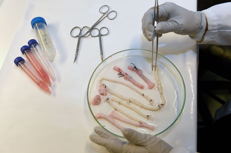
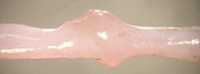
A new option might emerge in the future, however. Vienna University of Technology, in collaboration with the Vienna Medical University developed artificial blood vessels that were fabricated from specialized elastomer material that have excellent mechanical properties. After implantation, these artificial blood vessels are dissolved and replaced by the body’s own blood vessels. At the end of the healing process, natural, fully functional blood vessels are once again in place. The technique works quite well in tissue cultures systems, but now it has been shown to successfully regenerate blood vessels in laboratory animals, specifically rats.

Atherosclerotic vascular disorders, in which blood vessels are obstructed by cholesterol-filled plaques, are one of the most common causes of death in industrialized countries. Typically, patients are treated with a bypass operation, and for such procedures, blood vessels are extirpated from another part of the patient’s body and used to replace the damaged vessel. This creates a new wound and a new area of the body with less than optimal blood supply that must heal. Also, the transplanted vessel rarely has the properties necessary to thrive in its new location.

This new strategy to replace diseased blood vessels is the result of a fruitful collaboration between Vienna University of Technology (or TU Wien, which is short for Technische Universität Wien) and the Medical University of Vienna. Hopefully the success of this research will cause artificially manufactured vessels to be used more frequently in future.

To make an artificial blood vessel, the most important thing is to start with the right material. The material must be compatible with body tissue, and pliable enough to be formed into a small diameter tube that is not easily blocked by blood clots.

Extensive work at TU Wien has resulted in the development of new polymers. “These are so-called thermoplastic polyurethanes,” explains Robert Liska from the Institute of Applied Synthetic Chemistry of TU Wien.  “By selecting very specific molecular building blocks we have succeeded in synthesizing a polymer with the desired properties.”

In order to generate artificial blood vessels from their thermoplastic polyurethanes, TU Wien materials scientists spun polymer solutions in an electrical field. This allowed them to form very fine threads and that could be wound into a spool. “The wall of these artificial blood vessels is very similar to that of natural ones,” says Heinz Schima of the Medical University of Vienna. The thermoplastic polyurethanes form a polymer fabric that is slightly porous and allows a small amount of blood to leak through it. This also enriches the blood vessel wall with growth factors, which encourages the migration of endothelial progenitor cells. Martina Marchetti-Deschmann at TU Wien studied the interaction between the thermoplastic polyurethane material and blood by using spatially resolved mass spectrometry.

This new technology has already proven to successfully form functional blood vessels in rats. “The rats’ blood vessels were examined six months after insertion of the vascular prostheses,” says Helga Bergmeister of MedUni Vienna. “We did not find any aneurysms, thromboses or inflammation. Endogenous cells had colonized the vascular prostheses and turned the artificial constructs into natural body tissue.” In fact, the body’s own blood vessel-forming tissues re-grew significantly faster than expected, which shortened the degradation period of the plastic tubes and their replacement with the body’s own endothelial cells. TU Wein and the Medical University of Vienna are making further adaptations to the material.

A few more preclinical trials are necessary before the artificial blood vessels can be used in human clinical trials. However, based on the results so far, the research team is very confident that the new method will prove itself for use in humans in a few years’ time.

This project was recently awarded PRIZE prototype funding from Austria Wirtschaftsservice (AWS).

Amniotic Fluid Stem Cells Make Robust Blood Vessel Networks


The growth of new blood vessels in culture received in new boost from researchers at Rice University and Texas Children’s Hospital who used stem cells from amniotic fluid to promote the growth of robust, functional blood vessels in healing hydrogels.

These results were published in the Journal of Biomedical Materials Research Part A.

Engineer Jeffrey Jacot thinks that amniotic fluid stem cells are valuable for regenerative medicine because of their ability to differentiate into many other types of cells, including endothelial cells that form blood vessels. Amniotic fluid stem cells are taken from the discarded membranes in which babies are encased in before birth. Jacot and others combined these cells with an injectable hydrogel that acted as a scaffold.

In previous experiments, Jacot and his colleagues used amniotic fluid cells from pregnant women to help heal infants born with congenital heart defects. Amniotic fluids, drawn during standard tests, are generally discarded but show promise for implants made from a baby’s own genetically matched material.

“The main thing we’ve figured out is how to get a vascularized device: laboratory-grown tissue that is made entirely from amniotic fluid cells,” Jacot said. “We showed it’s possible to use only cells derived from amniotic fluid.”

Researchers from Rice, Texas Children’s Hospital and Baylor College of Medicine combined amniotic fluid stem cells with a hydrogel made from polyethylene glycol and fibrin. Fibrin is the proteins formed during blood clots, but it is also used for cellular-matrix interactions, wound healing and angiogenesis (the process by which new vessels are made). Fibrin is widely used as a bioscaffold but it suffers from low mechanical stiffness and is degraded rapidly in the body. When fibrin was combined with polyethylene glycol, the hydrogel became much more robust, according to Jacot.

Additionally, these groups used a growth factor called vascular endothelial growth factor to induce the stem cells to differentiate into endothelial cells. Furthermore, when induced in the presence of fibrin, these cells infiltrated the native vasculature from neighboring tissue to make additional blood vessels.

When mice were injected with fibrin-only hydrogels, thin fibril structures formed. However if those same hydrogels were infused with amniotic fluid stem cells that had been induced with vascular endothelial growth factor, the cell/fibrin hydrogel concoctions showed far more robust vasculature.

In similar experiments with hydrogels seeded with bone marrow-derived mesenchymal cells, once again, vascular growth was observed, but these vessels did not have the guarantee of a tissue match. Interestingly, seeding with endothelial cells didn’t work as well as the researchers expected, he said.

Jacot and others will continue to study the use of amniotic stem cells to build biocompatible patches for the hearts of infants born with birth defects and for other procedures.

Mesenchymal Stem Cells from Neonatal Thymus Helps Make New Blood Vessels


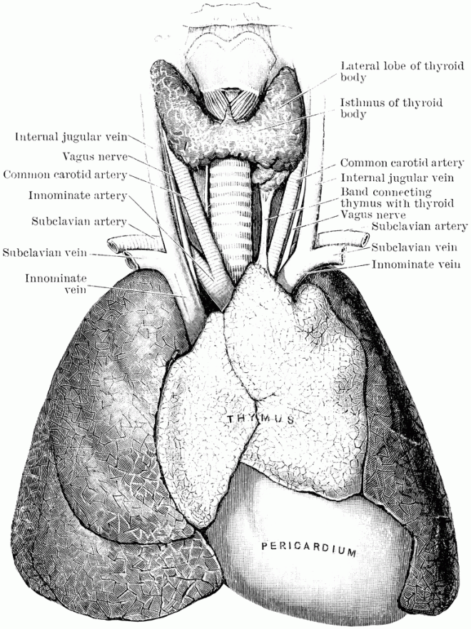
The thymus is an organ that sits over the top of the heart and it plays a pivotal role in the development of T-lymphocytes. The thymus is a very durable organ that can readily regenerate if it is injured. This regenerative ability is largely due to it high level of vascularization (lots of blood vessels). This vascularization is due to a robust population of resident mesenchymal stem cells that supports blood vessel formation in the damaged thymus. The process of blood vessel formation is called “angiogenesis.” The angiogenic potential of these thymus-based mesenchymal stem cells might hold excellent potential for regenerative therapies.

Thymus_lg

As it turns out, neonatal surgeries tend to generate thymus tissue that is usually thrown out as medical waste. Ming-Sing Si from Mott’s Children Hospital in Ann Arbor, Michigan and colleagues isolated mesenchymal stem cells from these surgically-derived neonatal thymuses and tested their ability to stimulate blood vessels in an experimental setting.

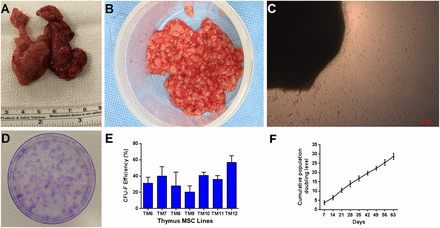
Discarded thymus tissue was obtained from the University of Michigan, and this tissue was minced, degraded with enzymes, and cultured. The mesenchymal stem cells (MSCs) moved from the thymus tissue onto the culture dishes. These thymus-based MSCs grew like gangbusters in culture and could be passaged over 30 times.

Discarded human neonatal thymus tissue is a source of mesenchymal stromal cells (MSCs). (A): Discarded human neonatal thymus tissue during pediatric cardiac surgery. (B): Minced thymus tissue prior to plating. (C): Cells migrating from thymus tissue fragments during explant culture at 10 days. (D): Clonogenicity of thymus MSCs at 2 weeks (representative of 7 donors). (E): Colony-forming efficiency of thymus MSCs. (F): Averaged cumulative population doubling of thymus MSCs (n = 4) over 9 weeks of culture. Abbreviation: CFU-F, fibroblastic colony-forming unit.

When these thymus-based MSCs were combined with human umbilical vein endothelial cells, within one day, the cells formed an extensive network of blood vessels.

Thymus mesenchymal stromal cells (MSCs) cooperate with human umbilical vein endothelial cells (HUVECs) to form a network in a two-dimensional angiogenesis assay. (A): Monolayer appearance of HUVECs after 48 hours of culture on fibrin hydrogel. (B): Thymus MSCs clustered together after 24 hours of culture on fibrin hydrogel. (C): Combining HUVECs with thymus MSCs (2:1) resulted in the appearance of interconnected tubules at 24 hours. Scale bars = 100 μm. Results are representative of two independent experiments.

Gene expression studies showed that culturing thymus MSCs with human umbilical vein endothelial cells (HUVECs) caused the HUVECs to express a variety of blood vessel-specific genes.  These thymus-based MSCs were also able to induce blood vessels if the cells were wadded up into a ball (spheroids).

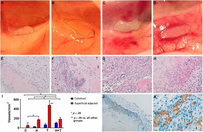
To top it all off, Si and others implanted thymus-based MSCs underneath the skin of nude mice.  They used hydrogels with no cells, hydrogels plus HUVECs, hydrogels plus thymus-based MSCs, and hydrogels with thymus-based MSCs plus HUVECs.  The control implants and the HUVEC implants showed no blood vessels.  HUVECs make very good blood vessels, but they have to be directed to do so.  Both the thymus-based MSCs and the MSCs plus HUVECs showed extensive integration into the host tissue with lots of blood vessels.

Thymus mesenchymal stromal cells (MSCs) incite angiogenesis in vivo. Fibrin constructs without spheroids (control) or with 500 spheroids with 600 human umbilical vein endothelial cells (HUVECs) per spheroid, 200 thymus MSCs per spheroid, or 600 HUVECs plus 200 thymus MSCs per spheroid were generated (n = 3 per group) and were implanted subcutaneously for 14 days in NOD-SCID mice. Explanted constructs were photographed (edges traced in A–D) and processed for histology. (A): Controls did not manifest local reaction. (B): HUVEC constructs appeared avascular. (C): Thymus MSC constructs were integrated and caused increased adjacent vascularization. (D): HUVEC plus thymus MSC constructs were integrated and surrounded by a host vascular response and appeared to have vessels within. (E–H): Construct hematoxylin and eosin staining. Scale bars = 50 μm. (E): Avascular tissue invasion of control construct. Scale bar = 100 μm. (F): HUVEC construct with adjacent cellularity and vascularity between panniculus carnosus muscle layer (∗) and construct. “Ghost” (†) of the prior locations of spheroid and necrotic spheroid (‡) were present in internal regions of all constructs with spheroids. (G): Thymus MSC construct with increased adjacent cellularity and vascularity. (H): HUVEC plus thymus MSC construct with increased vascularization within the construct. (I): Manual measurement of vessel density demonstrates significant differences by two-way analysis of variance. Control and HUVEC constructs had minimal adjacent vascularization. Thymus MSC constructs promoted the greatest adjacent response, whereas HUVEC plus thymus MSC constructs contained the greatest vessel density within the construct. (J, K): Immunohistochemical staining with human-specific CD31 monoclonal antibody revealed that only constructs with HUVEC plus thymus MSCs contained CD31-positive luminal structures with blood cells. Scale bar = 20 μm. Abbreviations: C, controls; H, human umbilical vein endothelial cell constructs; T, thymus mesenchymal stromal cell construct.

These MSCs show low expression of human leukocyte antigen class I, which, translated, means that these cells are unlikely to be recognized by the patient’s immune system.  Therefore, these cells could be donated to patients whose resident MSCs are of poor quality or do not have enough of their own MSCs for therapeutic processes.

This paper shows that discarded neonatal thymus contains large numbers of resident MSCs that can be isolated and cultured by a standard explant method.  These MSCs have all the characteristics of traditional MSCs, but have more robust growth characteristics in culture.  These thymus MSCs also possess outstanding proangiogenesis qualities that should be further tested and considered as promoters of tissue and organ regeneration in tissue engineering strategies.

Stem Cells from Fat Improve Blood Vessel Responses after Injury


When tissues are injured, the blood vessels that feed them are often shocked and damaged as well. “Vasoactivity” refers the ability of blood vessels to dilate or constrict. When tissues are harmed, blood vessels tend to shrink in order to squelch blood loss at the site of damage. This same response, however, and deprive the damaged tissues of much-needed oxygen and lead to “ischemia,” which is the insufficient supply of blood and oxygen to an organ.

James B. Hoying and his colleagues at the University of Louisville in Kentucky used the “stromal vascular fraction” or SVF from fat in order to treat damaged blood vessels to determine if they could mitigate the decrease in vasoactivity as a result of injury.

The SVF refers to the stem fraction from fat after the fat has been minced, digested with enzymes, and centrifuged (it’s more complicated than that, but this is a short summary). The cells that remain include mesenchymal stromal cells, growth factors, immune cells, pre-fat cells and fat cells, blood-cell-making stem cells, and blood vessel-making cells (endothelial cells). The SVF, therefore, contains a cocktail of cell types and growth factors that are available for regenerative medicine.

Hoying and his team discovered that when fluorescent SVF cells were injected into a laboratory mouse, they cells distributed to a variety of tissues. Further and more detailed examinations showed that these cells were finding their ways into organs and tissues because they traveled through the circulatory system and could be found in the walls of blood vessels.

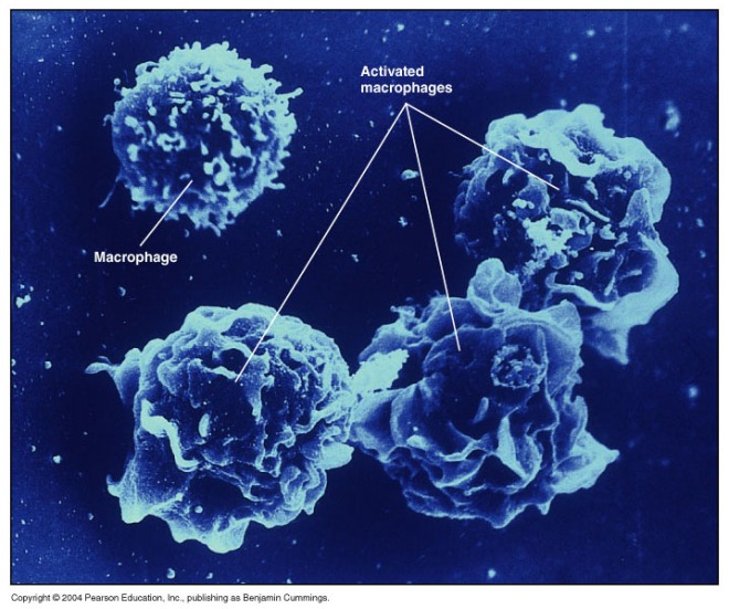
Next, the composition of the SVF was examined. About 25% of the cells in the SVF were endothelial cells, 22% were various types of blood cells, 20% were “CD11b” cells, which means that these cells had a protein called CD11b on their cell surfaces. That protein was formerly canned “Mac-1” and is was normally found on the surfaces of phagocytic cells called macrophages. Therefore, this CD11b faction could very well be macrophages, but other cell types have this protein on their surfaces as well.

Macrophages

Next, Hoying and others injected these SVF-derived cells into the large leg vein (saphenous) of the leg. Such injections consistently caused these vessels to relax and dilate. Secondly, the SVF-derived cells caused the vessels to relax in a CD11b-dependent manner. In other words, the more CD11b cells there were in the SVF preparation, the greater the amount of vasoactivity they induced. If fractions were depleted of their CD11b, they could not induce vasoactivity.

When Hoying and others examined the SVF-treated vessels, they saw CD11b+ cells lining the inner layer of the vessels. Thus these cells were getting right up against the inside of the vessel and signaling to the underlying smooth muscle to relax.

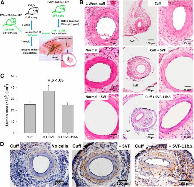
Finally, Hoying and others clamped the saphenous veins of laboratory mice. Such clamping will induce tissue ischemia and inflammation in the vessels. Can SVF cells calm the inflammation and make the vessels more vasoactive? The answer is an unqualified yes.  See below.  The veins from SVF-treated animals show signficantly greater dilation than those from untreated or CD11b-depleted SVF-treated animals.

SVF cells relax vasomotor tone in inflamed saphenous arteries. (A): Schematic of the experimental plan involving the cell treatment of locally inflamed (cuffed) saphenous arteries of mice injected with syngeneic adipose SVF cells constitutively expressing luciferase and GFP reporter transgenes or SVF cells depleted of CD11b+ cells. Also shown is a gross view and a histological cross-section of a cuffed saphenous artery. (B): Hematoxylin and eosin-stained histological cross-sections of normal (noncuffed) and cuffed mouse saphenous arteries untreated or injected with SVF cells or SVF-11bΔ cells. Rightmost panels: Higher magnification images of the adjacent images. Scale bars = 25 μm in the left and right columns and 100 μm in the middle column. (C): Lumen diameters of untreated (n = 9) and cell-injected cuffed saphenous arteries measured from histological sections. Cell treatments included complete SVF cell isolates (C + SVF, n = 7) or SVF isolates depleted of CD11b+ cells (C + SVF-11bΔ, n = 7). Data are shown as the mean ± SEM; ∗, p < .05, determined by one-way analysis of variance. (D): Visualization of luciferase-positive SVF cells within histological paraffin sections of cuffed saphenous arteries from untreated, SVF-injected, and SVF-11bΔ-injected mice via immunostaining for luciferase. Brown stain indicates positive luciferase immune-staining and the presence of SVF cells. Tissues were harvested 1 week after cell delivery. Scale bars = 100 μm. Abbreviations: C, cuff; GFP, green fluorescent protein; PE, polyethylene; SVF, stromal vascular fraction; SVF-11bΔ, CD11b+ cell-depleted adipose SVF cells.

This an interesting and exciting finding not only because of the ability of these fat-based cells to maintain vasoactivity even under pro-inflammatory conditions, but because it is the macrophage cell population that is doing the work.  In most stem preparations, macrophages are excluded.  This paper shows that macrophages have greater therapeutic capabilities than previously thought, and should also be tested for sanative properties.

Preconditioning Your Way to Better Stem Cells


When stem cells are implanted into injured tissues, they often face a hostile environment that is inimical to their survival. A stroke, for example, can produce brain tissue without ample blood flow, low oxygen levels, and lots of cell debris and inflammation. The same can be said for the heart after a heart attack. If stem cells are going to help anyone we have to find a way for them to survive.

The first hints came in the form of genetically-engineered stem cells that expressed a host of genes that can help cells survive in low oxygen, high stress environments. However, the FDA is unlikely to approve genetically engineered cells for therapeutic purposes. Therefore, a more “user-friendly” way to precondition cells was sought, and found. Instead of loading cells up with extra genes, all you had to do was grow the cells under low oxygen, high stress conditions, and they would adapt and survive when implanted into damaged tissue. This, however, has a drawback: if you want to treat a patient, you do not always have the time it takes for extract and isolate their cells, grow them in culture over a week or two, and then implant them. Is there a better way?

The answer turns out to be yes. Treating cells with particular compounds or growth factors can induce resistance to low-oxygen, high-stress conditions, and two papers show us how it’s done.

The first paper is from the laboratory of Ling Wei at Emory University School of Medicine in Atlanta who has shown in the past that low-oxygen adaptation of mesenchymal stem cells from bone marrow made them better able to treat acute heart attacks in laboratory animals. In this paper, Wei and her colleagues exploited a biochemical pathway known to induce resistance of low-oxygen conditions known as the HIF-1 pathway. The HIF-1 pathway consists of two proteins that work as a pair; HIF1alpha and HIF1beta. HIF1beta is made all the time and HIF1alpha is oxygen sensitive. In the presence of oxygen, enzymes called prolyl hydroxylases modify HIF1alpha, marking it for destruction. In the absence of oxygen, the prolyl hydroxylases do not have enough oxygen to modify HIF1alpha and the HIF1alpha/beta complex activates the expression of a host of genes necessary for increased tolerance to low oxygen levels. Therefore, to make cells more tolerant to low oxygen levels, we need to turn on the HIF1 pathway and to do that we need to inhibit the prolyl hydroxylases.

This turns out to be pretty straight forward. A small molecule called dimethyloxalyglycine or DMOG can effectively inhibit prolyl hydroxylase and induce survival in low-oxygen, high-stress environments. Therefore Wei and her group used DMOG to treat cells and test them out.

In culture, the DMOG-treated cells made proteins known to be important for the establishment of new blood vessels and for survival. When they were compared to cultured stem cells that had not been treated with DMOG, the DMOG-treated cells expressed significantly more of VEGF, Glut-1 and HIF1alpha, all of which are important for surviving in low-oxygen environments. In a Matrigel assay, the DMOG-treated cells also made more blood vessels that were longer than their non-DMOG-treated counterparts.

When used in laboratory animals that had suffered heart attacks, the DMOG-treated cells distinguished themselves once again. They survived better than the control cells and hearts that had received the DMOG-treated cells had much smaller heart scars after heart attacks. Functional assays of heart function illustrated that the DMOG-treated cells helped their heart perform above and beyond what was shown observed in the animals implanted with stem cells that had not bee treated with DMOG.

Thus it is possible to precondition cells without long culture periods or genetic engineering. One compound can accomplish it and the cells only needed to be exposed to DMOG for 24 hours.

In a similar vein, Genshan Ma and others from Zhongda Hospital in Nanjing, China used a small peptide called bradykinin to precondition human umbilical cord endothelial progenitor cells (EPCs); the cells that form blood vessels. In this paper, Ma and colleagues used bradykinin-treated EPCs to treat heart attacks in mice. One nice aspect of this paper is the large number of controls they ran with their experimental runs.

The bradykinin-treated cells outperformed their untreated counterparts when it came to the size of the heart scar, the number of dead cells in the heart, and heart performance parameters. Cell culture experiments established that the bradykinin-treated cells expressed the Akt kinase at high levels, and expressed higher levels of VEGF, the blood vessel-inducing growth factor. Bradykinn-treated cells also were more resistant to being starved for oxygen, and survived better under unusual culture conditions. All of these benefits could be abrogated by inhibiting the activity of the Akt kinase by treating cells with LY294002, a compound that specifically inhibits the activator of Akt.

In this case, cells were treated with bradykinin for 10 minutes to 12 hours.

Two papers, two success stories. Stem cell preconditioning certainly works in laboratory animals. Since stem cell trials have been completed in human patients, it might be time to try preconditioned stem cells in human patients.

Mesenchymal Stem Cell Transplantation Improves Atherosclerotic Lesions


Several animal studies have shown that transplantation of mesenchymal stem cells from several different sources is beneficial in myocardial infarction and hind limb ischemic. However, can these cells improved atherosclerosis, otherwise known as hardening of the arteries?

Shih-Chieh Hung and colleagues from National Yang-Ming University in Taipei, Taiwan tested this very hypothesis.

Hung and others used to lines of experimentation to address this question. First, they used cultured endothelial cells that had been treated with oxidized low-density lipoprotein particles. Secondly, they fed mice mutant for ApoE-deficient a high-fat diet.  ApoE-deficient humans and mice develop atherosclerotic plaques rather quickly.

In the cultured endothelial cells, oxidized LDL turned off the production of nitric oxide (NO). NO is a signaling molecule produced by several cell types, but in particular, endothelial cells use NO to dilate blood vessels. NO also is a good signal of endothelial health. Therefore, when oxidized LDL causes cultured endothelial to decrease NO production, it is affecting endothelial cell health. However, when cultured endothelial cells that had been treated with oxidized LDL were cocultured with mesenchymal stem cells, NO production and the enzymes that synthesize NO increased precipitously. Thus in a cultured system, MSCs have the ability to prevent the deleterious of oxidized LDL.

In ApoE-deficient mice fed a high fat diet, the arteries of the mice showed extensive plaque formation. However, if these animals were implanted with bone-marrow-derived mesenchymal stem cells, plaque formation was greatly decreased. Further work showed that a protein secreted by mesenchymal stem cells called macrophage inflammatory protein-2 (MIP-2) was responsible for these ameliorative effects. If MIP-2 was applied without mesenchymal stem cells, plaque formation was limited, and if antibodies that neutralize MIP-2 were co-administered with mesenchymal stem cells, the cells failed to reduced plaque formation.

Thus, this interesting study shows that transplantation of mesenchymal stem cells can limit plaque formation in atherosclerotic animals and they do this through secretion of MIP-2. Secondly, mesenchymal stem cells can improve the health of endothelial cells, which are the cells that form the inner layer of blood vessels, which are so adversely affected by atherosclerosis. By utilizing the encore of proteins secreted by mesenchymal stem cells, scientists should be able to develop a cocktail of proteins that can ameliorate atherosclerosis in human patients.

Young Blood Vessels Rejuvenate Aged Insulin-Producing Beta Cells


Professor Per-Olof Berggren Rolf Luft at the Research Center for Diabetes and Endocrinology at Karolinska Institutet has led an important study in collaboration with Alejandro Caicedo at University of Miami Miller School of Medicine and Hong Gil Nam at DGIST from the Republic of Korea that demonstrates that young capillary vessels can rejuvenate aged pancreatic islets. This study was published in the Proceedings of the National Academy of Sciences, USA and challenges prevailing views on the causes of age-dependent impaired glucose balance regulation, which often, in older patients, develops into type 2 diabetes mellitus.  These results suggest that treating inflammation and fibrosis in the small blood vessels of the pancreatic islets might provide a new way to treat age-dependent dysregulation of blood glucose levels.

“This is an unexpected but highly important finding, which we expect will have a significant impact on diabetes research in the future. The results indicate that beta cell function does not decline with age, and instead suggest that islet function is threatened by an age-dependent impairment of vessels that support them with oxygen and nutrients”, says Berggren.

Pancreatic beta cells are in the pancreatic islets and secrete the hormone insulin, which regulates blood glucose levels and also is one of the most important anabolic hormones of the human body.  Ageing may lead to a progressive decline in glucose regulation which may contribute to the onset of diabetes.  Generally, it has been assumed that this is due to reduced capacity of the beta cell to secrete insulin or multiply.

“In the study we challenged the view that the age-dependent impairment in glucose homeostasis is solely due to intrinsic, dysfunction of islet cells, and hypothesized that it is instead affected by systemic aging factors”, says first author Joana Almaça at the Diabetes Research Institute, University of Miami.

Even though the common wisdom in modern medicine is that insulin-producing pancreatic beta cell lose function through the constant demands on them,  Berggren and his collaborators showed that mouse and human beta cells are fully functional at advanced age.  When they replaced the islet vasculature in aged islet grafts with young capillaries, the investigators found that the islets were rejuvenated and glucose homeostasis fully restored.

“While expanding beta cell mass may still be desirable for future diabetes therapy, improving the local environment of the otherwise healthy aged beta cell could prevent age-associated deterioration in glucose homeostasis and thereby promote healthy ageing, which is conceptually novel and highly exciting”, says Per-Olof Berggren.

Mesenchymal Stem Cells Make Blood Vessel Cells and Improve Wound Healing


Mesenchymal stem cells from umbilical cord have the ability to differentiate into cartilage cells, fat cells, bone cells, and blood vessels cells. These cells also are poorly recognized by the immune system of the patient and are at a low risk of being rejected by the patient’s immune system.

Valeria Aguilera and her colleagues from the laboratory of Claudio AguayoWe at the University of Concepción, Chilee have evaluated the use of mesenchymal stem cells from umbilical cord in the formation of new blood vessels in damaged tissues. Wharton’s jelly mesenchymal stem cells of hWMSCs were used to potentially accelerate tissue repair in living animals.

Aguilera and her co-workers began by isolating mesenchymal stem cells from human Wharton’s jelly (a connective tissue in umbilical cord). Then they grew these cells in culture for 14 or 30 days. Interestingly, the longer the WMSCs grew in culture, the more they looked like blood vessel cells. They began to express blood vessel-specific genes and proteins. WMSCs cultured for 30 days were even more like blood vessels than those grown in culture for 14 days.

When these cells were injected in the mice with damaged skin, the results showed that the WMSCs cultured for 30 days significantly accelerated wound healing compared with animals injected with either undifferentiated hWMSCs or with no cells.

Effect of hWMSCs and endothelial-differentiated hWMSC transplantation in a wound-healing model. A) Representative images of wounds at day 1 (top panels) and 12 (lower panels) after injury and subcutaneous injection of hWMSCs, hWMSC trans-differentiated into endothelial cells for 14 days (hWMSC-End14d) or 30 days (hWMSC-End30d), or control (PBS). B) Wound healing quantified in PBS (○), hWMSC (•), hWMSC-End14d (□) or hWMSC-End30d (▪) treated mice (n = 5 independent experiments, in duplicate). Values are expressed as mean±S.E.M, +P<0.05 in hWMSC-End30d v/s hWMSC, hWMSC-End14d, at the corresponding time; **P<0.03 in hWMSC-End30d v/s PBS; *P<0.001 in hWMSC-End30d v/s PBS; # P<0.01 in hWMSC-End30d v/s PBS.
Effect of hWMSCs and endothelial-differentiated hWMSC transplantation in a wound-healing model.
A) Representative images of wounds at day 1 (top panels) and 12 (lower panels) after injury and subcutaneous injection of hWMSCs, hWMSC trans-differentiated into endothelial cells for 14 days (hWMSC-End14d) or 30 days (hWMSC-End30d), or control (PBS). B) Wound healing quantified in PBS (○), hWMSC (•), hWMSC-End14d (□) or hWMSC-End30d (▪) treated mice (n = 5 independent experiments, in duplicate). Values are expressed as mean±S.E.M, +P


The wounds of mice treated with the WMSCs cultured for 30 days looked healthier, but they had many more blood vessels.

Histologic analysis of wounds in the wound-healing model. A) Representative photographs of wounds (hematoxilin/eosin staining) 12 days after injury and subcutaneous injection of PBS, hWMSCs, hWMSC-End14d or hWMSC-End30d. Quantification of histological images, for blood vessels area (B) and histological score (C) for each group of mice. Values are mean ± S.E.M (n = 5 independent experiments, in duplicate), *P<0.001 in hWMSC-End30d or hWMSC-End14d v/s MSC; +P<0.05 in hWMSC-End30d or hWMSC-End14d v/s hWMSC. Magnification x40 (-). Ep, epidermis; D, dermis; H, hypodermis.
Histologic analysis of wounds in the wound-healing model.
A) Representative photographs of wounds (hematoxilin/eosin staining) 12 days after injury and subcutaneous injection of PBS, hWMSCs, hWMSC-End14d or hWMSC-End30d. Quantification of histological images, for blood vessels area (B) and histological score (C) for each group of mice. Values are mean ± S.E.M (n = 5 independent experiments, in duplicate), *P

When laboratory animals received the culture medium from the WMSCs cultured for 30-days also showed significant acceleration of their healing, which suggests that these cells secrete a host of healing molecules that induced the formation of new blood vessels.  One might also conclude that the implanted WMSCs did not contribute to the formation of new blood vessels, but simply directed the formation of new blood vessels by secreting healing molecules.  However, when WMSCs were detected in the healed tissue, they were predominantly found in the walls of new blood vessels.

Immunohistochemical detection of human mesenchymal cells in a wound-healing model. A. Immunohistochemical staining of human mitochondria was performed in permeabilized tissue sections obtained after 12 days of subcutaneous injection of PBS, hWMSCs, hWMSC-End14d or hWMSC-End30d in mice. Cell nuclei were stained with hematoxyline. In B. Number of positive cells per vessel. Representative images of 5 independent experiments, in duplicate. Magnification x40 and insert 100x. Bars 50 µm.
Immunohistochemical detection of human mesenchymal cells in a wound-healing model.
A. Immunohistochemical staining of human mitochondria was performed in permeabilized tissue sections obtained after 12 days of subcutaneous injection of PBS, hWMSCs, hWMSC-End14d or hWMSC-End30d in mice. Cell nuclei were stained with hematoxyline. In B. Number of positive cells per vessel. Representative images of 5 independent experiments, in duplicate. Magnification x40 and insert 100x. Bars 50 µm.

These results, which were published in PLOS ONE, demonstrate that mesenchymal stem cells isolated from umbilical cord connective tissue or Wharton’s jelly can be successfully grown in culture in the laboratory and trans-differentiated into blood vessels-forming cells (endothelial cells).  These differentiated hWMSC-derived endothelial cells seem to promote the formation of new networks of blood vessels, which augments tissue repair in laboratory animals through the secretion of soluble pro-blood vessel-making molecules and, occasionally, by contributing to the formation of new vessels, themselves.

A Faster Way to Make Blood Vessels


Suchitra Sumitran-Holgersson and Michael Olausson from the Sahlgrenska Academy have designed a new way to make blood vessels that takes only seven days and a few tablespoons of blood.

Thanks to their new procedure, the ability to make new tissues from stem cells has taken a huge stride forward. Three years ago, a patient at Sahlgrenska University Hospital received a blood vessel transplant grown from her own stem cells. Suchitra Sumitran-Holgersson, Professor of Transplantation Biology at Sahlgrenska Academy, and Michael Olausson, Surgeon/Medical Director of the Transplant Center and Professor at Sahlgrenska Academy, came up with the idea, planned and carried out the procedure.

Now Sumitran-Holgersson and Olausson have published a new study in the journal EBioMedicine based on two other transplants that were performed in 2012 at Sahlgrenska University Hospital. The patients in this procedure were two young children who were afflicted with the same condition as in the first patient. All three patients were missing the vein that goes from the gastrointestinal tract to the liver.

“Once again we used the stem cells of the patients to grow a new blood vessel that would permit the two organs to collaborate properly,” Professor Olausson says.  This time, however, Sumitran-Holgersson, found a way to extract stem cells that did not necessitate taking them from the bone marrow. “Drilling in the bone marrow is very painful,” she says. “It occurred to me that there must be a way to obtain the cells from the blood instead.”

The extreme youth of these patients motivated Sumitran-Holgersson find a new way to extract these stem cells. She came upon the extraction of stem cells from 25 millilitres (approximately 2 tablespoons) of blood, which is the minimum quantity of blood needed to obtain enough stem cells.

Then they used a novel technique to generate transplantable vascular grafts by using decellularized allogeneic vascular scaffolds that were then populated with peripheral whole blood and then grown in a bioreactor.  Circulating, VEGFR-2 +/CD45 + and a smaller fraction of VEGFR-2 +/CD14 + cells largely repopulated the graft to form new vessels for transplantation.

Fortunately, her idea worked out better than she could have ever expected, and worked perfectly the first time. “Not only that, but the blood itself accelerated growth of the new vein,” Professor Sumitran-Holgersson says. “The entire process took only a week, as opposed to a month in the first case. The blood contains substances that naturally promote growth.”

Olausson and Sumitran-Holgersson have treated three patients so far, two children and one adult. Two of the three patients have recovered well and have veins that are functioning normally. In the third case the child is under medical surveillance and the outcome is less certain.

The technology for creating new tissues from stem cells has taken a giant leap forward. Two tablespoons of blood are all that is needed to grow a brand new blood vessel in just seven days. This is shown in a new study from Sahlgrenska Acadedmy and Sahlgrenska University Hospital published in EBioMedicine.

These studies show that it is feasible to avoid taking painful blood marrow samples to extract stem cells for blood vessel production, and that it is equally feasible to produce those blood vessels in a matter of a week.

“We believe that this technological progress can lead to dissemination of the method for the benefit of additional groups of patients, such as those with varicose veins or myocardial infarction, who need new blood vessels,” Professor Holgersson says. “Our dream is to be able to grow complete organs as a way of overcoming the current shortage from donors.”

New Stem Cell Technology to Form Blood Vessels and Treat Peripheral Artery Disease


How to make new blood vessels for patients who need them? Researchers at the University of Indiana University School of Medicine have developed a new therapy for illnesses such as peripheral artery disease. Diseases such a peripheral artery disease can lead to skin problems, gangrene and sometimes amputation.

Our bodies have the ability to repair blood vessels and creating new ones, because of a cell type called “endothelial colony-forming cells.” Unfortunately, these cells tend to lose their ability to proliferate and form new blood vessels as patients age or develop diseases like peripheral arterial disease, according to Mervin C. Yoder Jr., M.D., who is the Richard and Pauline Klingler Professor of Pediatrics at IU and leader of the research team.

Physicians can prescribe drugs that improve blood flow to patients with peripheral artery disease, but if the blood vessels are reduced in number or function, the benefits from such drugs are minimal. A better treatment might be to introduce “younger,” more effective endothelial colony forming into the affected tissues. In this case, such a treatment would jump-start the creation of new blood vessels. Gathering such cells, however is rather difficult, since endothelial colony-forming cells are somewhat difficult to find in adults, especially in those with peripheral arterial disease. Fortunately, endothelial colony-forming cells are rather numerous in umbilical cord blood.

Yoder and his colleagues published their work in the journal Nature Biotechnology, and they have reported that they have developed a potential therapy by using patient-specific induced pluripotent stem cells (iPSCs). Induced pluripotent stem cells are pluripotent stem cells that are derived from normal adult cells by means of genetic engineering and cell culture techniques. Once an iPSC line has been derived from a patient, they can potentially be differentiated into any adult cells type, including endothelial colony-forming cells.

In this paper, Yoder and his research team developed a novel methodology to differentiate iPSCs into cells with the characteristics of the endothelial colony-forming cells that are found in umbilical cord blood. These laboratory-generated endothelial colony-forming cells were injected into mice, and they proliferated and generated human blood vessels that nicely restored blood flow to damaged tissues in mouse retinas and limbs

Another problem addressed in this paper was growing endothelial colony-forming cells from umbilical cord in culture so that they can achieve sufficient numbers for therapies. In this paper, Yoder and his team designed a cell culture system that was able to dramatically expand these iPSC-derived endothelial colony-forming cells in culture from one founding cell to 100 million new cells in a little less than three months.

“This is one of the first studies using induced pluripotent stem cells that has [sic] been able to produce new cells in clinically relevant numbers — enough to enable a clinical trial,” Dr. Yoder said. According to Yoder, the next steps will be to reach solidify an agreement with a facility approved to produce cells for use in human testing. Additionally, Yoder would like to treat more than just peripheral artery disease, since he and his colleagues are evaluating the potential uses of these cells to treat diseases of the eye and lungs that involve blood flow problems.

Stem Cells Make Heart-Like Cuff to Improve Venous Blood Flow


Researchers at George Washington University have used stem cells to make a new organ that help return blood from defective veins back to the heart. This mini-organ consists of heart muscle cells that surround the vein and act as a miniature heart that pulsates that aids blood flow through venous segments. This mini-cuff is made from the patient’s own stem cells (induced pluripotent stem cells), which eliminates the possibility of immunological rejection.  See video here.

Beating Venous Engineered Heart Tissue Cuff made from an excised segment of a rat posterior tibial vein
Beating Venous Engineered Heart Tissue Cuff made from an excised segment of a rat posterior tibial vein.

“We are suggesting for the first time, to use stem cells to create, rather than just repair damaged organs,” said Narine Sarvazyan, professor of pharmacology and physiology at the GW School of Medicine and Health Sciences. “We can make a new heart outside of one’s own heart, and by placing it in the lower extremities, significantly improve venous blood flow.”

Such a mini-pump might provide relief to patients who suffer from chronic venous insufficiency. which is a pervasive disease in developed countries. Chronic venous insufficiency occurs in about 20-30 percent of people over 50 years of age and is responsible for approximately 2% of health costs. A long-term condition, chronic venous insufficiency decreases the efficiency of the veins to send blood from the legs back to the heart. Chronic vein insufficiency can result from partial blockage or poorly functional, leaky valves in the veins.

Chronic venous insufficiency patients may suffer from varicose veins and ulcers on their legs and ankles. They may need surgery to remove varicose veins – if the condition causes skin sores and leg pain – and using compression stockings to decrease swelling.

The treatment option for chronic venous insufficiency outlined in a recently published paper in the Journal of Cardiovascular Pharmacology and Therapeutics, is a masterful advance in tissue engineering since it moves from organ repair to organ creation. Sarvazyan, together with members of her team, has demonstrated the feasibility of this novel approach in culture and they are working toward testing these devices in a living animal.

Because studies with valvular reconstruction or transplantation of excised autologous valves have shown that replacement of even a single valve can lead to significant improvements in the affected limb, Sarvazyan is hopeful that this treatment can provide genuine, durable relief in human patients some day.

Vascular Progenitors Made from Induced Pluripotent Stem Cells Repair Blood Vessels in the Eye Regardless of the Site of Injection


Johns Hopkins University medical researchers have reported the derivation of human induced-pluripotent stem cells (iPSCs) that can repair damaged retinal vascular tissue in mice. These stem cells, which were derived from human umbilical cord-blood cells and reprogrammed into an embryonic-like state, were derived without the conventional use of viruses, which can damage genes and initiate cancers. This safer method of growing the cells has drawn increased support among scientists, they say, and paves the way for a stem cell bank of cord-blood derived iPSCs to advance regenerative medical research.

In a report published Jan. 20 in the journal Circulation, Johns Hopkins University stem cell biologist Elias Zambidis and his colleagues described laboratory experiments with these non-viral, human retinal iPSCs, that were created generated using the virus-free method Zambidis first reported in 2011.

“We began with stem cells taken from cord-blood, which have fewer acquired mutations and little, if any, epigenetic memory, which cells accumulate as time goes on,” says Zambidis, associate professor of oncology and pediatrics at the Johns Hopkins Institute for Cell Engineering and the Kimmel Cancer Center. The scientists converted these cells to a status last experienced when they were part of six-day-old embryos.

Instead of using viruses to deliver a gene package to the cells to turn on processes that convert the cells back to stem cell states, Zambidis and his team used plasmids, which are rings of DNA that replicate briefly inside cells and then are degraded and disappear.

Next, the scientists identified and isolated high-quality, multipotent, vascular stem cells that resulted from the differentiation of these iPSC that can differentiate into the types of blood vessel-rich tissues that can repair retinas and other human tissues as well. They identified these cells by looking for cell surface proteins called CD31 and CD146. Zambidis says that they were able to create twice as many well-functioning vascular stem cells as compared with iPSCs made with other methods, and, “more importantly these cells engrafted and integrated into functioning blood vessels in damaged mouse retina.”

Working with Gerard Lutty, Ph.D., and his team at Johns Hopkins’ Wilmer Eye Institute, Zambidis’ team injected these newly iPSC-derived vascular progenitors into mice with damaged retinas (the light-sensitive part of the eyeball). The cells were injected into the eye, the sinus cavity near the eye or into a tail vein. When Zamdibis and his colleagues took images of the mouse retinas, they found that the iPSC-derived vascular progenitors, regardless of injection location, engrafted and repaired blood vessel structures in the retina.

“The blood vessels enlarged like a balloon in each of the locations where the iPSCs engrafted,” says Zambidis. Their vascular progenitors made from cord blood-derived iPSCs compared very well with the ability of vascular progenitors derived from fibroblast-derived iPSCs to repair retinal damage.

Zambidis says that he has plans to conduct additional experiments in diabetic rats, whose conditions more closely resemble human vascular damage to the retina than the mouse model used for the current study, he says.

With mounting requests from other laboratories, Zambidis says he frequently shares his cord blood-derived iPSC with other scientists. “The popular belief that iPSCs therapies need to be specific to individual patients may not be the case,” says Zambidis. He points to recent success of partially matched bone marrow transplants in humans, shown to be as effective as fully matched transplants.

“Support is growing for building a large bank of iPSCs that scientists around the world can access,” says Zambidis, although large resources and intense quality-control would be needed for such a feat. However, Japanese scientists led by stem-cell pioneer Shinya Yamanaka are doing exactly that, he says, creating a bank of stem cells derived from cord-blood samples from Japanese blood banks.

Stem Cell Treatments for Aortic Aneurysms


The aorta is the largest blood vessel in our bodies and it emerges from the left ventricle of the heart, takes a U-turn, and swings down toward the legs (descending or dorsal aorta). There are several branches of the aorta as it sharply turns that extend towards the head and upper extremities.

Aorta structure

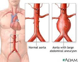
Sometimes, as a result of inflammation of the aorta or other types of problems, the elastic matrix that surrounds and reinforces the aorta breaks down.  This weakens the wall of the aorta and it bulges out.  This bulge is called an aortic aneurysm and it is a dangerous condition because the aneurysm can burst, which will cause the patient to bleed to death.

Aortic Aneurysm

If an aneurysm is discovered through medical imaging techniques, drugs are given to lower blood pressure and take some of the pressure off the aorta.  Also, drugs that prevent further degradation of the elastic matrix are also used.  Ultimately, for large or fast-growing aneurysms, surgical repair of the aorta is necessary.  For aneurysms of the abdominal aorta, a surgical procedure called abdominal aortic aneurysm open repair is the “industry standard.”  For this surgery, the abdomen is cut open, and the aneurysm is repaired by the use of a long cylinder-like tube called a graft.  Such grafts are made of different materials that include Dacron (textile polyester synthetic graft) or polytetrafluoroethylene (PTFE, a nontextile synthetic graft).  The surgeon sutures the graft to the aorta, and connects one end of the aorta at the site of the aneurysm to the other end.

A “kinder, gentler” way to fix an aneurysm is to use a procedure called endovascular aneurysm repair (EVAR).  EVAR uses these devices called “stents” to support the wall of the aorta.  A small insertion is made in the groin and the collapsed stent is inserted through the large artery in the leg.  Then the stent, which is long cylinder-like tube made of a thin metal framework and covered with various materials such as Dacron or polytetrafluoroethylene (PTFE), is inserted into the aneurysm.  Once in place, the stent-graft will be expanded in a spring-like fashion to attach to the wall of the aorta and support it.  The aneurysm will eventually shrink down onto the stent-graft.

In some cases, the patient is too weak for surgery, and is not a candidate for EVAR.  A much better option would be to non-surgically repair the elastic support framework that surrounds the aorta, and stem cells are candidates for such repair.

To repair the elastic mesh work that surrounds the wall of the aorta, smooth muscle cells that secrete the protein “elastin” must be introduced into the wall of the aorta.  Also, using the patient’s own stem cells offers a better strategy at this point, since this circumvents such issues as immune rejection of implanted tissues and so on.  The sources of stem cells for smooth muscle cells include bone marrow stem cells, fat-based stem cells, and stem cells from peripheral blood.  All three of these stem cell sources have problems with finding enough cells in the body and expanding them to high enough numbers in order to properly treat the aneurysm.

Fortunately, the use of induced pluripotent stem cells, which are made from a patient’s mature cells and have many, though not all of the characteristics of embryonic stem cells, can provide large quantities of elastin-secreting smooth muscle cells.  Also, one laboratory in particular has reported differentiating human induced pluripotent stem cells into smooth muscle cells (Lee TH, Song SH, Kim KL, et al. Circ Res 106:120–128).  While there are challenges to making functional elastin, there are possibilities that many of these can be overcome.

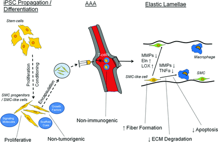
Ideal characteristics and expected roles of iPSCs and differentiated SMC-like derivatives for treating AAAs. Shown are several of the necessary properties for expansion/differentiation in culture, delivery to the AAA, and elastogenesis within the tunica media microenvironment. Abbreviations: AAA, abdominal aortic aneurysm; ECM, extracellular matrix; Eln, elastin; iPSC, induced pluripotent stem cell; LOX, lysyl oxidase; MMPs, matrix metalloproteinases; SMC, smooth muscle cell; TNFα, tumor necrosis factor-α.

In addition to induced pluripotent stem cells, other laboratories have examined umbilical cord mesenchymal stem cells and their ability to decrease the inflammation within the aorta that leads to aneurysms.  The researchers discovered that all the indicators of inflammation decreased, but the synthesis of new elastin was not examined.  However, a Japanese laboratory used mouse mesenchymal stem cells from bone marrow and found that not only did these cells shut down enzymes that tend to degrade elastin, but also initiated new elastin synthesis in culture.  The same study also showed that MSCs implanted into the vessel walls of an aorta that was experiencing an aneurysm stabilized the aneurysm by inhibiting the elastin-degrading enzymes, and increasing the elastin content of the vessel wall.  This had the net effect of stabilizing the aneurysms and preventing them from growing further (see Hashizume R, Yamawaki-Ogata A, Ueda Y, et al. J Vasc Surg 54:1743–1752).  

These experiments show that stem cell treatments for abdominal aneurysms are feasible and would definitely be a much-needed nonsurgical treatment option for the high-risk elderly demographic, which is rapidly growing in the developed world.

For more information on this interesting topic, see Chris A. BashuraRaj R. Raob and Anand Ramamurthia. Perspectives on Stem Cell-Based Elastic Matrix Regenerative Therapies for Abdominal Aortic Aneurysms.  Stem Cells Trans Med June 2013 vol. 2 no. 6 401-408.

Placental Stem Cell Provides Model System for Pregnancy Complications


Preeclampsia occurs during pregnancy, and is characterized by a gradual rise in blood pressure to dangerous levels. It usually presents after the 20th week of pregnancy, and can even persist after delivery.

How common is preeclampsia? In the United States, preeclampsia affects 5-8% of all births. Among the women of Canada, the United States, and Western Europe, the births affected by preeclampsia range from 2-5%. (5,6) In the developing world, the percentage of births affected by preeclampsia range from 4% of all deliveries to as high as 18% in parts of Africa. In Latin America, preeclampsia is the number one cause of maternal death.

Globally, ten million women develop preeclampsia each year, and 76,000 pregnant women die each year from preeclampsia and related disorders. The number of babies who die from these disorders is thought to be on the order of 500,000 per year.

In developing countries, a woman is seven times more likely to develop preeclampsia than a woman in a developed country, and between 10-25% of those cases will result in the death of the mother.

Now that I’ve hopefully convinced you that preeclampsia is a problem, how do we address it? Research in laboratory mice have told us a great deal about preeclampsia and other disorders that arise during pregnancy, but finding a sound model system that can be used to develop effective and safe treatments requires something closer to humans.

To that end, Hanna Mikkola and her research team and the University of California, Los Angeles Eli and Edythe Broad Center of Regenerative Medicine and Stem Cell Research (that’s a mouthful), have identified a type of progenitor cell that is key to the growth of a health placenta.

Work in laboratory mice has shown that preeclamsia often arises because of a malformed placenta. This poorly-formed placenta does not provide enough oxygen and nutrients for the growth needs of the baby at the fetal stages of development, and the mother’s body responds by increasing the mother’s blood pressure in order to increase blood flow through the placenta.

The work by Mikkola and her colleagues have provided physicians and developmental biologists with a new “tool box” for understanding the development of the placenta and the different cell types that compose it. Hopefully, various complications during pregnancy might be due to malfunctions of these particular cell types and the progenitor cells that produce them.

Mikkola and others started with laboratory mice, since it is possible to label single cells in mouse embryos and track exactly where those cells and their progeny go and what they do. The powerful genetic tools available in laboratory mice also allows scientists to identify the various biochemical signaling pathways that cells use to communicate with other cells during placental development. Also, if something goes wrong with particular cell signaling pathways, the mouse model allows scientists to precisely characterize the developmental consequences of much dysfunction.

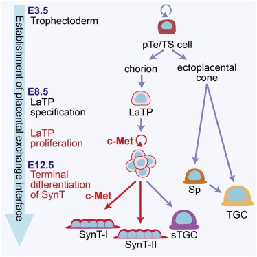
Through their work in the mouse, Mikkola and her co-workers identified a placental progenitor cells called the Epcamhi labyrinth trophoblast progenitor or LaTP. The LaTP is like a multipotent adult of tissue-specific stem cell that can become many of the cells required to make the placenta.

Mikkola and her group also showed that the “c-Met” signaling pathway was required to sustain the growth of LaTPs during placental development and that this same signaling pathway was required to form a specific group of cells (syncytiotrophoblasts) that form the interface between the placenta and the mother’s endometrium. Elimination of c-Met signaling completely compromised the growth of the fetus and its development.

g abs7

This new cell type should provide a wealth of opportunities to examine complications during pregnancy like preeclampsia and others and design treatments that can save the lives of mothers and their babies.

Human Neural Stem Cells Heal Damaged Limbs


The term “ischemia” refers to conditions under which a part of your body, organ, or tissue is deprived of oxygen. Without life-giving cells begin to die. Therefore, ischemia is usually a very bad thing.

Critical limb ischemia or CLI results when blood vessels to the legs, feet or arms are severely obstructed. The results of CLI are never pretty, and CLI remains a medical condition that presents few treatment options.

A study from a research team and the University of Bristol’s School of Clinical Sciences has used stem cells in a trial that uses laboratory mice to treat CLI. The success of this study provides a new direction and new hope for procedures that relieve symptoms and prolong the life of the limb.

Autologous stem cells treatments, or those stem treatments that utilize a patient’s own stem cells care subject to clear limitations. After collection from bone marrow, fat, or other source, the stem cells must be expanded in culture after stimulation with chemicals called cytokines. After growth in culture, the cells typically contain a collection of different types of stem cells of variable quality and potency. Also, if the patients has had a heart attack or has diabetes, then the quality and potency of their own stem cells are seriously compromised.

To circumvent this problem, Paulo Madeddu and his team at the Bristol Heart Institute have used an immortalized human neural stem cell line called CTX to treat animals who suffered from diabetes mellitus and CLI.

The CTX cell line comes from a biotechnology company called ReNeuron. This company is using this cell line in a clinical trial for stoke patients, and wants to use the CTX cell line in a clinical trial for CLI patients in the future.

When CTX cells are injected into the muscle of diabetic mice with CLI, the cells promote recovery from CLI. The CTX cells do so by promoting the growth of new blood vessels.

Madeddu said, “There are not effective drug interventions to treat CLI. The consequences are a very poor quality of life, possible major amputation and a life expectancy of less than one year from diagnosis in 50 percent of all CLI patients.”

Dr. Madeddu continued: “Our findings have shown a remarkable advancement towards more effective treatments for CLI and we have also demonstrated the importance of collaborations between universities and industry that can have a social and medical impact.”

Stem Cell Treatments to Improve Blood Flow in Angina Patients


Angina pectoris is defined as chest pain or discomfort that results from poor blood flow through the blood vessels in the heart and is usually activated by activity or stress.

In Los Angeles, California, physicians have initiated a double-blind, multicenter Phase III clinical trial that uses a patient’s own blood-derived stem cells to restore circulation to the heart of angina patients.

This procedure utilizes state-of-the-art imaging technology to map the heart and generate a three-dimensional image of the heart. These sophisticated images will guide the physicians as they inject stem cells into targeted sites in the heart.

This is a double-blinded study, which means that neither the patients nor the researcher will know who is receiving stem-cell injections and who is receiving the placebo.

The institution at which this study is being conducted, University of Los Angeles (UCLA), is attempting to establish evidence for a stem cell treatment that might be approved by the US Food and Drug Administration for patients with refractory angina. The subjects in this study had received the standard types of care but did not receive relief. Therefore by enrolling in this trial, these patients had nothing to lose.

Dr. Ali Nasir, assistant professor of cardiology at the David Geffen School of Medicine and co-principal investigator of this study, said: “We’re hoping to offer patients who have no other options a treatment that will alleviate their severe chest pain and improve their quality of life.”

Before injecting the stem cells or the placebo, the team examined the three-dimensional image of the heart and ascertained the health of the heart muscle and voltage it generated. Damaged areas of the heart fail to produce adequate quantities of voltage and show low levels of energy.

Jonathan Tobis, clinical professor of cardiology and director of interventional cardiology research at Geffen School of Medicine, said: “We are able to tell by the voltage levels and motion which area of the [heart] muscle is scarred or abnormal and not getting enough blood and oxygen. We then targeted the injections to the areas just adjacent to the scarred and abnormal heart muscle to try to restore some of the blood flow.”

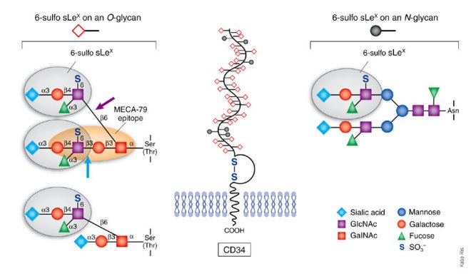
What did they inject? The UCLA team extracted bone marrow from the pelvic bones and isolated CD34+ cells. CD34 refers to a cell surface protein that is found on bone marrow stem cells and mediates the adhesion of bone marrow stem cells to the bone marrow matrix. It is found on the surfaces of hematopoietic stem cells, placental cells, a subset of mesenchymal stem cells, endothelial progenitor cells, and endothelial cells of blood vessels. These are not the only cells that express this cell surface protein, but it does list the important cells for our purposes. Once the CD34+ cells were isolated, the were injected into the heart through a catheter that was inserted into a vein in the groin.

CD34

The team hopes that these cells (a mixture of mesenchymal stem cells, hematopoietic stem cells, and endothelial progenitor cells) will stimulate the growth of new blood vessels (angiogenesis) in the heart, and improve blood flow and oxygen delivery to the heart muscle.

“We will be tracking patients to see how they’re doing,” said William Suh MD, assistant clinical professor of medicine in the division of cardiology at Geffen School of Medicine.

The goal of this study is to enroll 444 patients nation-wide, of which 222 will receive the stem cell treatment, 111 will receive the placebo, and 111 who will be given standard heart care.

Adult Stem Cells Help Build Human Blood Vessels in Engineered Tissues


University of Illinois researchers have identified a protein expressed by human bone marrow stem cells that guides and stimulates the construction of blood vessels.

Jalees Rehman, associate professor of cardiology and pharmacology at the University of Illinois at Chicago College of Medicine and lead author of this paper, said: “Some stem cells actually have multiple jobs.”

As an example, stem cells from bone marrow known as mesenchymal stem cells can form bone, cartilage, or fat, but they also have a secondary role in that they support other cells in bone marrow.

Rehman and others have worked on developing engineered tissues for use in cardiac patients, and they noticed that mesenchymal stem cells were crucial for organizing other cells into functional stem cells.

Workers from Rehman’s laboratory mixed mesenchymal stem cells from human bone marrow with endothelial cells that line the inside of blood vessels. The mesenchymal stem cells elongated and formed a kind of scaffold upon which the endothelial cells adhered and organized to form tubes.

“But without the stem cells, the endothelial cells just sat there,” said Rehman.

When the cell mixtures were implanted into mice, blood vessels formed that were able to support the flow of blood. Then Rehman and his colleagues examined the genes expressed when their stem cells and endothelial cells were combined. They were aided in this venture by two different bone marrow stem cell lines, one of which supported the formation of blood vessels, and the other of which did.

Their microarray experiments showed that the vessel-supporting mesenchymal stem cells expressed high levels of the SLIT3 protein. SLIT3 is a blood vessel-guidance protein that directs blood vessel-making cells to particular places and induces them to make blood vessels. The cell line that do not stimulate blood vessel production made little to no SLIT3.

Rehman commented, “This means that not all stem cells are created alike in terms of their SLIT3 production and their ability to encourage blood vessel formation.”

Rehman continued: “While using a person’s own stem cells for making blood vessels is ideal because it eliminates the problem of immune rejection, it might be a good idea to test a patient’s stem cells to make sure they are good producers of SLIT3. If they aren’t, the engineered vessels may not thrive or even fail to grow.

Mesenchymal stem cells injections are being evaluated in clinical trials to see if their can help grow blood vessels and improve heart function in patients who have suffered heart attacks.

So far, the benefits of stem cell injection have been modest, according to Rehman. Evaluating the gene and protein signatures of stem cells from each patient may allow for a more individualized approach so that every patient receives mesenchymal stem cells that are most likely to promote blood vessel growth and cardiac repair. Such pre-testing might substantially improve the efficacy of stem cell treatments for heart patients.