The Society for Neuroscience Meeting Continued


Glymphatics is a new subdiscipline in neuroscience that was essentially discovered by a Danish neuroscientist named Maiken Nedergaard. Dr. Nedergaard gave a fine seminar on this subject on Sunday.

Glymphatics consists of the system that removes waste products from the brain. Dr. Nedergaard showed movies that showed how the cerebrospinal fluid that bathes the periphery of the brain pulsates as it moves over the brain. When die molecules are injected into the cerebrospinal fluid, these dyes wend up in the blood system. How does this happen?

Nedergaard reasoned that diffusion of the fluid was far too slow for the dye to get to the blood system as fast as it does. Instead, she suspected that fluid moves by means of a “convection current.” How does this work? The blood vessels that feed the brain are surrounded by cells known as astrocytes. These astrocytes prevent molecules from entering the brain unless they can properly negotiate their way across these astrocytes, and this forms the basis for the blood-brain barrier. Cerebrospinal fluid moves across the cells of the brain and is removed by the astrocyte-surrounded vessels. This sink for the cerebrospinal fluid essentially pulls the cerebrospinal fluid across the brain cells and serves as the means by which the brain is cleansed of waste products.

This system, however, is subject to regulation, since the flow of fluid from the cerebrospinal fluid depends on the size of the spaces between brain cells. As it turns out, the spaces between brain cells in larger during sleep than when we are awake. Therefore, sleep seems to be the means by which our bodies clear the rubbish from our brains.

The molecule that controls the space between brain cells is norepinephrine. How it does that remains uncertain, but this is the molecule that is released during sleep to help clear out the garbage in the brain.

Since Alzheimer’s disease, Parkinson’s disease, other neurodegenerative diseases include the accumulation of protein aggregates in the brain, the removal of waste products in the brain would seem to be a rather important process. Also, when there is a head injury, surgeons sometimes leave the skull cap open while the brain heals. This, however, hamstrings the glymphatic system and surgeons should replace the skull cap so that the glymphatic system can do its job. Secondly, if norepinephrine can regulate this system, then this might be a way to increase clearance of waste products from the brain to reduce or delay the accumulation of protein aggregates in the brain.

Remarkable isn’t it?
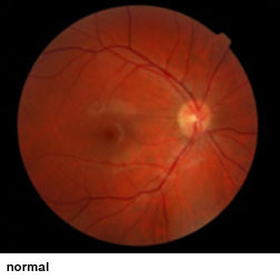
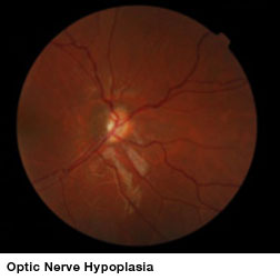
Tiny, Poorly-Controlled Study Shows No Benefit for Stem Cell Treatment in Children with Optic Nerve Hypoplasia


Optic nerve hypoplasia (ONH), an underdevelopment of optic nerves that occurs during fetal development, can appear as an isolated condition or as a part of a group of disorders characterized by brain anomalies, developmental delay, and endocrine abnormalities. ONH is a leading cause of blindness in children in North America and Europe and is the only cause of childhood blindness that shows increasing prevalence. No treatments have been shown to improve vision in these children.

RetinaRetina ONH

Because stem cells heal or even regenerate some tissues, some have considered stem cell treatments as an option for this condition.  However, a very small clinical study at Children’s Hospital Los Angeles found no evidence that stem cell therapies improve vision for children with optic nerve hypoplasia (ONH). Their results are reported in the Journal of the American Association for Pediatric Ophthalmology and Strabismus (AAPOS).

Families with a child that has ONH are traveling to China to undergo stem cell treatments that would be illegal in the United States. Because there are presently no viable treatment options available to improve vision in ONH children, such trips are often an act of desperation. The American Association for Pediatric Ophthalmology and Strabismus has also expressed its concern about these procedures, which are usually rather expensive, and have a dubious safety record.

Pediatric neuro-ophthalmologist Mark Borchert, MD, director of both the Eye Birth Defects and Eye Technology Institutes in The Vision Center at Children’s Hospital Los Angeles, realized that a controlled trial of sufficient size was needed to evaluate whether stem cell therapy is effective as a treatment for children with ONH. He agreed to conduct an independent study at the behest of Beike Biotech, which is based in Shenzhen, China and offers a stem cell treatment for ONH. This treatment uses donor umbilical cord stem cells and injects these cells into the cerebrospinal fluid.

Beike Biotech identified 10 children with bilateral ONH (ages 7 to 17 years) who had volunteered to travel to China for stem cell therapy. These patients gave their consent to participate in the study and Children’s Hospital found matched controls from their clinic. However, only two case-controlled pairs were evaluated because Beike Biotech was only able to recruit two patients.

Treatments consisted of six infusions over a 16-day period of umbilical cord-derived mesenchymal stem cells and daily infusions of growth factors. Visual acuity, optic nerve size, and sensitivity to light were to be evaluated one month before stem cell therapy and three and nine months after treatment.

Unfortunately no therapeutic effect was found in the two case-control pairs that were enrolled. “The results of this study show that children greater than 7 years of age with ONH may have spontaneous improvement in vision from one examination to the next. This improvement occurs equally in children regardless of whether or not they received treatment. Other aspects of the eye examination included pupil responses to light and optic nerve size; these did not change following treatment. The results of this research do not support the use of stem cells in the treatment of ONH at this time,” said lead author Cassandra Fink, MPH, program administrator at The Vision Center, Children’s Hospital Los Angeles.

However, confounding factors affect the interpretation of these results because the test subjects received additional alternative therapies (acupuncture, functional electrical stimulation and exercise) while receiving stem cell treatments. They were not supposed to receive such treatments. Additionally, the investigators could not determine the effect of these additional therapies on the subjects’ eyes.

“This study underscores the importance of scientifically testing these procedures to validate them and ensure their safety. Parents of afflicted children should be aware that the science behind the use of stem cell technology is unclear. This study takes a step toward testing this technology and finds no beneficial effect,” said William V. Good, MD, senior associate editor, Journal of AAPOS and Clinical Professor of Ophthalmology and Senior Scientist at the Smith-Kettlewell Eye Research Institute.

Basically, we have an incredibly small study that is also poorly controlled. Because the optic nerve forms during embryonic, fetal and postnatal development, using stem cells to make new nerves seems like a long shot as a treatment.  I better treatment strategy might be to increase the myelination of the optic nerve with neural stem cells, oligodendrocyte precursor cells (OPCs), or Schwann cells.  In general, this study does little to establish the lack of efficacy of such a stem cell treatment.

New Cell Type Derived from Embryonic Stem Cells for Possible Treatment of Brain Diseases


This story comes from my alma mater, UC Irvine. Go anteaters!! No really, UC Irvine’s mascot is the anteater, not to be confused with the aardvark.

Edwin Monuki at the Sue and Bill Gross Stem Cell Research Center with his graduate student Momoko Watanabe and other colleagues devised culture conditions to differentiate embryonic stem cells in “choroid plexus epithelial cells.” Monuki and he team were able to make choroid plexus epithelial cells (CPECs) from mouse and human embryonic stem cell (ESC) lines.

Now you are probably reading this and screaming, “what the heck are CPECs?” Calm down, we will explain:

The central nervous system is surrounded by a clear fluid known as cerebrospinal fluid or CSF. CSF flows all around the brain and spinal cord and also flows inside it. The CSF has several functions. These functions include buoyancy, protection, stability, and prevention of stroke (ischemia).

The CSF provides buoyancy to the brain, since the brain is large a potentially heavy. However, by filling the brain from within and around it with CSF decreases the density of the brain. CSF allows the brain to maintain a density that prevent it from collapsing under its own weight. If the brain were denser, then it would compact and cut off the blood supply of the cells in the lower part of the brain. This would kill off neurons.

Protection provided by the CSF comes during those times the head is struck. The CSF prevents the brain from coming into contact with the skull. The stability provided by the CSF

CSF flows throughout the inside of the brain through cavities known as “ventricles.” These ventricles provide reservoirs through which the CSF flows and is absorbed back into the bloodstream. This constant movement of the CSF through the CNS rinses it and removes metabolic wastes from the central nervous system. It also guarantees an even distribution of neural materials through the central nervous system.

CSF also helps prevent strokes, since the low pressure of the CSF in the skull translates into low intracranial pressure, which facilitates blood perfusion.

Now that have seen that CSF is very important in the life of the brain, where does it come from?  The answer is the CPECs make CSF as a fine filtrate from blood plasma. A minority of the CSF is also made by the walls of the brain ventricles. Nevertheless, CSF circulates from the lateral ventricles to the interventricular foramen, to the third ventricle, through the cerebral aqueduct, to the fourth ventricle, through the median aperture and lateral apertures to the subarachnoid space over the brain and spinal cord where arachnoid granulations return the CSF to the bloodstream.

Thus, CPECs make CSF, which helps remove metabolic wastes and other toxic compounds from the brain. In various neurodegenerative diseases, the choroid plexus and CPECs degenerate too and fail to efficiently remove debris and other rubbish from the central nervous system. Transplantation experiments in rodents have demonstrated that implanting a healthy set of CPECs can restore CSF function and slow down the damage done to the brain by neurodegenerative diseases (see Matsumoto et al., Neurosci Lett. 2010 Jan 29;469(3):283-8).  The problem is the lack of good cultures of CPECs.

Monuki and Watanabe and company seem to have fixed this problem. Monuki commented on his publication, “Our method is promising, because for the first time we can use stem cells to create large amounts of these epithelial cells (CPECs), which could be utilized in different ways to treat neurodegenerative diseases.” Monuki is an associate professor of pathology and laboratory medicine and cell and developmental biology at UC Irvine.

To make CPECs from ESCs, Monuki and his team differentiated cultured ESCs into neural stem cells. The neural stem cells were then differentiated into CPECs. This strategy makes a great deal of sense, since a neural stem cell population seems to exist in the CPECs (see Itokazu Y et al., Glia. 2006 Jan 1;53(1):32-42.

According to Monuki, there are three ways that these cells could be used to treat neurodegenerative diseases. First, the CPECs could be used to make more CSF and that would help flush out the proteins and other toxic compounds that kill off neurons. The down side of this is that it would also increase intracranial pressure, which is not optimal. Secondly,. the CPECs could be engineered into superpumps that transport high concentrations of therapeutic agents into the brain. Third, cultured CPECs could be used as a model system to screen drugs and other agents that shore up the endogenous CPECs in the patient’s brain.

Monuki’s next step is to develop an effective drug screening system in order conduct proof-of-concept studies to determine how CPECs afect the brain in mouse models of Huntington’s disease and other pediatric neurological diseases.