Stem Cell-Based Skin Graft for Severe Burns


Severe wounds are typically treated with full thickness skin grafts. Some new work by researchers from Michigan Tech and the First Affiliated Hospital of Sun Yat Sen University in Guangzhou, China might provide a way to use a patient’s own stem cells to make split thickness skin grafts (STSG). If this technique pans out, it would eliminate the needs for donors and could work well for large or complicated injury sites.

This work made new engineered tissues were able to capitalize on the body’s natural healing power. Dr. Feng Zhao at Michigan Tech and her Chinese colleagues used specially engineered skin that was “prevascularized, which is to say that Zhao and other designed it so that it could grow its own veins, capillaries and lymphatic channels.

This innovation is a very important one because on of the main reasons grafted tissues or implanted fabricated tissues fail to integrate into the recipient’s body is that the grafted tissue lacks proper vascular support. This leads to a condition called graft ischemia. Therefore, getting the skin to form its own vasculature is vital for the success of STSG.

STSG is a rather versatile procedure that can be used under unfavorable conditions, as in the case of patients who have a wound that has been infected, or in cases where the graft site possess less vasculature, where the chances of a full thickness skin graft successfully integrating would be rather low. Unfortunately, STSGs are more fragile than full thickness skin grafts and can contract significantly during the healing process.

In order to solve the problem of graft contraction and poor vascularization, Zhao and others grew sheets of human mesenchymal stem cells (MSCs) and mixed in with those MSCs, human umbilical cord vascular endothelial cells or HUVECs. HUVECs readily form blood vessels when induced, and growing mesenchymal stem cells tend to synthesize the right cocktail of factors to induce HUVECs to form blood vessels. Therefore this type of skin is truly poised to form its own vasculature and is rightly designated as “prevascularized” tissue.

Zhao and others tested their MSC/HUVEC sheets on the tails of mice that had lost some of their skin because of burns. The prevascularized MSC/HUVEC sheets significantly outperformed MSC-only sheets when it came to repairing the skin of these laboratory mice.

When implanted, the MSC/HUVEC sheets produced less contracted and puckered skin, lower amounts of inflammation, a thinner outer skin (epidermal) thickness along with more robust blood microcirculation in the skin tissue. And if that wasn’t enough, the MSC/HUVEC sheets also preserved skin-specific features like hair follicles and oil glands.

The success of the mixed MSC/HUVEC cell sheets was almost certainly due to the elevated levels of growth factors and small, signaling proteins called cytokines in the prevascularized stem cell sheets that stimulated significant healing in surrounding tissue. The greatest challenge regarding this method is that both STSG and the stem cell sheets are fragile and difficult to harvest.

An important next step in this research is to improve the mechanical properties of the cell sheets and devise new techniques to harvest these cells more easily.

According to Dr. Zhao: “The engineered stem cell sheet will overcome the limitation of current treatments for extensive and severe wounds, such as for acute burn injuries, and significantly improve the quality of life for patients suffering from burns.”

This paper can be found here: Lei Chen et al., “Pre-vascularization Enhances Therapeutic Effects of Human Mesenchymal Stem Cell Sheets in Full Thickness Skin Wound Re-pair,” Theranostics, October 2016 DOI: 10.7150/ thno.17031.
Cultured Skin-Based Stem Cells Regenerate Hair Follicles and Sebaceous Glands


It has been over ten years since the development of cultured skin substitutes or CSSs. CSSs consist of cultured epidermis from the patients and dermal fibroblasts that can form a good epidermal layer. However, CSSs do not form hair follicles or sebaceous glands which makes for mighty dry skin. Therefore, it is highly preferable to make at skin substitute that can form such epidermal appendages.

Fortunately the skin possesses several types of stem cells for use in regenerative medicine. Epidermal stem cells (Epi-SCs) in the basal layer of the epidermis that constantly provide new cells to the epidermis (the uppermost layer of the skin). Unfortunately, adult Epi-SCs cannot form hair follicles, but they can if they are combined with embryonic or newborn dermal cells. Dermal papilla (DP) cells can induce hair follicles, but the availability of DP cells has greatly limited work with them. Adult dermal skin also contains multipotent SKPs or skin-derived precursors. Injection of SKPs underneath the skin of mice, leads to the induction of new hair follicles (Biernaskie et al., 2009, Cell Stem Cell 5:610-623). This suggests that SKPs might be applicable in a clinical setting to induce hair follicles in cultured skin substitutes.

A new paper by Yaojiong Wu and coworker from the Shenzhen Key Laboratory of Health Sciences and Technology, in collaboration with Edward E. Tredget from University College Dublin has successfully induced the growth of hair follicles and sebaceous glands in a cultured skin substitute. They combined cultured Epi-SCs and SKPs, from mice and humans, and embedded them into a hydrogel. These stem cell-embedded hydrogels were then implanted into immunodeficient mice with substantial skin wounds.

The results were remarkable. The implants able to induce the formation of new epidermis with hair follicles. Furthermore, when Epi-SCs and SKPs taken from human scalps were used in these experiments, they worked just as well as those taken from mice.

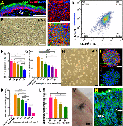
Hair neogenesis with cultured epidermal stem cells (Epi-SCs) and skin-derived precursors (SKPs). (A): Putative epidermal stem cells residing in the basal layer of neonatal mouse epidermis expressed CD49f (red) in immunofluorescence stain, and mature keratinocytes in the top layers of the epidermis expressed cytokeratin (CK)6 (green). Nuclei were stained with 4′,6-diamidino-2-phenylindole. (B–E): Cultured Epi-SCs derived from neonatal mice (B) were positive for CD49f (C) and CK15 (D) in immunofluorescence stain; fluorescence-activated cell sorting analysis of the Epi-SCs indicated high levels of surface CD29 and CD49f (E). (F): The expression level of CD49f decreased progressively upon successive passages (P) in culture as determined by immunofluorescence analysis (in relation to the fluorescence intensity of P0 cells). Triple wells were used for each of the above experiments, and each experiment was repeated three times with similar results (∗, p < .05; ∗∗, p < .01; ∗∗∗, p < .001). (G): Hair genesis of cultured Epi-SCs in different passages. Cultured Epi-SCs derived from neonatal mice in different passages (P0 to P5) were implanted into excisional wounds in nude mice in combination with freshly isolated neonatal dermal cells (fresh D) in Matrigel; dermal cells alone or freshly isolated neonatal epidermal cells plus dermal cells (fresh E+D) were used as controls. Hair shafts generated 20 days posttransplant were counted (n = 6; ∗, p < .05; ∗∗∗, p < .001). (H–J): SKPs derived from neonatal mice in spheroid culture (H) expressed nestin, fibronectin (I), and BMP6 (J) in immunofluorescence analysis. (K): Hair genesis of SKPs in different passages. SKPs in P0 to P5 were implanted into excisional wounds in nude mice in combination with freshly isolated neonatal mouse epidermal cells (fresh E), and freshly isolated neonatal mouse epidermal cells alone or in combination with freshly isolated neonatal mouse dermal cells (fresh E+D) were used as controls. Twenty days posttransplant, hairs generated were counted (n = 6; ∗, p < .05; ∗∗, p < .01; ∗∗∗, p < .001). (L-N): Cultured Epi-SCs and SKPs in hair genesis. Combinations of cultured neonatal mouse Epi-SCs (P0 to P3) and SKPs (P0 to P3) were engrafted into excisional wounds in nude mice, and the number of hairs generated were counted 20 days posttransplant (n = 3, ∗, p < .05). (L). A representative image of hairs generated 20 days after a transplantation of P1 Epi-SCs and SKPs (M). Immunofluorescence analysis of the skin tissue with hair genesis showed densely populated hair follicles and sebaceous glands (N). Scale bars = 50 μm. Abbreviations: BM, basement membrane; BMP6, bone morphogenetic protein 6; CK, cytokeratin; DAPI, 4′,6-diamidino-2-phenylindole; Derm, dermis; Epi, epidermis; Epi-SC, epidermal stem cells; FITC, fluorescein isothiocyanate; fresh D, freshly isolated neonatal dermal cells; fresh D+E, freshly isolated neonatal epidermal cells plus dermal cells; HF, hair follicle; HS, hair shafts; P, passage; PE, phycoerythrin.

Additionally, when the ability of Epi-SCs to differentiate into sebaceous glands was examined, Wu and others showed that Epi-SCs can form the precursors to sebaceous glands, sebocytes. Additionally, the oils secreted by these Epi-SC-derived sebocytes were chemically similar to sebaceous glands from native skin.

Thus, a combination of Epi-SCs and SKPs from human or mouse skin were sufficient to generate newly formed hair follicles and functional sebaceous glands. These results provide knowledge that is potentially transferable to clinical applications for regenerating damaged skin.

Two Proteins Safeguard Skin Stem Cell Function


Ever scraped your knees or elbows? It’s a good thing that they didn’t stay that way, since human skin readily renews, heals wounds, and regenerates the hair that covers it thanks to a resident population of stem cells. These cells continually produce new ones. Depending on someone’s age, the complete skin is renewed every 10-30 days.

A new study led by Salvador Aznar Benitah (Institute for Research in Biomedicine, Barcelona, Spain) has identified two proteins that are integral to the conservation of skin stem cells. Without these proteins these skin-based skin cells are lost.

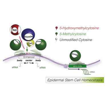
The proteins identified, Dnmt3a and Dnmt3b, trigger the first step of the genetic program that leads to stem cell renewal and regeneration of the skin. “Without them (i.e. Dnmt3a & Dnmt3b), this program is not activated and the stem cells collapse and disappear from the tissue,” said Benitah.

Dnmt3a & 3b are enzymes that attach methyl groups (-CH3) to the cytosines in DNA molecules.  The full name of these enzymes, DNA (cytosine-5)-methyltransferase 3A, catalyze the transfer of methyl groups to specific CpG structures in DNA.  This process is known as “DNA methylation.”  These particular DNA methyltransferases participate in de novo DNA methylation.  They must be distinguished from so-called “maintenance DNA methylation,” which ensures the fidelity of replication of inherited epigenetic patterns.

Epigenetics refers to cellular and physiological trait variations that result from external or environmental factors that switch genes on and off and affect how cells express genes, but do not involve changes in nucleotide sequences, but in chemical modifications to DNA or higher-order structures of DNA.

DNMT3A forms part of the family of DNA methyltransferase enzymes that includes DNMT1, DNMT3A and DNMT3B.  While de novo DNA methylation modifies the information passed on by parents to their progeny, it enables key epigenetic modifications essential for processes such as cellular differentiation and embryonic development, transcriptional regulation, heterochromatin formation, X-inactivation, imprinting and genome stability.

Lorenzo Rinaldi, a graduate student in Benitah’s laboratory who was also the first author of this study, has mapped the regions of the genome that houses the genes that encodes these two proteins. Rinaldi and others have shown that Dnmt3a & 3b affect gene expression by methylating “gene enhancers” and “superenhancers.” Gene enhancers and superenhancers are sequences that tend to be far away from genes but still have the ability to increase the speed of gene expression up to 200-fold.

“It was surprising to see that two proteins that have always been associated with gene repression through DNA methylation are activated in the most transcriptionally active regions of stem cells. We had never observed this activity because we were unable to study the global distribution of Dnmt3a and Dnmt3b at the genomic level. Thanks to advances in sequencing techniques, more researchers are observing the very mechanism that we have described,” said Rinaldi.

Of the 12,000 gene enhancers in the genome, about 300 are superenhancers related to stem cell activity. Dnmt3a & 3b activate expression of the approximately 1,000 genes that are required for the self-renewing capacity of stem cells. By methylating the superenhancer, these proteins trigger the first step of the machinery that leads to the amplified expression of these essential genes for the stem cell.  Dnmt3a and Dnmt3b clearly associate with the most active enhancers in human epidermal SCs.

Graphical_Done

The expression of Dnmt3a & 3b is also altered in cancer cells. Cancer cells tend to show altered DNA methylation and altered gene enhancers that affect gene expression. The mass sequencing of tumor cell genomes has provided these observations. Dnmt3a and Dnmt3b activities are altered in many types of tumor, including leukemias, and cancers of the lung and the colon.

“Each of these three components is associated with the development of various kinds of cancer. Given that these proteins activate gene expression enhancers through DNA methylation, we believe that it would be of interest to study them in cancer cells in order to determine whether they participate in tumor development,” said Benitah.

This work appeared in: Lorenzo Rinaldi et al., “Dnmt3a and Dnmt3b Associate with Enhancers to Regulate Human Epidermal Stem Cell Homeostasis,” Cell Stem Cell, July 2016 DOI: 0.1016/j.stem.2016.06.020.

Stem Cells that Control Skin and Hair Color


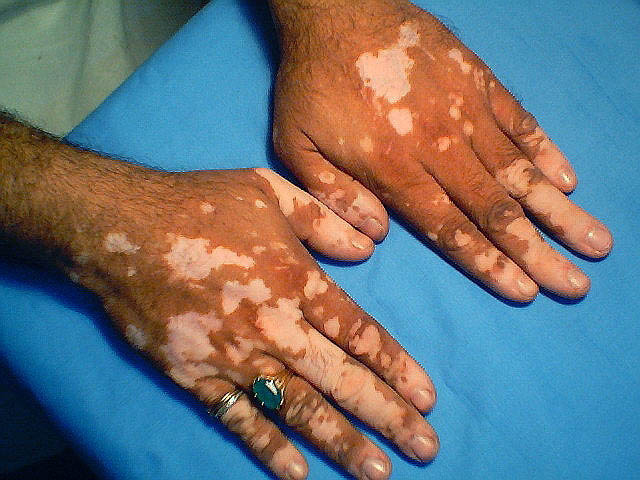
A research team at NYU Langone Medical Center has uncovered a pair of molecular signals that control the hair and skin color in mice and humans. Manipulation of these very signals may lead to therapies or even drugs to treat skin pigment disorders, such as vitiligo.

Vitiligo
Vitiligo

Vitiligo is somewhat disfiguring condition characterized by the loss of skin pigmentation, leaving a blotchy, white appearance. Finding ways to activate these two signaling pathways may provide clinicians with the means to mobilize the pigment-synthesizing stem cells that place pigment in skin structures and, potentially, repigment the pigment-bearing structures that were damaged in cases of vitiligo. Such treatment might also repigment grayed hair cells in older people, and even correct the discoloration that affects scars.

Workers in the laboratory of Mayumi Ito at the Ronald O. Perelman Department of Dermatology and the Department of Cell Biology showed that a skin-based stem cell population of pigment-producing cells, known as “melanocytes,” grow and regenerate in response to two molecular signals. The Endothelin receptor type B (EdnrB) protein is found on the surfaces of melanocytes. EdnrB signaling promotes the growth and differentiation of melanocyte stem cells (McSCs). Activation of EdnrB greatly enhances the regeneration of hair-based and epidermal-based melanocytes. However, EdnrB does act alone. Instead, the effect of EdnrB depends upon active Wnt signaling. This Wnt signal is initiated by the secretion of Wnt glycoproteins by the hair follicle cells.

This work was published in Cell Reports, April 2016 DOI: http://dx.doi.org/10.1016/j.celrep.2016.04.006.

Previous work on EdnrB has established that it plays a central role in blood vessel development. This work by Ito and his team is the first indication that pigment-producing melanocytes, which provide color to hair and skin, are controlled by this protein.

A lack of EdnrB signaling in mice caused premature graying of the hair. However, stimulating the EdnrB pathway resulted in a 15-fold increase in melanocyte stem cell pigment production, and by two months, the mice showed hyperpigmentation. In fact, wounded skin in these mice became pigmented upon healing.

Overexpression of Edn1 Promotes Upward Migration of McSCs and Generation of Epidermal Melanocytes following Wounding (A–D) Whole-mount image of X - gal - stained wound area of Dct-LacZ (control; A and B) and Tyr-CreER ; EdnrB fl/fl ; Dct-lacZ (C and D) at indicated days after re-epithelialization. (E–J) Double immunohistochemical staining of Dct and Ki67 in the bulge (E and H), upper hair follicle (F and I), and inter-follicular epidermis (G and J ) in control (E–G) and K14-rtTA ; TetO-Edn1-LacZ (Edn1; H–J) mice. (K–N) Whole - mount analyses of wound site (K and L) and de novo hair follicles (M and N) within wound site from control (K and M) and Edn1 mice (L and N) at 8 days after re-epithelialization. (O–R) Quantification of the number of Dct - LacZ+ cells in wound site (O), the percentage of Ki67+/Dct+ cells (P), the number of pigmented cells in wound site (Q), and the percentage of pigmented de novo hair (R), respectively. Dashed lines indicate periphery of wound site in (A) and (D) and boundary between epidermis and dermis in (E)–(J). Arrowheads show Dct - LacZ + cells in wound area in (A)–(D) and Ki67+/Dct+ cells (H)–(J). IFE, inter-follicular epidermis; UF, upper follicle. Data are presented as the mean ± SD. *p < 0.01; **p < 0.02; ***p < 0.05. The scale bar represents 1 mm in (A), 50 m m in (E), 200 m m in (K) and (L), and 100 m m in (M) and (N).
Overexpression of Edn1 Promotes Upward Migration of McSCs and Generation of Epidermal Melanocytes following Wounding (A–D) Whole-mount image of X-gal – stained wound area of
Dct-LacZ (control; A and B) and Tyr-CreER; EdnrB fl/fl; Dct-lacZ (C and D) at indicated days after
re-epithelialization. (E–J) Double immunohistochemical staining of Dct and Ki67 in the bulge (E and H), upper hair follicle (F and I), and inter-follicular epidermis (G and J) in control (E–G) and K14-rtTA;
TetO-Edn1-LacZ (Edn1; H–J) mice.  (K–N) Whole-mount analyses of wound site (K and L) and de novo hair follicles (M and N) within wound site from control (K and M) and Edn1 mice (L and N) at
8 days after re-epithelialization.  (O–R) Quantification of the number of Dct-LacZ+ cells in wound site (O), the percentage of Ki67+/Dct+ cells (P), the number of pigmented cells in wound site (Q),
and the percentage of pigmented de novo hair (R), respectively.  Dashed lines indicate periphery of wound site in (A) and (D) and boundary between epidermis and dermis in (E)–(J). Arrowheads show Dct-LacZ+ cells in wound area in (A)–(D) and Ki67+/Dct+ cells (H)–(J). IFE, inter-follicular epidermis; UF, upper follicle. Data are presented as the mean ± SD. *p < 0.01; **p < 0.02; ***p < 0.05.
The scale bar represents 1 mm in (A), 50 micrometer in (E), 200 micrometer in (K) and (L), and 100
micrometer in (M) and (N).

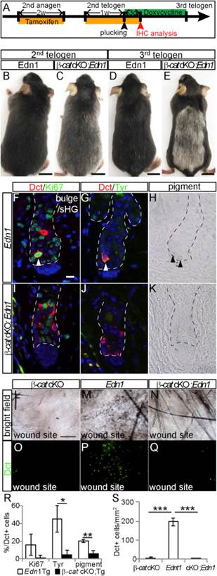
If the Wnt signaling pathway was blocked, stem cell growth and maturation sputtered and stalled and never got going, even when the EdnrB pathway was working properly. These mice had unpigmented fur (see E in figure below).

Loss of beta-catenin Function Suppresses Edn1-Mediated Effects on McSC Proliferation, Differentiation, and Upward Migration (A) Experimental scheme for treatment of Tyr-CreER ; b -catenin fl/fl ; K14-rtTA ; TetO-Edn1-LacZ ( b -cat cKO; Edn1 ) mice and control K14-rtTA ; TetO-Edn1- LacZ ( Edn1 ) mice. (B–E) Gross appearance of Edn1 (B and D) and b -cat cKO; Edn1 mice (C and E) at second (B and C) and third telogen (D and E). (F–K) Immunohistochemistry for indicated markers (F, G, I, and J) and bright- field image (H and K) of bulge/sHG region in skin sections from Edn1 mice (F–H) and b -cat cKO; Edn1 mice (I–K) at anagen II. (L–Q) Bright-field image (L–N) and Dct immunostaining of whole-mount wound site (O–Q) from Tyr-CreER ; b -catenin fl/fl ( b -cat cKO; L and O), Edn1 (M and P), and b -cat cKO; Edn1 mice (N and Q). (R and S) Quantification of the percentage of Dct+ cells positive for Ki67, Tyr, and pigmentation (R) and the number of Dct+ cells in wounded site (S). Dashed lines indicate border between hair follicle and dermis. Arrow- heads indicate double positive cells for indicated markers (F and G) and pigmented cells (H). Data are presented as the mean ± SD.*p<0.05;**p< 0.02; ***p < 0.001. The scale bar represents 1 cm in (B)–(E), 10 m min(F), and 200 m min(L). 1
Loss of beta-catenin Function Suppresses Edn1-Mediated Effects on McSC Proliferation, Differentiation, and Upward Migration
(A) Experimental scheme for treatment of
Tyr-CreER; beta-catenin fl/fl; K14-rtTA; TetO-Edn1-LacZ (beta-cat cKO; Edn1) mice and control K14-rtTA; TetO-Edn1-LacZ (Edn1) mice.
(B–E) Gross appearance of Edn1 (B and D) and
beta-cat cKO; Edn1 mice (C and E) at second (B and C) and third telogen (D and E). (F–K) Immunohistochemistry for indicated markers (F, G, I, and J) and bright-field image (H and K) of bulge/sHG region in skin sections from Edn1 mice (F–H) and beta-cat cKO; Edn1
mice (I–K) at anagen II. (L–Q) Bright-field image (L–N) and Dct immunostaining of whole-mount wound site (O–Q) from
Tyr-CreER; beta-catenin fl/fl (beta-cat cKO; L and O), Edn1 (M and P), and beta-cat cKO; Edn1 mice (N and Q). (R and S) Quantification of the percentage of Dct+ cells positive for Ki67, Tyr, and pigmentation (R) and the number of Dct+ cells in wounded site (S).
Dashed lines indicate border between hair follicle and dermis. Arrow-heads indicate double positive cells for indicated markers (F and G) and pigmented cells (H). Data are presented as the mean ± SD.*p<0.05;**p<
0.02; ***p < 0.001. The scale bar represents 1 cm in (B)–(E), 10 micrometers in (F), and 200 micrometer  in (L).

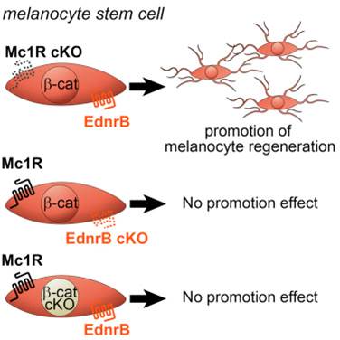
However, perhaps the most exciting finding for Ito and his colleagues was that Wnt-dependent, EdnrB signaling rescued the defects in melanocyte regeneration caused by loss of the Mc1R receptor. This is precisely the receptor that does not function properly in red-heads, which causes them to have red hair and very light skin that burns easily in the sun. These data suggest that Edn/EdnrB/Wnt signaling in McSCs can be used therapeutically to promote photoprotective-melanocyte regeneration in those patients with increased risk of skin cancers due to their very lightly colored skin.

Melanocyte Stem Cell Modeld

Plant Polyphenol May Help Improve Wound Healing By Activating Mesenchymal Stem Cells


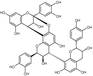
Akito Maeda and his coworkers from Osaka University in Osaka, Japan have discovered that a plant-based polyphenol promotes the migration of mesenchymal stem cells (MSCs) in blood circulation. This same plant polyphenol also causes MSCs to accumulate in damaged tissues and improve wound healing.

This compound, cinnamtannin B-1, might be a candidate drug for stem cell treatments for cutaneous disorders associated with particular diseases and lesions.

Cinnamtannin B-1
Cinnamtannin B-1

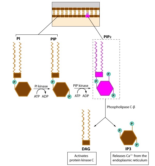
Cinnamtannin B-1, a flavonoid, seems to activate membrane-bound enzymes; specifically the Phosphatidylinositol-3-kinase enzyme, which is an integral enzyme in the phosphoinositol signal transduction pathway, which culminates in the mobilization of intracellular calcium stores and profoundly alters cell behavior and function.

Phosphoinositol pathway signaling

Flow cytometry analysis of mouse blood established that administration of cinnamtannin B-1 increased the release of MSCs from bone marrow. Laboratory experiments with cultured MSCs showed that cinnamtannin B-1 treatment activated MSC migration and recruitment to wounds. This seems to suggest that the enhanced healing caused by cinnamtannin B-1 treatment is due to enhanced MSC migration and homing to damaged tissues.

Imaging analysis of whole animals that had MSCs that expressed the firefly luciferase enzyme showed that cinnamtannin B-1 treatment increased the homing of MSCs to wounds and accelerated healing in a diabetic mouse model.

When Maeda and his colleagues treated MSCs with small molecules that inhibited phosphatidylinositol-3-kinase, those cells no longer responded to cinnamtannin B-1, which confirms the role of the phosphoinositol signal transduction pathway in cinnamtannin B-1 activation of MSCs.

Thus, cinnamtannin B-1 promotes MSC migration in culture and accelerates wound healing in mice. In addition, cinnamtannin B-1-induced migration of MSCs seems to be mediated by specific signaling pathways.

RepliCel Skin Rejuvenation and Tendon Repair Trials With Hair Follicle Stem Cells Underway


RepliCel Life Sciences has enrolled subjects for their skin rejuvenation and tendon repair trial.  The primary goal of these trials is to determine the safety of their cell therapeutic products.

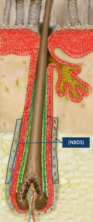
NBDS-Cells-Follicle

In the first trial will test a product called RCS-01, which consists of cells derived from non-bulbar dermal sheath (NBDS) cells, which are taken from the outer regions of hair follicles.  NBDS cells express type 1 collagen, a protein that is steadily degraded in aged skin (hence the formation of wrinkles).  Therefore, RepliCel is confident that RCS-01 injections underneath the skin has the potential to rejuvenate aged or damaged skin.  The trial will examine male and female subjects, between 50-65 years old, and will address the inherent deficit of active fibroblasts required for the production of type 1 collagen, elastin and other critical extracellular dermal matrix components found in youthful skin. The trial will be conducted at the IUF Leibniz-Institut für umweltmedizinische Forschung GmbH in Dusseldorf, Germany.  Originally, RepliCel wished to enrolled 15 men and 15 women, but the large number of female subjects and paucity of men persuaded the company to move forward with the trial despite only enrolling a few men and all the projected women.

The second trial will test the safety and efficacy of RCT-01 in the repair of damaged Achilles tendons.  RCT-01 also consists of NBDS cells and this trial is a phase 1/2 clinical trial that examines the ability of NBDS cells to treat chronic tendinosis caused by acute and chronic tensile overuse.  This trial will take place at the University of British Columbia in Vancouver, BC, and will only treat 10 subjects.  Even though RepliCell wishes to originally test 28 participants, the company shorted the trial in order to have safety data by the end of 2016.

Darrell Panich, RepliCel Vice President of Clinical Affairs, said that the company had a late start on its trials, and therefore truncated the recruitment process in order to have safety data for analysis by the end of 2016.  Despite the small size of these trials (and they are small), the company is hopeful that their safety data will provide the impetus for moving forward with larger phase 2 trials.

Panich said, “We have adjusted our plans for the RCT-01 clinical trial in part because it started later in 2015 and enrolled slower than originally anticipated. While the trial did not meet projected enrollment targets, we are confident the safety and preliminary efficacy data obtained by year-end will provide a signal of the product’s potential to regenerate chronically injured tendon that has failed to respond to other treatments. This will allow our teams to effectively plan larger phase 2 trials in 2017 which are powered to be statistically significant for clinical efficacy (evidence the product works as intended).”

“Future trials involving products from our non-bulbar dermal sheath (NBDS) platform will be designed to investigate the efficacy of these products at different dose levels and treatment frequencies while continuing to collect other data that will be used to support eventual RCS-01 and RCT-01 marketing applications by our commercial partners.”

“The delivery of clinical data when promised is important to management”, said R. Lee Buckler, President & CEO, RepliCel Life Sciences Inc. “We have made critical decisions to keep our commitment to the financial community and we believe the data from these trials will facilitate us closing a licensing and co-development deal on one or both of these products similar to the kind we have in place with Shiseido Company for our RCH-01 product,” he added.

RepliCel is confident that their NBDS fibroblast platform will address numerous indications where impaired tissue healing has been stalled due to a paucity of active fibroblasts, which are required for tissue remodeling and repair.  NBDS fibroblasts, isolated from the hair follicles of healthy individuals, are a rich source of fibroblasts and are unique in their ability to express high levels of type 1 collagen and elastin to push-start the healing process.

RepliCel is also developing products from this same platform to address larger commercial markets in the areas of musculoskeletal and skin-related conditions.

A Stem Cell Treatment for Hair Loss


Male and female pattern baldness involves the receding of the hair-line, hair loss and the thinning the hair covering on the scalp. Hair loss is also called alopecia and is a common problem among the elderly, those afflicted with certain diseases of the scalp, or those who take certain medicines. Hair life is not the end of life, but it can change someone’s appearance, affect their self-image, and affect someone’s emotional state. People with hair loss can use topical monoxidil (Rogaine), take an oral drug called finasteride (Propecia), or undergo a hair transplant. The drugs, however, must be used constantly or they stop working and hair transplants are horribly expensive.

Can stem cell-based treatment restore hair after it has been lost? Fortunately, a stem cell population called dermal papilla cells (DPCs), which are a type of mesenchymal stem cell population in hair follicles, have been identified and even characterized to some extent. DPCs are responsible for the formation of hair-follicles and play a very role in the process of hair cycling in which the hair shaft grows, is shed, is reestablished, and grows again. DPC might be useful for treating alopecia, but they do not survive when cultured outside the body, and this limitation has limited developing stem cell-based treatments for hair loss.

Now collaboration between two scientific research teams from Canada and China have resulted in a new way to use stem cells to treat hair loss.

A research team from the Nanfang Hospital of Southern Medical University, China, led by Zhi-Qi Hu and a Canadian team from the University of Manitoba, Canada, led by Malcolm Xing have designed a three-layered tunic that feeds and protects the DPCs and allows them to grow outside the body.

Xing describes this nutritive tunic as a “nutritious nano-clothing,” made of gelatin and alginate. These molecules can self-assemble and Hu and Xing and their coworkers encapsulated the cells within an inner layer of gelatin, a middle layer of alginate loaded with fibroblast growth factor-2, and an outer layer of gelatin. They call this method of encapsulation “layer-by-layer (LBL) nano-coating.”  This gelatin/alginate coating creates a protective microenvironment for cultured DPC and provides them with a significant source of a growth factor called fibroblast growth factor-2 (FGF2), which enhances proliferation of the DPCs and induces hair cell fates. The use of these three-layered tunics keeps the inductive signals close to the DPCs and circumvents the difficulties encountered in regenerating new hair follicles on bald skin.

DPC - hair follicle regeneration

When the Xing and Hu teams implanted these encased DPCs into the skin of nude mice, the implanted encapsulated cells generated the growth of abundant hair. The hair produced by these cells also was rooted in hair follicles that were normal in their appearance and function. The coating improvised by these teams greatly augmented the therapeutic capabilities of the DPCs by recapitulating the niche in which these cells are normally found. This stem cell niche induces the cells to secrete the native extracellular matrix that typically surrounds the cells and release the growth factors that keep the cells growing and in the proper stage of the cell cycle.

According to Xing, the most difficult part of this research project was “optimizing the concentrations of the coated polymers and manufacturing conditions to make the cells happy and healthy.”

Regenerative medicine researcher Oommen Varghese, from Uppsala University in Sweden, who was not involved in this work, said, “This is fascinating science that has enormous potential for clinical translational of stem cell based regenerative medicine. Such a coating could also protect cells from innate immunity, thereby improving the in vivo survivability. This is a major challenge in stem cell based translational research.”

Xing and his collaborators and colleagues would like to transform this technique from a laboratory bench to a clinical application that can be tested in human clinical trials.

Bone Marrow Mesenchymal Stem Cell Aggregates for Treating Deep Skin Wounds


Cuts, bruises, lacerations, abrasions, of the skin are some of the most common injuries. Deeper wounds that extend into the dermis are more susceptible to chronic inflammation and suffer greatly if they are bumped, knocked, or run into. Such wounds are also more difficult to heal, since a full-thickness cutaneous wound usually damages many different structures and cell lineages. Fortunately, the healing of these structures begins directly after production of the wound. Regeneration is largely orchestrated by growth factors such as Vascular Endothelial Growth Factor and Transforming Growth Factor-β. Since mesenchymal stem cells (MSCs) are a good source of these growth factors, they might be promising candidates for treating full-thickness wounds.

Yan Jin and his colleagues at the Research and Development Center for Tissue Engineering in Shaanxi, China have tested the ability of bone marrow-derived mesenchymal stem cells (BMMSCs) to accelerate the healing of deep skin wounds.

Jin and his colleagues, Yulin An, Wei Wei, Huan Jing, Leigo Ming, and Shiyu Lui used bone marrow from mice that had been genetically engineered to express Green Fluorescent Protein (GFP) in their bone marrow. After isolating mesenchymal stem cells from the bone marrow of these mice, they applied the cells to rats that had suffered full-layer skin cutaneous wounds. However, they tested several different ways of applying these cells to the wound.

In one group of rats, GFP+BMMSCs were grown in cell culture that used a growth medium that contained a steroid drug called dexamethasone and ascorbic acid phosphate. These chemicals caused the BMMSCs to grow in the form of clusters of cells called “cell aggregates” that could be scraped off mechanically and readily transplanted onto the wounds without the need for any scaffold.  These cell aggregates grow as sheets of cells that can act as a kind of patch made from healing MSCs that can be easily and readily applied to a wound or other lesion.  In a second group of rats, the same number of GFP+BMMSCs was topically administered around the wound. In the third group of rats, the BMMSCs were given intravenously through tail vein. All three groups of rats received the same number of BMMSCs. Samples from the bed of the wounds were taken at different time points, and the morphological, histological and molecular characteristics of the wounds were analyzed and compared.

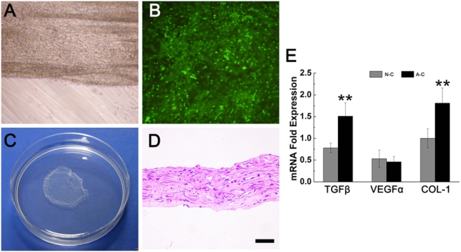
(A) The rim of aggregate curled a little on dish bottom. (B) GFP+BMMSCs in the aggregate gave green fluorescence under 509 nm excitation light. (C) The whole aggregate was scratched off the dish. (D) HE staining revealed a certain thickness of the aggregate with cells in it (Bar = 20 nm). (E) RT-PCR showed that BMMSC aggregate presented significantly higher expression of TGF-β and collagen I but had a similar VEGFα expression with normal cultured cells. (N-C: normal cultured cells; A-C: Aggregate cells; **p < 0.05 is considered statistically different.)

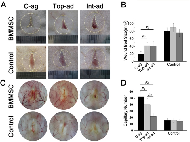
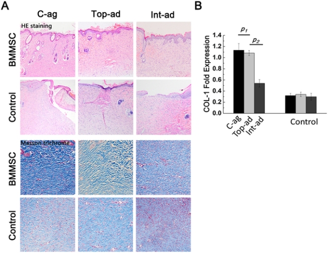
According to the results, the BMMSCs administered in cell-aggregates produced the highest expression of pro-healing genes than the other methods. Also animals treated with the BMMSC cell aggregates also showed better vascularization and more regular dermal collagen deposition than the other two groups of rats.

(A,B) Wound bed size and vascularization state in BMMSC-transplanted rats with each control at 4-week post-operation. Yellow dashed circle outside and the dashed line inside showed the original wound size and the left wound bed respectively; (C) Quantification of wound bed size revealed that rats of C-ag group had the smallest wound bed left at 4W. p1 = 0.000, p2 = 0.001, n = 14; (D) Quantification of capillary number revealed that C-ag group models enjoyed the highest capillary density followed by models of Top-ad group and Int-ad group (p1 = 0.001, p2 = 0.000, p3 = 0.000, n = 15).
(A) HE staining (top, 10×) of 4-week samples showed epithelialization in three BMMSC transplanted groups while not in all their controls. Masson trichrome staining (bottom, 20×) of dermal layer showed a superior collagen deposition with certain direction and thicker bundle for C-ag group and Top-ad group to that of Int-ad group, while the collagen disposition of control groups samples was short without certain direction; (B) RT-PCR confirmed that the samples of C-ag group and the Top-ad group presented the highest collagen I expression among groups and followed by Int-ad group (p1 > 0.05, p2 < 0.05).

Detection of inflammatory cells as ascertained by immunofluorescence staining of inflammatory cells also revealed that the duration of inflammation in the cell-aggregate-treated group was significantly shorter than the other two groups. These results were corroborated by RT-PCR experiments that measured the expression of pro-inflammatory genes in the wound tissue.

RT-PCR showed the expression profile of inflammatory cytokines TNF-α (A, p1  0.05, p2 > 0.05, p3 < 0.05) and immune-regulating gene iNOS (C, p1 < 0.05, p2 < 0.05). Wound bed tissues of C-ag and Top-ad group expressed lower level of TNF-α and IL-1β, which were significantly higher in Control groups. Tissue of C-ag group expressed highest level of iNOS among groups.
RT-PCR showed the expression profile of inflammatory cytokines TNF-α (A, p1 < 0.05, p2 < 0.05, p3 < 0.01) and IL-1β (B, p1 > 0.05, p2 > 0.05, p3 < 0.05) and immune-regulating gene iNOS (C, p1 < 0.05, p2 < 0.05). Wound bed tissues of C-ag and Top-ad group expressed lower level of TNF-α and IL-1β, which were significantly higher in Control groups. Tissue of C-ag group expressed highest level of iNOS among groups.

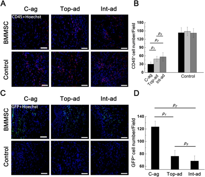
In situ immunofluorescence staining also demonstrated higher rates of GFP+-cell engraftment in the rats treated with BMMSC cell-aggregates than the other groups.

(A) Immunofluorescence staining on CD45+ lymphocytes on wound bed samples at 4W (counterstained with Hoechst 33342) (Bar = 50); (B) Quantification of CD45+ cell among groups. CD45+ cell infiltration in Top-ad and Int-ad wound bed tissue was heavier than that that in C-ag ones. p1 = 0.003, p2 = 0.000, p3 = 0.405, n = 6; (C) Immunofluorescence staining on GFP+ cell on wound bed samples at 4W (counterstained with Hoechst 33342) (Bar = 50 nm); (D) Quantification of GFP+ cell among groups indicated better engraftment for C-ag group than the other two cell transplanted groups, difference being significant. p1 = 0.001, p2 = 0.000, p3 = 0.135, n = 6.

These data show that not only are BMMSC cell aggregates safe, but they might also stimulate greater cutaneous regeneration for full layer cutaneous wounds than BMMSCs administered by other means.  These successful studies will hopefully be followed by large animal studies to confirm the expandability and efficacy of this technology in larger animals.

Wound Healing and Human Umbilical Cord Mesenchymal Stem Cells


Previous studies have shown that human bone marrow–derived mesenchymal stromal cells have potential to accelerate and augment wound healing. However, in the clinic, it is difficult to properly culture and then use bone marrow stem cells. Human umbilical cord blood–derived mesenchymal stromal cells (hUCB-MSCs) recently have been commercialized for cartilage repair as a cell-based therapy product that uses allogeneic stem cells.

Presently, current cell therapy products for wound healing utilize fibroblasts. Is it possible that hUCB-MSCs are superior to fibroblasts for wound healing? Seung-Kyu Han and his colleagues from the Department of Plastic Surgery at the Korea University College of Medicine in Seoul, South Korea used a cell culture system to compare the ability of hUCB-MSCs and fibroblasts to heal wounds.

For their study, Han and others used diabetic mice and isolated fibroblasts from normal and diabetic mice. Then they tested the ability of these cells to heal skin wounds in the very mice from which they were isolated. A third group of diabetic mice with skin wounds were treated with hUCB-MSCs. A comparison of all three groups examined the cell proliferation, collagen synthesis and growth factor (basic fibroblast growth factor, vascular endothelial growth factor and transforming growth factor-β) production and compared them among the three groups.

The results showed that hUCB-MSCs produced significantly higher amounts of vascular endothelial growth factor and basic fibroblast growth factor in comparison to both fibroblast groups. Human UCB-MSCs were better than diabetic fibroblasts but healthy fibroblasts in collagen synthesis, and there were no significant differences in cell proliferation and transforming growth factor-β production. Human UCB-MSCs produced significantly higher amounts of VEGF and bFGF when compared with both fibroblasts.

These results suggest that Human UCB-MSCs might be a better source for diabetic wound healing than either allogeneic or autologous fibroblasts. Larger animal studies will be needed, but this particular study seems like a good start.

Stem Cell Treatment Improves the Skin Quality of Children With inherited Skin Blistering Disease


A new stem cell-based therapy has shown some very promising results. This therapy was designed to treat a rare and debilitating skin condition that affects children, for which no cure currently exists. This cell-based therapy provided pain relief and reduced the severity of the skin condition for patients who participated in the clinical trial.

The clinical trial was led by scientists at King’s College London, who collaborated with researchers from the Great Ormond Street Hospital (GOSH). They recruited 10 children afflicted with a disease called recessive dystrophic epidermolysis bullosa (RDEB).

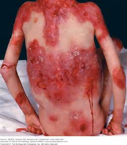
RDEB is a painful skin disease in which very minor skin injury leads to blisters and wounds that tend to heal very slowly or not at all. The skin of RDEB patients is quite fragile and it tends to scar, develops contractures, and is also prone to life-threatening skin cancers.

Dystrophic epidermolysis bullosa

This clinical trial, known as the EBSTEM trial, is a The Phase I/II trial whose results were published early online in the Journal of Investigative Dermatology. This study was designed to test the safety of infusions of stem cells and to determine if this treatment could help diminish the severity of the disease and improve quality of life for these patients.

During the first six months of the trial, participants were given three infusions of bone marrow- derived mesenchymal stromal cells from unrelated donors. Mesenchymal stem cells (MSCs) have been shown to home to wounded tissue and mediate wound healing in several previous studies. Although these infused stem cells do not survive permanently, they may still deliver therapeutic benefits.

The treated children were then monitored for a year after these cell infusions. Several different clinical tests failed to reveal any serious adverse effects in patients as a result of the stem cell treatment. When the pain levels of patients were measured, patients consistently reported lower pain levels after the treatment than before the treatment. Also the severity of their disease was also reported to have lessened following the stem cell infusions. Parents of these children reported better wound healing in their children and they also showed less skin redness and fewer blisters.

Overall, the outcomes of the trial are promising. However, this is an unblinded study of participants and may, therefore, contain positive biases in the way the information is reported. In interviews with families, participants reported a range of benefits from sleeping better, to the parents being able to return to work part-time because their children required less intense care. In fact, one family was actually able to plan their first vacation together.

Thus, further work is required to better understand the mechanisms that helped patients improve. Did the stem cells trigger the production of a growth factors and immune system regulators? Did these secreted compounds stimulate wound healing and reduce inflammation in the skin? Or did the presence of the cells somehow improve skin quality? Further studies are also required to confirm the efficacy of the treatment and establish the optimal dose of cells to give RDEB patients.

Fat-Based Stem Cells Speed the Healing of Bed Sores in Animals


Pressure ulcers, which are also knows as bedsores (or decubitus ulcers) are localized injuries to the skin that can also include the underlying tissue that usually occur as a result of pressure, or pressure in combination with rubbing or friction. They tend to occur some sort of bony prominence such as elbows, hips, shoulders, ankles, back of the head, and other such places. More than 2.5 million patients each year in the U.S. require treatment for pressure ulcers, and the elderly are at particularly high risk for these lesions. Currently, therapies for pressure ulcers consist of conservative medical management for shallow lesions and aggressive debridement and surgery for deeper lesions.

Jeffery Gimble and his colleagues from the Tulane University School of Medicine in New Orleans, Louisiana, used a mouse model for pressure ulcers to test the ability of fat-derived stromal/stem cell treatment to accelerate and enhance the healing of pressure ulcers.  The dorsal skin of both young (2 months old) and old (20 months old) C57BL/6J female mice was pinched between external magnets for 12 hours over 2 consecutive days. This treatment initiated a pressure ulcer, and one day after induction of the pressure ulcers, some of these mice were injected with fat-derived stromal stem cells that had been isolated from healthy mice that were of the same genetic lineage as the injured mice. However, the donor mice were genetically engineered to express a green fluorescent protein in all their tissues. Other mice were treated with injections of saline-treated controls.

The mice that were injected with fat-derived stromal/stem cells displayed a cell-concentration-dependent acceleration of wound closure. The cell-injected mice also showed improved epidermal/dermal architecture, increased fat deposition, and reduced inflammation at the sites of injury. Interestingly, these fat-derived stem cell-induced improvements occurred in both young and elderly mice. However, the gene expression profile of genes involved in the making of blood vessels, regulating the immune system, and tissue repair differed according to the age of the mice, with younger mice making more of these genes that their older counterparts. These results are consistent with clinical reports of the improved skin architecture after fat grafting in patients with thermal injuries.

This current proof-of-principle study sets the stage for clinical translation of the transplantation of fat-based stem cells as a treatment of pressure ulcers.

Using Cord Blood Stem Cells to “Re-educate” White Blood Cells and Treat Hair Loss


Alopecia areata (AA) is an autoimmune disease that targets the hair follicles. It affects the quality of life and self-esteem of patients because they lose their hair. Is there a way to treat this disease without suppressing the immune system?

Yong Zhao and from Tianhe Stem Cell Biotechnologies in Shandong, China and his collaborators used a so-called “Stem Cell Educator therapy” in which they took the patient’s blood and circulated it through a closed-loop system that separated mononuclear cells from the whole blood, and then allowed those cells to briefly interact with adherent human cord blood-derived multipotent stem cells (CB-SC). After this interaction, the mononuclear cells were returned to the patient’s circulation. This procedure uses the cord blood cells to “educate” the white blood cells of the patient to not attack the patient’s hair follicles.

In an open-label, phase 1/phase 2 study, nine patients with severe AA received one treatment with the Stem Cell Educator therapy. These patients were about 20 years old and had lost their hair, on the average, about 5 years ago.

All these patients experienced improved hair regrowth and quality of life after receiving Stem Cell Educator therapy.  Furthermore, analyses of immune cells from the blood of treated patients showed that the types of immune cells that attack tissues decreased and the number of cells that regulate the immune response increased. Also, investigations of hair follicles in the treated patients revealed that the restored hair follicles expressed a ring of transforming growth factor beta 1 (TGF-β1) around the hair follicles. TGF-β1 is a secreted molecule that down-regulates the immune response and prevents immune cells from attacking your own tissue. The fact that the hair follicles secreted all this TGF-β1 shows that the restored hair follicles had steeled them against the immune system.

How did the cord blood cells do this? By culturing white blood cells with cord blood cells in cell culture, Zhao and others showed that the human cord blood-derived multipotent stem cells induced white blood cells to increase their expression of molecules that are known to tame self-destructive white blood cells. Thus the cord blood stem cells secrete regulatory molecules that change the character of the immune cells so that they no longer attack the hair follicles.

These clinical data demonstrate the safety and efficacy of the Stem Cell Educator therapy for the treatment of AA. This is a very innovative approach that can produce lasting improvement in hair regrowth in subjects with moderate or severe AA.

Testing Stem Cell Quality


A new paper published in the journal EMBO Molecular Medicine by a team from the Lausanne University Hospital describes a protocol that can ensure the safety of adult epidermal stem cells before they are used as treatments for patients. The approach devised by this team takes cultivated, genetically modified stem cells and isolates single cells that are then used to make clonal cell cultures. These cloned cells are then rigorously tested to ensure that they meet the highest possible safety criteria. This protocol was inspired by approaches designed in the biotechnology industry and honed by regulatory authorities for medicinal proteins produced from genetically engineered mammalian cells.

“Until now there has not been a systematic way to ensure that adult epidermal stem cells meet all the necessary requirements for safety before use as treatments for disease,” says EMBO Member Yann Barrandon, Professor at Lausanne University Hospital, the Swiss Federal Institute of Technology in Lausanne and the lead author of the study. “We have devised a single cell strategy that is sufficiently scalable to assess the viability and safety of adult epidermal stem cells using an array of cell and molecular assays before the cells are used directly for the treatment of patients. We have used this strategy in a proof-of-concept study that involves treatment of a patient suffering from recessive dystrophic epidermolysis bullosa, a hereditary condition defined by the absence of type VII collagen which leads to severe blistering of the skin.”

Barrandon and co-workers have cultivated epidermal cells from patients who suffer from epidermolysis bullosa. These cells were then genetically engineered in order to insert a normal copy of the type VIII collagen gene. Then the genetically fixed cells were grown in culture so that they can be used to regenerate skin. Barrandon and others subjected these cells to an array of tests in order to determine which of the genetically engineered cells meet the requirements for safety and “stemness,” which refers to the stem cell characteristics that distinguish it from regular cells; its developmental immaturity and its ability to grow and self-renew. Clonal analysis revealed that the cultured, genetically engineered stem cells varied in their ability to produce functional type VII collagen. When the most viable, modified stem cells were selected and transplanted into the skin of immunodeficient mice, the cells regenerated skin and produced skin that did not blister in the mouse model system for recessive dystrophic epidermolysis bullosa. Furthermore, the cells produced functional type VII collagen. The safety of the cells was assessed by mapping the sites of integration of the viral vector. Because such viruses and produce gene rearrangements other mutations, the chosen cell lines were subjected to whole genome sequencing. Only the cells with insertions in benign locations were considered for use in their mouse model.

Barrandon concluded: “Our work shows that at least for adult epidermal stem cells it is possible to use a clonal strategy to deliver a level of safety that cannot be obtained by other gene therapy approaches. A clonal strategy should make it possible to integrate some of the more recent technologies for targeted genome editing that offer more precise ways to change genes in ways that may further benefit the treatment of disease. Further work is in progress in this direction.”

This work is certainly fascinating, but I think that using integrating viral vectors is asking for trouble. Certainly it should be possible to fix or replace the abnormal type VII collagen gene. Viruses that randomly insert genes into the genome can cause genetic problems, and even sequencing the genome may not properly address the safety concerns of the use of such viral vectors.

New Technology Reprograms Skin Fibroblasts


Fibroblasts are one of the main components of connective tissue, which is the main reason scientists typically exploit them for experiments. A collaborative team of scientists from the University of Pennsylvania, Boston University, and the New Jersey Institute of Technology have invented a way to reprogram fibroblasts without going through a pluripotent stage.

The senior author of this study, Xiaowei Xu, associate professor of pathology and laboratory medicine at the University of Pennsylvania School of Medicine, said, “Through direct reprogramming, we do not have to go through the pluripotent stem cell stage, but directly convert fibroblasts to melanocytes . So these cells do not have tumorigenicity” (the ability to cause tumors).

Melanocytes are found in the skin and they are responsible for the pigment in our skin. They are in the uppermost layer of the skin, known as the epidermis, and produce melanin, a brown pigment that helps screen against the harmful effects of UV light.

Turning a fibroblast into a melanocytes might seem trivial for a stem cell scientist; just reprogram the fibroblast into an induced pluripotent stem cells and then differentiate it into a melanocytes. However, this procedure utilized direct reprogramming, in which the fibroblast was converted into a melanocytes without traversing through the pluripotent stage. The difficultly with direct reprogramming is finding the right cocktail of genes and/or growth factors that will accomplish the deed.

Xu and his colleagues began their search by examining the genes that are specific to melanocytes. They found 10 different transcription factors that are important for melanocytes development. Next they screened these ten genes for their ability to convert a fibroblast into a melanocytes. They found that of the ten melanocytes-specific genes, three of them, Sox10, MITF, and PAX3 could do the job effectively. They called this gene combination “SMP3.”

When Xu and others tested SMP3 on mouse embryonic fibroblasts, they quickly expressed melanocytes-specific genes. When Xu’s group used SMP3 on human fetal dermal cells, once again, the cells rapidly differentiated into melanocytes. Xu and his team referred to these cells as hi-Mel, which is short for human, induced melanocytes.

When hi-Mel were grown in culture they produced melanin a plenty. When they were implanted into the skin of pigmentless mice, once again they rose to the challenge and made a great deal of pigment. Thus hi-Mel express the same genes as melanocytes and they behave for all intents and purposes as melanocytes.

Xu and his colleagues think that their procedure might be able to treat human patients with a condition called vitiligo in which the skin has patches that are devoid of pigment.

Another potential use of this technology is a way to effectively study melanoma, one of the most dangerous skin cancers known to human medicine. My good friend and SAU colleague died over a year ago from melanoma and having better ways to treat this monster would have been marvelous for Charlie, and his family, who miss him dearly. By generating melanocytes from the fibroblasts of melanoma patients, they can “screen not only to find why these patients easily develop melanoma, but possibly use their cells to screen for small compounds that can prevent melanoma from happening.”.

Also, because so the body contains so many fibroblasts in the first place, this reprogramming technique is well-suited for other cell-based treatments.

Hair Loss Cure Isn’t Here Yet, But Experimental Stem Cell Approach Looks Promising


While a cure for hair loss is some years away, a California research team has brought us much closer that such a treatment becoming a reality. Hair loss, a condition that affects 50 million men and 30 million women in the U.S. alone, might fall to stem cell treatments some day.

Dr. Alexey Terskikh led the team from the Sanford-Burnham Medical Research Institute in La Jolla, California that showed that stem cells derived from human skin can be used to grow hair in mice.

“The method is a marked improvement over current methods that rely on transplanting existing hair follicles from one part of the head to another,” Dr. Terskikh, who serves as an associate professor at the institute. “Our stem cell method provides an unlimited source of cells from the patient for transplantation and isn’t limited by the availability of existing hair follicles.”

Conventional hair transplantation and other hair restoration treatments that are presently in use must use whatever hair the patient has left. However a stem cell-based procedure could, in theory, grow all kinds of hair on the heads of completely bald men and women.

“If this approach is proven to work in humans, it will change existing treatments radically,” Dr. Nicole Rogers, a dermatologist and hair transplant surgeon in New Orleans, told The Huffington Post in an email.

Dr. Marie Jhin, a dermatologist in San Francisco and an adjunct clinical instructor at Stanford University, feels the same way about Terskikh’s results. If this treatment pans out, she said that it “absolutely would be a breakthrough.”

Rogers, however, tempered her excitement by advising caution and skepticism, since there have been many “fits and starts” over the years in the hair-restoration field. Rogers added that the Sanford-Burnham group must face many challenges in order to replicate their results in large-scale human trials.

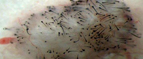
The technique exploits the ability of human pluripotent stem cells to differentiate into almost any other adult cell type in the body. Terskikh and his collaborators differentiated induced pluripotent stem cells made from reprogrammed skin cells into the dermal papilla cells that regulate the formation and growth of hair follicles. Furthermore, when they injected these cells into the lower layers of the skin of mice, they grew hair.

Close-up photograph showing new hair growth | Sanford-Burnham Medical Research Institute

Human dermal papilla cells are unsuitable for conventional hair transplants because quickly lose their hair-growing potency and cannot be obtained in necessary numbers for clinical purposes.

Terskikh wisely did not prognosticate when they would be able to extend their protocol to treat hairless humans. The next step, according to Terskikh is to secure a partner to fund future research into this area.

Fingernail Stem Cell Population Identified


The ability of fingernails to grow back, unlike other body parts seems to be the result of the presence of a resident stem cells population.

Researchers at the University of Southern California (USC), led by Krzysztof Kobielak has identified a new stem population in nails that can either self-renew or differentiate into other distinct cell types.

Identifying these cells was no small chore, and Kobielak used an ingenious new technique for attaching fluorescent dyes and other tags to mouse nail cells. while many cells in the nail divided and spread throughout the nail, another small population stayed at the base of the nail and divided slowly or not at all.

Localization of LRCs in the nail proximal fold. (A) Components of the mouse nail. Top view, horizontal sections of the fingertip before (C) and after (B and D) 4 wk of chase with Dox identifying a population of H2BGFP marked LRCs surrounding the nail structure. Side view, perpendicular sections of the digit tip before (E and G) and after (F) 4 wk of chase with Dox demonstrating the presence of upper LRCs in the upper PF. (H) Lower LRCs at the lower nail PF. GFP; green fluorescent protein; HF, hair follicle; L-LRCs, lower label-retaining cells; U-LRCs, upper label-retaining cells. Yellow box denotes region of interest in B, and red and blue boxes (G) denote representative U-PF and L-PF regions for orientation. (Scale bars: 50 μm.)

Kobielak and his team showed that these slow-dividing cells normally contribute to the growth of her nails and nearby skin. However, if the nail undergoes some kind of injury or physical insult, a signaling protein called bone morphogen protein or BMP signals to the nail bed stem cells to switch to exclusively repairing the nail. Thus this nail bed stem cell population has the flexibility to perform dual roles in the finger tips.

Nail proximal fold cells participate in nail regeneration in response to plucking injury and upon transplantation. (A) Whole-mount Tomato expression in regenerated nails 2 wk after plucking. Linear streams of Tomato+ cells (red) in regenerating nails (arrows) extending from the nail base toward the tip. (B) Schematic model representing the role of K15+ NPFSCs during nail regeneration. (C–C′′) K5 expression (green) in regenerating nails localizing the linear K15-derived, Tomato+ (red) cell streams emanating from the basal K5+ Mx extending upward (arrows) into the overlying differentiated NP; yellow box denotes region of interest in (C′ and C′′). (D) H2BGFP+ nail LRCs persist in the finger following nail removal. (E) Nail LRCs are quiescent, whereas the nail Mx contains actively proliferating cells marked by BrdU incorporation. (F) Upon NP removal, LRCs become activated, indicated by Ki67 coexpression. (G) Nail LRCs transplantation strategy. H2BGFP+ nail cells contribute to the nail structure 17 d after transplantation (H), sectioning of 22-d chased transplant (I), demonstrating the presence of remaining LRCs from the transplant. d, day; Epi, epidermis; GFP, green fluorescent protein; L-LRCs, lower label-retaining cells; U-LRCs, upper label-retaining cells. DAPI counterstaining (blue) was used to localize cell nuclei in immunofluorescent images. (Scale bars: A and H, 500 μm; C–F and I, 50 μm.)

The members of the Kobielak laboratory are also interested in other types of signals and what they might do to these nail bed stem cells. For example, could they induce them to differentiate into additional cell types besides skin and nail? Could they aid in amputation repair and the repair of severe skin injuries?

Kobielak said: “That was very surprising discovery [sic], since the dual characteristics of these nail stem cells to regenerate both the nail and the skin under certain physiological conditions is quite unique and different from other skin stem cells, such as those of the hair follicle or sweat gland.”

Treating A Genetic Skin Disorder with Induced Pluripotent Stem Cells


Dystrophic epidermolysis bullosa (RDEB) is an inherited skin disease that causes fragile skin. RDEB is caused by mutations in the gene that encoded a protein called type VII collagen. Because collagen is a major structural component of skin, collagen mutations result in fragile skin and mucous membranes that blister easily if they are subjected to even slight mechanical stresses. There are no cures for such diseases, but skin creams and palliative care can decrease the severity of the symptoms.

Induced pluripotent stem cells (iPSCs) have the ability to treat such genetic diseases. In order to provide proof of principle of the applicability of iPSCs for the treatment of RDEB, Daniel Wenzel and his colleagues in the laboratory of Arabella Meixner from the Institute of Molecular Biotechnology of the Austrian Academy of Sciences in Vienna, Austria made iPSCs from mice that harbored mutations in the gene that encodes type VII collagen (Col7a1) and exhibited skin fragility and blistering. The symptoms displayed by these Col7a1-mutant mice resembled human RDEB.

Wenzel and his coworkers then genetically repaired the Col7a1 mutations in these iPSCs, and then differentiated these cells into functional fibroblasts that expressed and secreted normal type VII collagen. When implanted, the genetically-repaired iPSC–derived fibroblasts did not form tumors, and could be successfully traced up to 16 weeks after intradermal injection. Therapy with iPSC-derived fibroblasts also resulted in faithful and long-term restoration of type VII collagen deposition at the epidermal-dermal junction of Col7a1 mutant mice, and restored the resistance of the skin to mechanical stresses.

Thus, intradermal injection of genetically repaired iPSC-derived fibroblasts restored the mechanical resistance of the skin to blistering in RDEB mice. These data demonstrate that, at least in principle, RDEB skin can be effectively and safely repaired using a combination of gene therapy and iPSC-based cell therapy.

A similar study examined another type of epidermolysis bullosa.  Noriko Umegaki-Arao and her colleagues in the laboratory of Angela Christiano from Columbia University used iPSCs to treat mice with a distinct type of epidermolysis bullosa that resulted from mutations in COL17A1 gene, which encodes type XVII collagen (Col17).  In this case, however, the mutation has been observed to revert or fix itself in patients.  Patients tend to have patches of skin that are normal in a sea of abnormal skin.

Therefore, Umegaki-Arao and her coworkers derived iPSCs from Col17-mutant mice, differentiated them into skin cells (keratinocytes) and then cultured them, examining individual clones for reversion to normal Col17, which was fairly easy to do as it turns out.  Once revertant-iPSC keratinocytes were properly secured, and then used them to reconstitute human skin in mutant mice.  Thus, revertant keratinocytes can be a viable source of spontaneously gene-corrected cells for developing iPSC-based therapeutic approaches in the treatment of epidermolysis bullosa.

Mesenchymal Stem Cells Make Blood Vessel Cells and Improve Wound Healing


Mesenchymal stem cells from umbilical cord have the ability to differentiate into cartilage cells, fat cells, bone cells, and blood vessels cells. These cells also are poorly recognized by the immune system of the patient and are at a low risk of being rejected by the patient’s immune system.

Valeria Aguilera and her colleagues from the laboratory of Claudio AguayoWe at the University of Concepción, Chilee have evaluated the use of mesenchymal stem cells from umbilical cord in the formation of new blood vessels in damaged tissues. Wharton’s jelly mesenchymal stem cells of hWMSCs were used to potentially accelerate tissue repair in living animals.

Aguilera and her co-workers began by isolating mesenchymal stem cells from human Wharton’s jelly (a connective tissue in umbilical cord). Then they grew these cells in culture for 14 or 30 days. Interestingly, the longer the WMSCs grew in culture, the more they looked like blood vessel cells. They began to express blood vessel-specific genes and proteins. WMSCs cultured for 30 days were even more like blood vessels than those grown in culture for 14 days.

When these cells were injected in the mice with damaged skin, the results showed that the WMSCs cultured for 30 days significantly accelerated wound healing compared with animals injected with either undifferentiated hWMSCs or with no cells.

Effect of hWMSCs and endothelial-differentiated hWMSC transplantation in a wound-healing model. A) Representative images of wounds at day 1 (top panels) and 12 (lower panels) after injury and subcutaneous injection of hWMSCs, hWMSC trans-differentiated into endothelial cells for 14 days (hWMSC-End14d) or 30 days (hWMSC-End30d), or control (PBS). B) Wound healing quantified in PBS (○), hWMSC (•), hWMSC-End14d (□) or hWMSC-End30d (▪) treated mice (n = 5 independent experiments, in duplicate). Values are expressed as mean±S.E.M, +P<0.05 in hWMSC-End30d v/s hWMSC, hWMSC-End14d, at the corresponding time; **P<0.03 in hWMSC-End30d v/s PBS; *P<0.001 in hWMSC-End30d v/s PBS; # P<0.01 in hWMSC-End30d v/s PBS.
Effect of hWMSCs and endothelial-differentiated hWMSC transplantation in a wound-healing model.
A) Representative images of wounds at day 1 (top panels) and 12 (lower panels) after injury and subcutaneous injection of hWMSCs, hWMSC trans-differentiated into endothelial cells for 14 days (hWMSC-End14d) or 30 days (hWMSC-End30d), or control (PBS). B) Wound healing quantified in PBS (○), hWMSC (•), hWMSC-End14d (□) or hWMSC-End30d (▪) treated mice (n = 5 independent experiments, in duplicate). Values are expressed as mean±S.E.M, +P


The wounds of mice treated with the WMSCs cultured for 30 days looked healthier, but they had many more blood vessels.

Histologic analysis of wounds in the wound-healing model. A) Representative photographs of wounds (hematoxilin/eosin staining) 12 days after injury and subcutaneous injection of PBS, hWMSCs, hWMSC-End14d or hWMSC-End30d. Quantification of histological images, for blood vessels area (B) and histological score (C) for each group of mice. Values are mean ± S.E.M (n = 5 independent experiments, in duplicate), *P<0.001 in hWMSC-End30d or hWMSC-End14d v/s MSC; +P<0.05 in hWMSC-End30d or hWMSC-End14d v/s hWMSC. Magnification x40 (-). Ep, epidermis; D, dermis; H, hypodermis.
Histologic analysis of wounds in the wound-healing model.
A) Representative photographs of wounds (hematoxilin/eosin staining) 12 days after injury and subcutaneous injection of PBS, hWMSCs, hWMSC-End14d or hWMSC-End30d. Quantification of histological images, for blood vessels area (B) and histological score (C) for each group of mice. Values are mean ± S.E.M (n = 5 independent experiments, in duplicate), *P

When laboratory animals received the culture medium from the WMSCs cultured for 30-days also showed significant acceleration of their healing, which suggests that these cells secrete a host of healing molecules that induced the formation of new blood vessels.  One might also conclude that the implanted WMSCs did not contribute to the formation of new blood vessels, but simply directed the formation of new blood vessels by secreting healing molecules.  However, when WMSCs were detected in the healed tissue, they were predominantly found in the walls of new blood vessels.

Immunohistochemical detection of human mesenchymal cells in a wound-healing model. A. Immunohistochemical staining of human mitochondria was performed in permeabilized tissue sections obtained after 12 days of subcutaneous injection of PBS, hWMSCs, hWMSC-End14d or hWMSC-End30d in mice. Cell nuclei were stained with hematoxyline. In B. Number of positive cells per vessel. Representative images of 5 independent experiments, in duplicate. Magnification x40 and insert 100x. Bars 50 µm.
Immunohistochemical detection of human mesenchymal cells in a wound-healing model.
A. Immunohistochemical staining of human mitochondria was performed in permeabilized tissue sections obtained after 12 days of subcutaneous injection of PBS, hWMSCs, hWMSC-End14d or hWMSC-End30d in mice. Cell nuclei were stained with hematoxyline. In B. Number of positive cells per vessel. Representative images of 5 independent experiments, in duplicate. Magnification x40 and insert 100x. Bars 50 µm.

These results, which were published in PLOS ONE, demonstrate that mesenchymal stem cells isolated from umbilical cord connective tissue or Wharton’s jelly can be successfully grown in culture in the laboratory and trans-differentiated into blood vessels-forming cells (endothelial cells).  These differentiated hWMSC-derived endothelial cells seem to promote the formation of new networks of blood vessels, which augments tissue repair in laboratory animals through the secretion of soluble pro-blood vessel-making molecules and, occasionally, by contributing to the formation of new vessels, themselves.

Skin Tissue Grown From Human Stem Cells


A research team from King’s College, London, in collaboration with the San Francisco Veteran Affairs Medical Center has succeeded in growing the epidermal layer of skin in culture, this cultured skin has many of the mechanical and biological properties of actual human skin.

The outermost layer of the skin, known as the epidermis forms a protective barrier between the external environment and the body. It protects against water loss and prevents the entry of microorganisms.

Tissue engineers have been able to grow skin cells (keratinocytes) in culture, but getting them to organize into an organ that resembles biological skin has proven rather difficult. However, the ability to test drugs on cultured skin that greatly mimics human skin has been the goal of such research for several years.

For this present project, keratinocytes were made from induced pluripotent stem cells that were derived from skin cells obtained from biopsies. These keratinocytes made from induced pluripotent stem cells (iPSCs) were very similar to keratinocytes made from embryonic stem cells and primary keratinocytes isolated from skin biopsies.

To form a three-dimensional structure like skin, the keratinocytes were cultured in a high-to-low humidity environment and they assembled into a layer structure that looked like human skin. When this cultured skin was compared with skin made from embryonic stem cell-derived keratinocytes or from keratinocytes isolated from skin biopsies, there were no significant structural differences.

Scientists hope to use this cultured skin to study congenital skin diseases like ichthyosis (characterized by dry, flaky skin) or atopic dermatitis. Growing large quantities of skin in culture will also allow drugs and cosmetics to be effectively tested for safety without the use of expensive and sometimes highly variable animal models.

This technology would also allow different laboratories to grow skin from different ethnic groups that have distinct types of skin with variable biological properties.

Sweat Glands Are A Source of Stem Cells for Wound Healing


Stem Cells from human sweat glands serve as a remarkable source for wound healing treatments according to a laboratory in Lübeck, Germany.

Professor Charli Kruse, who serves as the head of the Fraunhofer Research Institute for Marine Biotechnology EMB, Lübeck, Germany, and his colleagues isolated cultured pancreatic cells in the course of their research to look into the function of a protein called Vigilin. When the pancreatic cells were grown in culture, they produced, in addition to other pancreatic cells, nerve and muscle cells. Thus the pancreas contains a stem cell population that can differentiate into different cell types.

Kruse and his group decided to investigate other glands contained a similar stem cell population that could differentiate into other cell types.

Kruse explained: “We worked our way outward from the internal organs until we got to the skin and the sweat glands. Again, this yielded the same result: a Petri dish full of stem cells.”

Up to this point, sweat glands have not received much attention from researchers. Mice and rats only have sweat glands on their paws, which makes them rather inaccessible. Human beings, on the other hand, have up to three million sweat glands, predominantly on the soles of out feet, palms of the hand, armpits, and forehead.

Ideally, a patient could have stem cells taken from her own body to heal an injury, wound, or burn, Getting to these endogenous stem cell populations, however, represents a challenge, since it requires bone marrow biopsies or aspirations, liposuction, or some other invasive procedure.

Sweat glands, however, are significantly easier to find, and a short inpatient visit to your dermatologist that extracts three millimeters of underarm skin could provide enough stem cells to grow in culture for treatments.

Stem cells from sweat glands have the capacity to aid wound healing. Kruse and his group used sweat gland-based stem cells in laboratory animals. The Kruse group used skin biopsies from human volunteers and separated out the sweat gland tissues under a dissecting scope. Then the sweat gland stem cells were grown in culture and induced to differentiate into a whole host of distinct cell types.

Then Kruse’s team grew these sweat gland stem cells in a skin-like substrate that were applied to wounds on the backs of laboratory animals. Those animals that had received stem cell applications healed faster than those that received no stem cells.

If the stem cells were applied to the mice with the artificial substrate, the cells moved into the bloodstream and migrated away from the site of the injury. In order to help heal the wound the cells had to integrate into the skin and participate in the healing process.

“Not only are stem cells from sweat glands easy to cultivate, they are extremely versatile, too,” said Kruse.

Kruse and his team are already in the process of testing a treatment for macular degeneration using sweat gland-based stem cells. “In the long-term, we could possibly set up a cell bank for young people to store stem cells from their own sweat glands/ They would then be available for use should the person need new cells, following an illness,l perhaps, or in the event of an accident,” Kruse said.