Nerve Growth Factor-Secreting Mesenchymal Stem Cells To Treat Huntington’s Disease


Vicki Wheelock at the UC Davis Medical Center has registered clinical trial number NCT01937923, which is otherwise known as “PRE-CELL.” This clinical trial will use various imaging techniques, laboratory tests, and clinical evaluations of Huntington’s disease (HD) patients to map the disease progression over 12-18 months. This trial will then hopefully identify candidates for a new trial in which these patients will be implanted with mesenchymal stem cells that secrete nerve growth factors. This represents one of the first clinical trials to examine the use of mesenchymal stem cells in the treatment of HD

The rationale for this study comes from a 2012 study in mice. Ofer Sadan, Eldad Melamed, and Daniel Offen from the Rabin Medical Center in Tel Aviv University, Israel, used R6/2 mice to test the efficacy of nerve growth factor-secreting mesenchymal stem cells isolated from bone marrow . In this paper, Sadan and others isolated mesenchymal stem cells from the bone marrow of healthy human volunteers and mice and then cultured them in special growth media that induces these cells to secrete special nerve growth factors. These so-called NTF+ cells were then transplanted into the striatum of R6/2 mice.

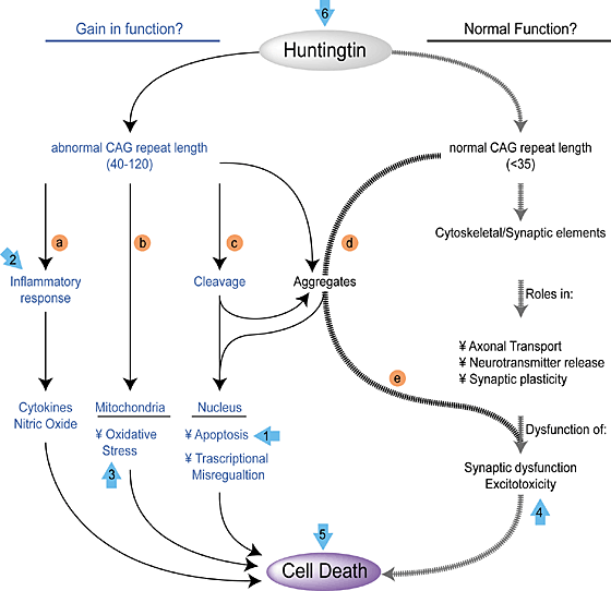
R6/2 mice express part of the human HTT gene; specifically the part that causes HD. Since HD is an inherited disease, there is a specific gene responsible for the vast majority of HD cases, and that gene is the human HTT gene, which encodes the Huntington protein. The function of the Huntington protein is uncertain, but it is found at high levels in neurons, even though it is found in other tissues as well, and dysfunctional Huntington protein affects neuron health.

Huntingtin Function

The HTT gene in HD patients contains the insertion of extra copies of the CAG triplet. The more CAG triplets are inserted into the HTT gene, the more severe the HD caused by the mutation. The hitch is that normal copies of the HTT gene has multiple copies of this CAG repeat. CAG encodes the amino acid glutamine, and Huntington contains a stretch of glutamine residues that seem to allow the protein to interact with other proteins found in neurons. When this glutamine stretch becomes too long, the protein is toxic and it begins to kill the cells. How long is too long? Research has pretty clearly shown that people whose HTT genes contain less than 28 CAG virtually never develop HD. People with between 28–35 CAG repeats, are usually unaffected, but their children are at increased risk of developing HD. People whose HTT genes contain 36–40 CAG repeats may or may not show HD symptoms, and those who have over 40 copies almost always are afflicted with HD.

hunt_gene_big

Now, back to R6/2 mice. These animals contain a part of the human HTT gene that has 150 CAG triplets. These mice show the characteristic cell death in the striatum and have behavioral deficits. In short R6/2 mice are pretty good model systems to study HD.

Sadan and others implanted MSCs that had been conditioned in culture to express high levels of nerve growth factors. Then these cells were transplanted into the striatum of R6/2 mice. R6/2 mice were also injected with buffer as a control.

The results showed that injections of NTF+ MSCs before the onset of symptoms did little good. The mice still showed cell death in the brains and behavioral deficits. However, NTF+ MSCs injected later (6.5 weeks), resulted in temporary improvement in the ability of the R6/2 mice to move and these cells also extended their life span. These results were published in the journal PLoS Currents (2012 Jul 10;4:e4f7f6dc013d4e).

Other work, also by Sadan and others, showed that injected MSCs tended to migrate to the damaged areas. When the injected cells were labeled with iron particles, they could be robustly observed with MRIs, and MRIs clearly showed that the injected cells migrated to the damaged areas in the brain (Stem Cells 2008; 26(10):2542-51). Another paper by Sadan and others also demonstrated that the striatum of NTF+ MSC-injected mice show less cell death than control mice (Sadan, et al. Exp Neurol. 2012; 234(2): 417-27). Other workers have also shown that implanted MSCs can provide improve symptoms in R6/2 mice and that they primary means by which they do this is by the secretion of nerve growth factors (Lee ST, et al. Ann Neurol 2009; 66(5): 671-81).

Thus, there is ample reason to suspect the PRECELL trial may lead to a stem cell-based clinical trial that will yield valuable clinical information. The animal data shows definite value in using preconditioned MSCs as a treatment for HD, and if the proper patients are identified by the PRE-CELL trials, then hopefully it will lead to a “CELL” trial in which HD patients are treated with NTF+ MSCs.

Mind you, this treatment will only delay HD at best and buy them time. Such treatments will not cure them. The NTF+ MSCs survive for a finite period of time in the hostile environment of the striatum of the HD patient, and the relief they will provide will be temporary. MSCs do not differentiate into neurons in this case, and they do not replace dead neurons, but they only help spare living neurons from suffering the same fate.

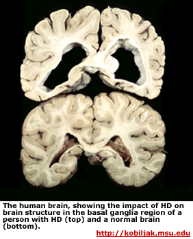
Huntington disease striatum

There is an MSC cell line that does make neurons, and if this cell line were used in combination with NTF+ MSCs, then perhaps neural replacement could be a possibility.  Also neural precursor cells could be used in combination with NTF+ MSCs to increase their survival.  Even then, as long as diseased neurons are producing toxic products, until gene therapy is perfected to the point that the actual genetic lesion in the striatal neurons is fixed, the deterioration of the striatum is inevitable. However, treatments like this could, potentially, delay this deterioration. This clinical trial should give us more information on exactly that question.

Two more points are worth mentioning.  When fetal striatal grafts were implanted into the brains of HD patients, the grafts underwent disease-like degeneration, and actually made the patients worse (see Cicchetti et al. PNAS 2009; 106(30): 12483-8 and Cicchetti F, et al. Brain 2011; 134(pt 3): 641-52).  Straight fetal implants do not seem to work.  Please let’s put the kibosh on these gruesome experiments.  Secondly, when neuronal precursor cells differentiated from human embryonic stem cells were implanted into HD rodents, the implanted cells formed some neurons and improved behavior to some extent, but non-neuronal differentiation remained a problem (Song J, et al., Neurosci Lett 2007; 423(1): 58-61).  Having non-brain cells in your brain is a significant safety problem.  Thus, embryonic stem cell-derived neuronal precursor cells do not seem to be the best bet to date either.  So, this present clinical trial seems to be making the most of what is presently safely available.
Stem Cell Transplant Repairs the Damage that Results from Inflammatory Bowel Disease


A source of stem cells from the digestive tract can repair a type of inflammatory bowel disease when transplanted into mice has been identified by British and Danish scientists.

This work resulted from a collaboration between stem cell scientists at the Wellcome Trust-Medical Research Council/Cambridge Stem Cell Institute at Cambridge University, and the Biotech Research and Innovation Centre (BRIC) at the University of Copenhagen, Denmark. This research paves the way for patient-specific regenerative therapies for inflammatory bowel diseases such as ulcerative colitis.

All tissues in out body probably contain a stem cell population of some sort, and these tissue-specific stem cells are responsible for the lifelong maintenance of these tissues, and, ultimately, organs. Organ-specific stem cells tend to be restricted in their differentiation abilities to the cell types within that organ. Therefore, stem cells from the digestive tract will tend to differentiate into cell types typically found in the digestive tract, and skin-based stem cells will usually form cell types found in the skin.

When this research team examined developing intestinal tissue in mouse fetuses, they discovered a stem cell population that differed from the adult stem cells that have already been described in the gastrointestinal tract. These new-identified cells actively divided and could be grown in the laboratory over a long period of time without terminally differentiating into adult cell types. When exposed to the right conditions, however, these cells could differentiate into mature intestinal tissue.

Fordham_CellStemCell_GraphicalAbstract

Could these cells be used to repair a damaged bowel? To address this question, this team transplanted these cells into mice that suffered from a type of inflammatory bowel disease, and within three hours the stem cells has attached to the damaged areas of the mouse intestine. integrated into the intestine, and contributed to the repair of the damaged tissue.

“We found that the cells formed a living plaster (British English for a bandage) over the damaged gut,” said Jim Jensen, a Wellcome Trust researcher and Lundbeck Foundation fellow, who led the study. “They seemed to response to the environment they had been placed in and matured accordingly to repair the damage. One of the risks of stem cell transplants like this is that the cells will continue to expand and form a tumor, but we didn’t see any evidence of that with this immature stem cell population from the gut.”

Because these cells were derived from fetal intestines, Jensen and his team sought to establish a new source of intestinal progenitor cells.  Therefore, Jensen and others isolated cells with similar characteristics from both mice and humans, and  made similar cells similar cells by reprogramming adult human cells in to induced pluripotent stem cells (iPSCs) and growing them in the appropriate conditions.  Because these cells grew into small spheres that consisted of intestinal tissue, they called these cells Fetal Enterospheres (FEnS).

Established cultures of FEnS expressed lower levels of Lgr5 than mature progenitors and grew in the presence of the Wnt antagonist Dkk1 (Dickkopf).  New cultures can be induced to form mature intestinal organoids by exposure to the signaling molecule Wnt3a. Following transplantation in a model for colon injury, FEnS contributed to regeneration of the epithelial lining of the colon by forming epithelial crypt-like structures that expressed region-specific differentiation markers.

“We’ve identified a source of gut stem cells that can be easily expanded in the laboratory, which could have huge implications for treating human inflammatory bowel diseases. The next step will be to see whether the human cells behave in the same way in the mouse transplant system and then we can consider investigating their use in patients,” Jensen said.

Misfolded Protein Can Transmit Parkinson’s Disease from Cell to Cell


A research team led by Virginia Lee, who works as a neurobiologist at the University of Pennsylvania in Philadelphia has provided a mechanism for how misfolded proteins cause Parkinson’s disease. Lee’s group has resurrected an old treatment strategy that was discarded long ago that just might to slow the progression of this neurological disease.

Alpha-synuclein is a strange name for a protein, but it is a specifically found in the nervous system. Alpha-synuclein protein can compose as much as 1% of all the protein in the cytoplasm of a neuron. It is found all over the brain. What this protein actually does is a bit of a mystery, but the latest data suggests that alpha-synuclein helps traffic proteins from membranes to other places in the cell (Cooper et al., (2006). Science 313: 324–328).

In the brains of patients with Parkinson’s disease, neurons accumulate protein aggregates known as Lewy bodies. A major component of Lewy bodies is alpha-synuclein that has folded in an aberrant manner. These aggregations of misfolded alpha-synuclein cause a variety of problems inside cells that culminate in the death of the neuron.

This is the story of Parkinson’s disease so far, but Lee and her colleagues injected a misfolded synthetic version of α-synuclein into the brains of normal mice and saw the key characteristics of Parkinson’s disease develop and progressively worsen. While that is not a surprise, what Lee and co-workers found when they examined the brains of the injected laboratory animals astounded them. This study, which was published in the journal Science, shows that the injected misfolded alpha-synuclein was able to spread from one nerve cell to another. Therefore, the malformed protein did not just take up residence inside neurons, but instead was able to travel from one neuron to another.

Apparently, cells affected by misfolded alpha-synuclein are able to secrete it into the areas that surround them and this secreted protein is taken up by healthy cells. Once taken up, the misfolded alpha-synuclein induces the normal copies of the alpha-synuclein protein snap into the misfolded conformation. This eventually kills off the once-healthy neuron and also turns it into a new factory for the secretion of misfolded alpha-synuclein, which them goes on to damage other neurons.

This finding, however, raises the possibility that an antibody that binds the misfolded α-synuclein could potentially bind the protein and prevent it from passing between nerve cells. “It’s very hard to ask antibodies not only to get inside the brain, but to get inside cells,” says Lee. “But now you have the possibility of stopping the spreading. And if you stop the spreading, perhaps you can slow the progression of the disease.”

The tendency of the pathology of Parkinson’s disease to spread from neuron to neuron by a rogue protein was actually suggested in 2008. Fetal neural tissue transplants were used to treat Parkinson’s patients, but upon post-mortem examination of the transplanted fetal tissue, it was quickly recognized that these transplants has developed the characteristic Lewy bodies associated with Parkinson’s disease. This indicated that the nearby diseased cells were able to infect the transplanted tissue with Parkinson’s disease. Subsequent studies have shown that misfolded alpha-synuclein does spread between neighboring cells and induce cell death (Desplats, P. et al. Proc. Natl Acad. Sci. USA 106, 13010–13015 (2009).). The neurons, apparently, can release vesicles filled with misfolded alpha-synuclein in the same way they release neurotransmitters. This release bathes the nearby cells in misfolded alpha-synuclein, but there are still questions as to whether or not the misfolded alpha-synuclein is responsible for the cascade of brain damage seen in Parkinson’s.

Lee says that her team has now captured the full consequences of runaway α-synuclein in the brain. “We knew this transfer from one cell to another can happen, but whether it could play a significant role in the disease was still open,” says Tim Greenamyre, director of the Pittsburgh Institute for Neurodegenerative Diseases in Pennsylvania, who was not involved in the latest work.

Besides Lewy bodies, the brains of patients with Parkinson’s disease also show a dramatic loss of those neurons that produce the chemical messenger dopamine. When Lee’s team injected the misfolded α-synuclein into a part of the mouse brain rich in dopamine-producing cells, Lewy bodies began to form, followed by the death of dopamine neurons. Nerve cells linked to those near the injection site also developed Lewy bodies, which showed that cell-to-cell transmission was occurring.

Greenamyre says that is possible, but hasn’t yet been proved. “All of the cells affected in this paper were those directly in contact with the injection site,” he says. But, within six months of the injection, coordination of movement, grip strength and balance had all deteriorated in the mice, which is a recapitulation of what occurs in people with Parkinson’s disease.

“It’s really pretty extraordinary,” says Eliezer Masliah, a neuroscientist at the University of California, San Diego. “We have been trying that experiment for a long time in the lab and we have not seen such dramatic effects.” According to Masliah, Lee’s work provides the impetus for that handful of biotechnology companies that are sponsoring clinical trials of alpha-synuclein antibodies for as therapeutic agents for Parkinson’s disease. Masliah hopes that this will also motivate neuroscientists to examine exactly how the protein enters and exits cells.

There is still one mystery that has not been addressed to data: why do the Lewy bodies appear in the first place? “Parkinson’s disease is not a disorder in which somebody injects synuclein into your brain,” notes Ted Dawson, director of the Institute for Cell Engineering at Johns Hopkins University in Baltimore, Maryland. “So what sets it in motion?” Clearly some mutations in the gene that encodes alpha-synuclein increase the tendency for this protein to spontaneously misfold. But this also suggests that there are particular triggers that lead to such events. The nature of these triggers will certainly be the subject of future work.

Human Neural Stem Cell Line From StemCells Inc. Makes Myelin in Shiverer Mice


Physicians and research scientists at the Oregon Health Science University in Portland, Oregon have used banked neural stem cells to make myelin in mice that have a severe disease that prevents the synthesis and deposition of myelin around nerves. This proof-of-concept experiment shows that such a treatment strategy is feasible for human patients.

In previous posts on this blog site, I have discussed the importance of the myelin sheath that surrounds and insulates certain nerves. I will not reiterate those points here, but simply refer you to those older posts.

In humans, myelin loss is not noticed until the patient begins to show symptoms.  Myelin disorders are quite disabling and even fatal in some cases.  Such disorders include cerebral palsy in children born prematurely, multiple sclerosis, and others.

Myelin loss has also been found to play an important role in age-related senility.  Researchers at the Oregon Health and Science University Doernbecher Children’s Hospital used very advance Magnetic Resonance Imaging to study myelin (white matter) in the brains of adults of all stages.  They discovered that widespread changes in the white matter, and damage to the myelin of the brain were highly correlated with progressive senility (See Black SA, et al., “White matter lesions defined by diffusion tensor imaging in older adults.” Annals of Neurology, 2011 Volume 70, Issue 3, pages 465–476).

Stephen Back and his colleagues at OHSU examined the ability of human stem cells to make myelin and heal the sick animals.  To test this possibility, Back used a mouse called the “Shiverer immunodeficient” mouse, that develops progressive neurological damage because of its inability to make myelin.  Remember that small regions of demyelination that cover particular segments or even patches of nerves are followed by repair, regeneration, and complete recovery of neural function.  However, extensive demyelination or myelin loss is typically followed by degeneration of the axon (the extension of the neuron that conducts nerve impulses to other neurons) and also the neuron cell body.  Neuron death and axonal death are often irreversible.

The use of the Shiverer mouse presented some unique challenges.  Most neural stem cell experiments utilize newborn rather than adult mice.  Back explained:  “Typically, new-born mice have been studied by other investigators because stem cells survive very well in the newborn brain.  We, in fact, found that the stem cells preferentially matured into myelin-forming cells as opposed to other types of brain cells in both newborn mice and older mice.  The brain-derived stem cells appeared to be picking up on cues in the white matter that instructed the cells to become myelin-forming cells.”

Back collaborated with StemCells Inc., to make use of their proprietary neural stem cell line.  His initial experiments showed that implanting these neural stem cells made myelin sheaths in presymptomatic newborn animals.  However, these experiments did not indicate whether or not these stem cells would survive after transplantation into older animals that were already showing symptoms and declining in health.  Black, therefore, wanted to perform a much more difficult experiment by transplanting the neural stem cells into very sick adult animals that showed the horrific symptoms of demyelination.

MRI studies confirmed that implanted neural stem cells did in fact make new myelin within weeks after transplantation.  However, the detection of something such as myelin in mice usually requires the use of dyes of some other agent that fills the thing you want to detect in order to see it.  Many of these Shiverer animals are so sick that they cannot survive MRI experiments.  Therefore OHSU used a very sophisticated piece of equipment to solve this problem:  ultra-high field MRI scanners that could detect myelin without the use of dyes.

Back further explained:  “This is an important advance because it provides proof of principle that MRI can be used to track the transplants as myelin is being made.  We actually confirmed that the MRI signal in the white matter was coming from human myelin made by the stem cells.”

This study is in combination with a clinical study at the University of California, San Francisco that examines the use of this same neural stem cell to myelinate the nerves of children with severe demyelination diseases.  Back’s group provides the crucial pre-clinical work that serves as the foundation for this clinical study.

Back noted:  “These findings provide us with much greater confidence that going forward, a wide variety of myelin disorder might be candidate for therapy.  Of course, each condition varies in terms of severity, how fast it progresses and the degree of brain injury it causes.  This must all be taken into consideration as neurologists and stem cell biologist [sic] work to make further advances for these challenging brain disorders.”

Huntington Disease therapy fails


Huntington Disease in a fatal, inherited disease that causes degeneration of the central nervous system. It clinically manifests itself as severe movement and cognitive problems, and the patient gradually loses control of their body in a slow, painful slide to death that is difficult to watch.

A treatment for Huntington Disease that generated a far amount of hope in the 1990s was to transplant healthy neural tissue from fetuses. In particular, the striatum — the brain region most severely affected in Huntington disease was replaced by fetal neural tissue. Unfortunately, this tissue came from babies who were killed by selective abortion. Unfortunately, clinical follow-up of this approach has shown that technique does not work (Cicchetti, F. et al. Proc. Natl Acad. Sci. USA advance online publication doi:10.1073/pnas.0904239106 (2009).

Source: http://www.glaucoma.org/uploads/eye-anatomy-2012_650.gif

University of South Florida neurosurgeon Thomas Freeman and his colleagues have conducted a post-mortem analysis of the brains of three people with Huntington’s disease who received fetal striatal-tissue transplants a decade before they died. The results were rather clear – instead of slowing or stopping the progression of the disease, the grafts degenerated even more severely than the patients’ own tissue.

Early results for this procedure generated some hope.  Animal experiment in rats (Kendall, A. L. et al. Nature Med. 4, 727-729 (1998) and non-human primates (Isacson, O. et al. Nature Med. 1, 1189-1194 (1995) showed that transplanted tissue could replace lost striatal neurons and improve behavioural symptoms.  Also, early clinical results tended to support the efficacy of this technique.  Patients who had received these striatial grafts showed modest improvements and autopsies showed that the grafts of fetal neural tissue had survived and integrated into the brain (see Hauser, R. A. et al. Neurology 58, 687-695 (2002), and Bachoud-Lévi, A.-C. et al. Lancet Neurol. 5, 303-309).

Unfortunately, this more recent examination shows that the animal models were deceptive.  The animals were treated with chemicals that destroyed the striatum but left the rest of the brain intact.  In the case of human patients, the entire brain is diseased, and dying neurons release extensive amounts of neurotransmitters that kill neurons by overdosing them on these neurotransmitters.  The transplanted tissue is killed by neurotransmitter overdose.

This has implications for stem cell treatments of Huntington Disease.  Transplanted stem cells or neural progenitor cells will be subjected to this same cocktail of death.  Therefore another strategy is needed.

Fortunately, some cells can surround transplanted cells and protect them from death by neurotransmitter overdose.  For example, co-transplantation of testicular Sertoli cells with neural grafts not only produce an area of localized immuno-suppression (due to local secretion of GDNF by the Sertoli cells) but they can push stem cells into dopaminergic neurons, which are killed in Parkinson Disease (see Halberstadt C, Emerich DF, Gores P. Expert Opin Biol Ther. 4, (2004): 813-25).  Therefore, some new thinking on this front might provide a new treatment scheme.