Transplantation of Neurons Made from Bioreactor-Grown Human Neural Precursor Cells Restores Brains of Rats With Huntington’s Disease


Huntington’s disease (HD) is an inherited disorder of the central nervous system characterized by progressive dementia, involuntary movements, and emotional deterioration.  The brain is affected in patients with HD and the part of the brain that takes the biggest beating is the “neostriatum.”

The term “neostriatum” is almost certainly not a word that you hear terribly often in conversation.  Therefore I will try to explain what it is.  The outer layers of the brain are known as the cerebral cortex and they are composed of so-called “grey matter.”  The cerebral cortex consists of grey matter because it is loaded with cells known as neurons.  Beneath the cortical layer of the brain, lies a whole host of extensions of these neurons that reside in the cerebral cortex.  These extensions are called “axons,” and beneath the cerebral cortex lies white matter, which consists, largely, of bundles of axons.  Think of the neurons are plugs and the axons as extension cords.  The neurons are plugged into each other by means of extension cords that extend from the cerebral cortex.

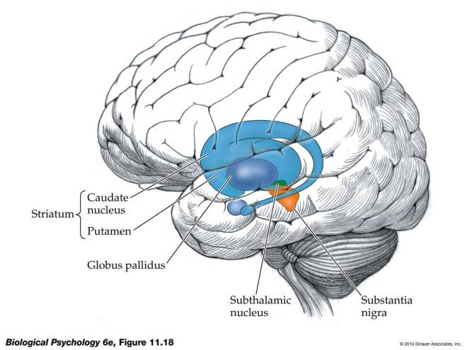
Now the cerebral cortex is not the only game in town.  There are also clusters of neurons that lie beneath the cerebral cortex called “nuclei.”  One of these nuclei beneath the cerebral cortex plays an extremely important role in voluntary motion, and this structure is called the “basal ganglia.”  Here’s a picture to make things a little clearer:

Basal ganglia

As you can see in the figure, the striatum consists of two structures: the putamen and the caudate nucleus.  The striatum or striate nucleus receives neural inputs from the cerebral cortex and inputs this neural information to the basal ganglia.

From a functional perspective, the striatum helps coordinate motivation with body movement.  It facilitates and balances motivation with both higher-level and lower-level functions – for example, inhibiting one’s behavior in a complex social interaction and fine-motor functions involved in inhibiting small voluntary movement.

Some of the neural outputs from the striatum are excitatory – they stimulate other neurons.  Other signals are inhibitory – they prevent the neurons to which they are connected from becoming stimulated.  Inhibitory neurons release a chemical called “GABA.”  These GABA-using neurons are very important for the work of the striatum, and it is exactly these neurons that die off at the greatest rate in patients with HD.  Therefore, treatments for patients with HD have focused on replacing or protecting these GABA-using neurons.

Experimentally, you can induce an HD-like disease in rodents if you inject a chemical into their brains called quinolinic acid.  Quinolinic acid causes many of the GABA-using neurons in the striatum to pack up and die, and for this reason, this chemical is heavily used in the laboratories of scientists who study HD and HD treatments.

In a paper by Marcus McLeod and others who did their work in the laboratory of Ivar Mendez, who was at Dalhousie University in Nova Scotia, Canada, but has since moved to the University of Saskatchewan, GABA-using neurons were made from cultured human neural precursor cells (hNPCs) and then implanted into the brains of rats that had been injected with quinolinic acid.  The results were spectacularly successful.  This work was published in the journal Cell Transplantation.

A definite twist with this particular paper is the way the GABA-using neurons were grown in culture; they were grown in bioreactors.  Bioreactors are devices that support biological cells, processes, or organisms.  They keep the environment of the cells constant, and provide a far superior way to grow cells or tissues in the laboratory.  McLeod and his colleagues used human neural progenitor cells and grew them to large numbers in bioreactors.  These expanded hNPCs were then differentiated them into GABA-using neurons and then injected into the brains of rats who has been treated with quinolinic acid.

The rat model allows the scientist to inject only one side of the brain with quinolinic acid.  This leaves the intact side of the brain as a control tissue that can be compared with the injected one.  The injected rats showed the characteristic death of the GABA-using neurons and the behavioral features that result from the death of these neurons.  Such animals do not walk normally when they are led through a cylinder, and they have trouble finding their way through a maze.  The animals that received the transplantations of the GABA-using neurons, however, performed almost as well in these tests as normal rats; not quite as well, but almost as well.  The rats treated with quinolinic acid did quite poorly, as expected.

Upon post-mortem examination, the rats transplanted with GABA-using neurons shows a host of new GABA-using neurons in their striatums.  These cells also underwent further maturation after transplantation, and they also made connections with other neurons.

Now this paper shows that the injected cells not only survived the transplantations, but they also matured, made connections and promoted recovery of many of the behavioral symptoms of HD.  This procedure certainly has promise.

Having said all that, there are two caveats to these experiments.  The rodent model is a good model as far as it goes, but it seems clear that the actual human disease turns the environment of the brain into a very inhospitable place.  Transplanted cells in the case of human HD patients do not usually survive terribly well.  It seems to me that treatments like this must be coupled with other treatments that seek to improve the actual cerebral environment.  The second caveat to this experiment is that the neural progenitor cells were taken 10-week-old from aborted fetuses.  While these scientists did not perform the abortions that ended the lives of these babies, it is more than little troubling that this research was done using the corpses of those babies whose lives were prematurely ended.

Nevertheless, despite these caveats, this paper represents a definite advance in the regenerative strategies available to treat HD patients.
Researchers Transplant Regenerated Esophagus


Paolo Macchiarini and a research consortium from the Karolinska Institutet in Stockholm, Sweden have made a tissue engineered scaffold for the esophagus from esophagi that were extracted from rats.

After the cells were stripped from the rat esophagi, bone marrow mesenchymal stromal cells were seeded onto the decellularized esophagi and grown in a perfusion bioreactor. A variety of experiments demonstrated that these mesenchymal stromal cells differentiated into esophageal epithelial cells and smooth muscle cells. Macchiarini and his group used several gene expression and functional assays to confirm that these cells had in fact differentiated into these esophageal-specific cell types.

Next, Maccharini and others transplanted these esophagi into rats. The transplanted rats survived 14 days after the transplantations and ate and gain weight. Because the cells used to reconstitute the esophagi came from the rats into which they were transplanted, immunosuppressive drugs were neither used nor needed.

When the recipients of the transplanted esophagi were sacrificed after fourteen days, tissue examinations showed that all the major cell and tissue components of the esophagus including the inside covering of the esophagus (epithelium), muscle fibers, nerves, and blood vessels had nicely regenerated.

This successful bioproduction and transplantation of a tissue-engineered esophagus represents a significant step towards the clinical application of bioengineered esophagi.

Think of it: children and adults with tumors, congenital malformations of the esophagus of traumatic injuries to the esophagus may have new hope and possibilities because of advances in tissue engineering like this.

Making Heart Muscle from Skeletal Muscle Stem Cells


Several experiments in animals and a few clinical trials in human patients have shown that implanting skeletal muscle cells isolated from muscle biopsies into the heart after a heart attack can help the heart to some degree, but the implanted skeletal muscle cells do not integrate into the existing heart muscle mass and the skeletal muscle cells do not differentiate into heart muscle cells.

Experiments like those mentioned above utilized muscle satellite cells. Muscle satellite cells are a resident stem cell population that respond to muscle damage and divide to form skeletal muscle cells form new muscle. Satellite cells are a perfect example of a unipotent stem cell, which is to say a cell that makes one type of terminally differentiated cell type.

Skeletal muscles, however, have another cell population called muscle-derived stem cells or MDSCs. MDSCs express an entirely different set of cell surface proteins than satellite cells, and have the capacity to differentiate into skeletal muscle, smooth muscle, bone, tendon, nerve, endothelial and hematopoietic cells. MDSCs grow well in culture, tolerate low oxygen conditions quite well, and show excellent regenerative potential.

Other laboratories have managed to culture MDSCs in collagen and produce beating heart muscle cells. Others have observed MDSCs forming a proper myocardium under certain conditions. Several studies have established the ability to MDSCs to treat laboratory animals that have suffered a heart attack. The most recent work from Sekiya and others has established that cell sheets made from MDSCs can reduce dilation of the left ventricle, increased capillary density, and promoted recovery without causing erratic heat beat patterns.

Despite their obvious efficacy. MDSCs remain difficult to isolate in high enough numbers to therapeutic purposes. None of the cell surface molecules sported by MDSCs are unique to those cells. Therefore, getting clean cultures of MDSCs remains a challenge. Still, these cells represent some of the best hopes for regenerative medicine in the heart. These cells do form heart muscle cells and heal ailing hearts. They can be grown in bioreactors to high numbers and can also be combined with engineered materials to shore up a damaged heart and mediate its regeneration. While the use of MDSCs is still in its infancy, the promise certainly is there.

Scientists Grow Small Chunks of Brain Tissue From Induced Pluripotent Stem Cells


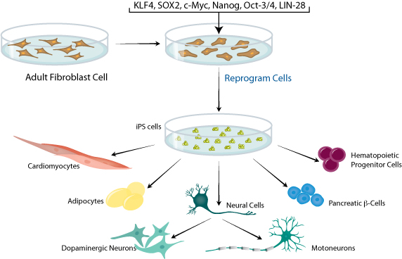
Induced pluripotent stem cells are made from adult cells by means of genetic engineering techniques that introduce into the cells a combination for four different genes that drive the cells to de-differentiate into a cell that has many of the characteristics of embryonic stem cells without the destruction of embryos.

A new study from the laboratory of Juergen Knoblich at the Institute of Molecular Biotechnology in Vienna has mixed induced pluripotent stem cells (iPSCs) to form structures of the human brain. He largely left the cells alone to allow them to form the brain tissue, but he also placed them in a spinning bioreactor that constantly circulates the culture medium and provides nutrients and oxygen to the cells. One other growth factor he supplied to the cells was retinoic acid, which is made by the meninges that surround our brains. All of this and the cells not only divided, differentiated and assembled, but they formed brain structures that had all the connections of a normal brain. These brain-like chunks of tissue are called “mini-brains” and the recent edition of the journal Nature reports their creation.

“It’s a seminal study to making a brain in a dish,” says Clive Svendsen, a neurobiologist at the University of California, Los Angeles. Svendsen was not involved in this study, but wishes he was. Of this study, Svendsen exclaimed, “That’s phenomenal” A fully formed artificial brain is still years and years away, but the pea-sized neural clumps developed in Knoblich’s laboratory could prove useful for researching human neurological diseases.

Researchers have previously used pluripotent human stem cells to grow structures that resemble the developing eye (Eiraku, M. et al. Nature 472, 51–56 (2011), and even tissue layers similar to the cerebral cortex of the brain (Eiraku, M. et al. Cell Stem Cell 3, 519–532 (2008). However, this latest advance has seen bigger and more complex neural-tissue clumps by first growing the stem cells on a synthetic gel that resembled natural connective tissues found in the brain and elsewhere in the body. After growing them on the synthetic gel, Knoblich and his colleagues transferred the cells to a spinning bioreactor that infuses the cells with nutrients and oxygen.

“The big surprise was that it worked,” said Knoblich. The clump formed structures that resembled the brains of fetuses in the ninth week of development.

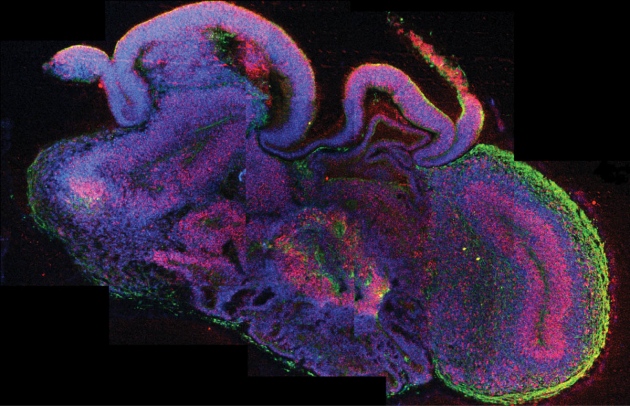
Under a microscope, the blobs contained discrete brain regions that seemed to interact with one another. However, the overall arrangement of the different proto-brain areas varied randomly across tissue samples. These structures were not recognizable physiological structures.

A cross-section of a brain-like clump of neural cells derived from human stem cells.

“The entire structure is not like one brain,” says Knoblich, who added that normal brain maturation in an intact embryo is probably guided by growth signals from other parts of the body. The tissue balls also lacked blood vessels, which could be one reason that their size was limited to 3–4 millimeters in diameter, even after growing for 10 months or more.

Despite these limitations, Knoblich and his collaborators used this system to model key aspects of microcephaly, which is a condition that causes extremely stunted brain growth and cognitive impairment. Microcephaly and other neurodevelopmental disorders are difficult to replicate in rodents because the brains of rodents develop differently than those of humans.

Knoblich and others found that tissue chunks cultured from stem cells derived from the skin of a single human with microcephaly did not grow as large as clumps grown from stem cells derived from a healthy person. When they traced this effect, they discovered that it was due to the premature differentiation of neural stem cells inside the microcephalic tissue chunks, which depleted the population of progenitor cells that fuels normal brain growth.

The findings largely confirm prevailing theories about microcephaly, says Arnold Kriegstein, a developmental neurobiologist at the University of California, San Francisco. But, he adds, the study also demonstrates the potential for using human-stem-cell-derived tissues to model other disorders, if cell growth can be controlled more reliably.

“This whole approach is really in its early stages,” says Kriegstein. “The jury may still be out in terms of how robust this is.”

Skin Cells Used to Make Personalized Bone Substitutes


Patient-specific bone substitutes have been produced by a team of scientists from the New York Stem Cell Foundation. Darja Marolt and Giuseppe Maria de Peppo from the New York Stem Cell Foundation (NYSCF) led the study that demonstrated that customizable, three-dimensional bone grafts that can be produced on-demand for patients from their own cells.

Marolt and de Peppo and their co-workers used skin grafts from their patients to isolate skin fibroblasts that were reprogrammed to induced pluripotent stem cells (iPSCs). Because iPSCs are made from the patient’s own cells, they have the same profile of cell surface proteins as the patient’s own tissues. Therefore, they are very unlikely to be rejected by the patient’s immune system. Also, iPSCs have the ability to differentiate into any cell type found in the adult body, and therefore, can be used to form bone cells.

iPS cells

After deriving iPSCs from patient skin cells, de Peppo, Marolt, and colleagues coaxed the cells to form osteoblasts (the cells that form bone), and seeded them onto a scaffold that mimicked three-dimensional bone structure. These structures were grown in a bioreactor that fed the cells oxygen and nutrients.

According to Marolt, “Bone is more than a hard mineral composite, it is an active organ that constantly remodels. Blood vessels shuttle important nutrients to healthy cells and remove waste; nerves provide connection to the brain; and, bone marrow cells form new blood and immune cells.”

Previous studies have demonstrated that cells from other sources also possess bone-forming potential. However, these same studies have revealed serious shortcomings of the clinical potential of such cells. A patient’s own bone marrow stem cells can form bone and cartilaginous tissue, but not the accompanying underlying vasculature and nerve compartments. Also, embryonic stem cell derived bone may prompt an immune rejection. Therefore, the use of iPSCs can overcome many of these limitations.

As de Peppo noted: “No other research group has published work on creating fully viable functional three-dimensional bone substitutes from humans iPS cells. These results bring us closer to achieving our ultimate goal, to develop the most promising treatments.”

Since bone injuries and defects are often treated with bone grafts that are taken from other parts of the body or a tissue bank. Alternatively, synthetic alternatives can also be used, but none of these possibilities provide the means for complex reconstruction and they may also be rejected by the immune system, or fail to integrate with surrounding connective tissue. n the case of trauma patients who suffer from shrapnel wounds or vehicular injuries , the traditional treatments provide only limited functional and cosmetic improvements.

To access the integrity of the bioreactor-made bone, the NYSCF team implanted them into animals. Implantation of undifferentiated iPSCs formed tumors, but transplantation of the iPSC-derived bone produced no tumors, but also produced grafts that effectively integrated into the bones, connective tissues and blood vessels of the animals.

Susan Solomon, CEO of NYSCF, said of this work, “Following from these findings, we will be able to create tailored bone grafts, on demand, for patients without any immune rejection issues. She continued: “it is the best approach to repair devastating damage or defects.”

The therapeutic relevance of this work aside, these adaptive bone substitutes can also serve as models for bone development and various bone pathologies. Such bone exemplars could serve as models for drug testing and drug development.