Positive Results from Mesoblast’s Phase 2 Trial of Cell Therapy in Diabetic Kidney Disease


Mesoblast Limited has announced results from its Phase 2 clinical Trial that evaluated their Mesenchymal Precursor Cell (MPC) product, known as MPC-300-IV, in patients who suffer from diabetic kidney disease. In short, their cell product was shown to be both safe and effective. The results of their trial were published in the peer-reviewed journal EBioMedicine.  Researchers from the University of Melbourne, Epworth Medical Centre and Monash Medical Centre in Australia participated in this study.

The paper describes a randomized, placebo-controlled, and dose-escalation study that administered to patients with type 2 diabetic nephropathy either a single intravenous infusion of MPC-300-IV or a placebo.

All patients suffered from moderate to severe renal impairment (stage 3b-4 chronic kidney disease for those who are interested).  All patients were taking standard pharmacological agents that are typically prescribed to patients with diabetic nephropathy.  Such drugs include angiotensin-converting enzyme inhibitors (e.g., lisinopril, captopril, ramipril, enalapril, fosinopril, ect.) or angiotensin II receptor blockers (e.g., irbesartan, telmisartan, losartan, valsartan, candesartan, etc.).  A total of 30 patients were randomized to receive either a single infusion of 150 million MPCs, or 300 million MPCs, or saline control in addition to maximal therapy.

Since this was a phase 2 clinical trial, the objectives of the study were to evaluate the safety of this treatment and to examine the efficacy of MPC-300-IV treatment on renal function.  For kidney function, a physiological parameter called the “glomerular filtration rate” or GFR is a crucial indicator of kidney health.  The GFR essentially indicates how well the individual functional units within the kidney, known as “nephrons,” are working.  The GFR indicates how well the blood is filtered by the kidneys, which is one way to measure remaining kidney function.  The decline or change in glomerular filtration rate (GFR) is thought to be an adequate indicator of kidney function, according to the 2012 joint workshop held by the United States Food and Drug Administration and the National Kidney Foundation.

nephronanatomy

Diabetic nephropathy is an important disease for global health, since it is the single leading cause of end-stage kidney disease.  Diabetic nephropathy accounts for almost half of all end-stage kidney disease cases in the United States and over 40% of new patients entering dialysis treatment.  For example, there are almost 2 million cases of moderate to severe diabetic nephropathy in 2013.

Diabetic nephropathy can even occur in patients whose diabetes is well controlled – those patients who manage to keep their blood glucose levels at a reasonable level.  In the case of diabetic nephropathy, chronic infiltration of the kidneys by inflammatory monocytes that secrete pro-inflammatory cytokines causes endothelial dysfunction and fibrosis in the kidney.

Staging of chronic kidney disease (CKD) is based on GFR levels.  GFR decline typically defines the progression to kidney failure (for example, stage 5, GFR<15ml/min/1.73m2).  The current standard of care (renin-angiotensin system inhibition with angiotensin converting enzyme inhibitors or angiotensin II receptor blockers) only delays the progression to kidney failure by 16-25%, which leaves a large residual risk for end-stage kidney disease.  For patients with end-stage kidney disease, the only treatment option is renal replacement (dialysis or kidney transplantation), which incurs high medical costs and substantial disruptions to a normal lifestyle.  Due to a severe shortage of kidneys, in 2012 approximately 92,000 persons in the United States died while on the transplant list.  For those on dialysis, the mortality rate is high with an approximately 40% fatality rate within two years.

The main results of this clinical trial were that the safety profile for MPC-300-IV treatment was similar to placebo.  There were no treatment-related adverse events.  Secondly, patients who received a single MPC infusion at either dose had improved renal function compared to placebo, as defined by preservation or improvement in GFR 12 weeks after treatment.  Third, the rate of decline in estimated GFR at 12 weeks was significantly reduced in those patients who received a single dose of 150 million MPCs relative to the placebo group (p=0.05).  Finally, there was a trend toward more pronounced treatment effects relative to placebo in a pre-specified subgroup of patients whose GFRs were lower than 30 ml/min/1.73m2 at baseline (p=0.07).  In other words, the worse the patients were at the start of the trial, the better they responded to the treatment.

The lead author of this publication, Dr David Packham, Associate Professor in the Department of Medicine at the University of Melbourne and Director of the Melbourne Renal Research Group, said: “The efficacy signal observed with respect to preservation or improvement in GFR is exciting, especially given that this trial was not powered to show statistical significance. Patients receiving a single infusion of MPC-300-IV showed no evidence of developing an immune response to the administered cells, suggesting that repeat administration is feasible and may in the longer term be able to halt or even reverse progressive chronic kidney disease. I hope that this very promising investigational therapy will be advanced to rigorous Phase 3 clinical trials to test this hypothesis as soon as possible.”

Patients who received s single IV infusion of MPC-300-IV cells showed no evidence of developing an immune response to the administered cells.  This suggests that repeated administration of MPCs is feasible and might even have the ability to halt, or even reverse progressive chronic kidney disease.

Packham and his colleagues hope that this cell-based therapy can be advanced to a rigorous Phase 3 clinical trial to further test this treatment.

Stem Cell-Derived Smooth Muscle Cells Help Restructure Urethral Sphincter Muscles in Rats


Stress urinary incontinence affects 25%-50% of the female population and is defined as the leakage of the bladder upon exertion. The exertions that can cause the bladder to leak can be as simple as laughing, coughing, sneezing, hiccups, yelling, or even jumping up and down. Stress urinary incontinence costs Americans some $12 billion a year and also causes a good deal of embarrassment and compromises quality of life. Unsurprisingly, stress urinary incontinence also is associated with an increased incidence of anxiety, stress, and depression.

In most cases of stress urinary incontinence, injury to the internal sphincter muscles of the urethra or to the nerves that innervate these muscles (both smooth and voluntary muscles) significantly contribute to the condition. Conservative management of stress urinary incontinence can work at first, but can fail later on. The other option is corrective surgery that reconstructs the urethral sphincter and increases urethral support. However, even though such surgeries can and often do work, recurrence of the incontinence is rather common. Is there a better way?

Yan Wen from Stanford University School of Medicine and colleagues and collaborators from College of Medicine of Case Western Reserve in Cleveland, Ohio, Southern Medical University in Guangzhou, China, and Montana State University have used a novel stem cell-based technique to treat laboratory Rowett nude rats that had a surgically-induced form of stress urinary incontinence. While the results are not overwhelming, they suggest that a stem cell-based approach might be a step in the right direction.

Wen and others used a human embryonic stem cell line called H9 and two different types of induced pluripotent stem cell lines to make, in culture, human smooth muscle progenitor cells (pSMCs). Fortunately, protocols for differentiating pluripotent stem cells into smooth muscle cells is well worked out and rather well understood. These pSMCs were also tagged with a firefly luciferase gene that allowed visualization of the cells after implantation.

Six groups of rats were treated in various ways. The first group had stress urinary incontinence and were only treated with saline solutions. The second group of animals also had stress urinary incontinence and were treated with cultured human pSMCs that were derived from human bladders. The third group of animals also had stress urinary incontinence and were treated with pSMCs made from H9 human embryonic stem cells. The next two groups also had stress urinary incontinence and were treated with two different induced pluripotent stem cell lines; one of which was induced with a retroviral vector and the second of which was made with episomal DNA. Both lines were originally derived from dermal fibroblasts. The final group of rats did not have stress urinary incontinence and were used as a control group.

The cells were introduced into the mice by means of injections into the urethra under anesthesia. Two million cells were introduced in each case, three weeks after the induction of stress urinary incontinence. All animals were examined five weeks after the cells were injected into the animals.

Because the cells were tagged with firefly luciferase, the animals could be given an injection of luciferin, which is the substrate for luciferase. Luciferase catalyzes a reaction with luciferin, and the cells glow. This glow is easily detected by means of a machine called the Xenogen Imaging System. Such experiments showed that the injected cells did not survive terribly well, and by 9 days after the injections, they were usually not detectable. Two rats that had been injected with retrovirally-induced induced pluripotent stem cell-derived pSMCs lasted until 35 days after injection, but these rats were the exception and not the rule.

Did the cells integrate into the urethral sphincter by the signal is too low to be detected using luciferase? The answer to this question was certainly yes, but the amount of integration was nothing to write home about. Small patches of cells showed up in the urethra sphincters that expressed human gene products, and therefore, had to be derived from the injected cells.

In vivo survival of transplanted pSMCs in RNU rats. (A): The RV-iPSC pSMCs were periurethrally injected into the rats and monitored with BLI. (B): At day 12, a small number of the transplanted cells were detected in the proximal rat urethra. The transplanted human cells were determined by positive staining of HuNuclei and smoothelin. (C): Gene expression of human ERV-3 in rat urethras 5 weeks after cell transplantation. Y-axis on left shows the scale for ERV-3 copy numbers from tissue samples. Each human cell contains one copy of ERV-3 transcript; hence, the number of copies is equal to the number of cells. Y-axis on right shows the ERV-3 copy numbers of the standard cell samples. The cell numbers in the standard graph are 0.5 × 10³, 1 × 10³, 2 × 10³, 4 × 10³, 8 × 10³, and 10 × 103 (red dots). ERV-3 amplifications in all pSMC-treated groups were very low. Abbreviations: BLI, bioluminescent imaging; bSMC, bladder smooth muscle cell; DAPI, 4′,6-diamidino-2-phenylindole; Epi, episomal plasmid; ERV-3, endogenous retrovirus group 3; H&E, hematoxylin and eosin; Hu, human; HuNuclei, human nuclei; iPSC, induced pluripotent stem cell; pSMCs, smooth muscle progenitor cells; RNU, Rowett Nude; RV, retrovirus vector.

The exciting part about these results, however, was that when Wen and others examined the rat urethral sphincters for the presence of things like elastin and other proteins that make for a healthy urethral sphincter, there was a good deal of elastin, but it was not human elastin but rat elastin. Therefore, this elastin synthesis was INDUCED by the implanted cells even though it was not made by the implanted cells. Instead, the implanted cells seemed to signal to the native cells to beef up their own production of sphincter-specific gene products, which made from a better sphincter. This was not the case in animals that received injections of human pSMCs derived from human bladders.

Assessment of elastin fibers in the proximal urethra of the rat. (A): Representative images of cross-section of proximal urethra with Weigert’s Resorcin-Fuchsin’s elastin and van Gieson’s collagen staining. Elastic fiber shown as dark blue, collagen as red pink, and other tissue elements as yellow. Scale bars = 100 µm. (B): Quantification of elastin fibers was assessed using Image-Pro Plus software and expressed as a percentage of the arbitrary ROIs. Each bar represents the mean value ± SEM. Abbreviations: bSMC, human bladder smooth muscle cells; H9-pSMC, surgery plus H9-pSMC injection; Hu-bSMC, surgery plus injection of human bladder smooth muscle cells; pSMCs, smooth muscle progenitor cells; Pure control, no surgery and no treatment; ROIs, regions of interest; Sham saline, surgery plus saline injection.

Because these mice were sacrificed five weeks after the injections, Wen and others could not assess the urethral function of these animals. Therefore, it is uncertain if the improved tissue architecture of the urethral sphincter properly translated into improved function even though it is reasonable to assume that it would. Having said that, it is possible that the experiments that detected the presence of increased amounts of elastin and collagen in the sphincters of these rats was complicated by the presence of bladder tissue in the preparations. Since bladder tissue was included in all trials of this experiment, it is unlikely that bladder tissue is the sole cause of increase elastin and collagen in the stem cell-treated rats. Secondly, rat regenerative properties may not properly match the regenerative properties in older human patients. Here again, unless such an experiment is attempted in larger animal models and then in human patients, we will never know if this procedure is viable for regenerative treatments in the future.

For now, it is an interesting observation, and perhaps a promising start to might someday become a viable regenerative treatment for human patients.

This paper appeared in Stem Cells Translational Medicine, vol 5, number 12, December 2016, pp. 1719-1729.

USC Researchers Isolate Human Nephon Progenitor Cells – Future Possibilities for Kidney Regeneration


Researchers at the Saban Research Institute of Children’s Hospital of Los Angeles and the University of Southern California (USC) have reported the isolation of human nephron progenitor (NP) cells. These results, which were published in the journal Stem Cell Translational Medicine, might very well elucidate how progenitor cells differentiate into become renal cells and then develop into kidneys. Such insights could, possibly provide new strategies to promote renal regeneration after chronic kidney failure or acute kidney injury.

Kidneys are composed of about a million tiny filtration units known as “nephrons.” These diminutive structures filter waste and concentrate those wastes into urine, which is leaked into the bladder. In humans, approximately 500,000 to 1,000,000 nephrons are generated before week 34 – 36 of fetal gestation. However, at this point in development, the NP cells are exhausted and kidney development (known as “nephrogenesis”) effectively ceases. If the kidney loses a large enough quantity of nephrons after this time period, such losses may lead to irreversible kidney failure, since no further cell repair or regeneration is possible.

kidney_nephron_civyrose

In past studies, NPs were made from induced pluripotent stem cells, or by utilizing animal models. Scientists at USC and Children’s Hospital of Los Angeles (CHLA), chose a different tactic; they designed an efficient protocol by which they could directly isolate human NPs. To accomplish this, Dr. Laura Perin and her colleagues used RNA-labeling probes to obtain cells that expressed the SIX2 and CITED1 genes. Cells expressing both of these genes are almost certainly NPs, since SIX2 and CITED1 are master regulatory genes that promote renal development.

Dr. Perin, co-director of CHLA’s GOFARR Laboratory for Organ Regenerative Research and Cell Therapeutics in Urology, added, “In addition to defining the genetic profile of human NP, this system will facilitate studies of human kidney development, providing a novel tool for renal regeneration and bioengineering purposes.”

On a rather sanguine note, Perin noted that these experiments, which constitute proof-of-concept work, may create new applications to researchers who might be able to use her laboratory’s techniques to isolated progenitor cells for other organs, the pancreas, heart, or lung. “This technique provides a ‘how to’ of human tissue during development,” said Perin.

“It is an important tool that will allow scientists to study cell renewal and differentiation in human cells, perhaps offering clues to how to regulate such development,” added first author of this paper, Stefano Da Sacco.

Scientists Reprogram Adult Skin Cells to Make Mini Kidneys


Japanese and Australian researchers have used induced pluripotent stem cell (iPSC) technology to reprogram human skin cells to make the most mature human kidneys yet to be grown in a culture. These mini kidneys have hundreds of filtering units (nephrons) and blood vessels and appear to be developing just as kidneys would in an embryo.

“The short-term goal is to actually use this method to make little replicas of the developing kidney and use that to test whether drugs are toxic to the kidney,” said lead researcher Professor Melissa Little, of the Murdoch Children’s Research Institute. “Ultimately we hope we might be able to scale this up so we can … maybe bioengineer an entire organ.”

In other previous research, Professor Little and her co-workers generated cells that self-organized into the nephrons and collecting ducts needed for the kidney to filter blood and produce urine. They used a precise combination of called growth factors to direct embryonic stem cells to develop into the different cell types.

In the journal Nature, Professor Little and her collaborators report they have made a developing kidney from a type of skin cell called a fibroblast. Little and her team reprogrammed adult fibroblasts to become “induced pluripotent stem cells,” which act like embryonic stem cells, and can become any cell in the body. By adopting their growth factor recipe, Little and others were able to grow these cells into larger and more complex, three-dimensional kidneys than previously made.

“These kidneys have something like 10 or 12 different cell types in them … all from the one starting stem cell,” said Professor Little. “What we had previously were little flat structures over the surface of a dish … Now we have an organoid that is about 5-6 millimetres across, has about 100 filtering units in it, and is starting to form blood vessels. It’s starting to mature and the cell types are starting to do more of the functions of the final kidney.”

Scientists in Little’s laboratory demonstrated that the genes expressed in the mini kidneys as they formed faithfully recapitulated the expression of those same genes in a developing kidney in a first trimester embryo.

“It is actually mirroring what is happening in human development,” said Professor Little.

Little and her group also found that the laboratory-grown kidney was damaged when it was treated with known renal toxins. Little suggested that the iPSCs cells they had created were functioning as a kidney, but further tests would be required to demonstrate that.

It might be possible to use these bioengineered kidneys to test the renal toxicity of drugs. Likewise, the production of mini kidneys using cells from kidney patients might provide a way to study inherited forms of kidney disease.

“You can take a fibroblast [from someone with inherited kidney disease], make a stem cell out of it, generate a little kidney and use that as our model for their disease,” said Professor Little.

Perhaps most exciting, laboratory-generated kidneys might one day provide rejection-free transplants for patients, and gene editing could be used to fix the genetic defect that caused an inherited kidney disease.

Professor Jamie Davies of the University of Edinburgh, who was not involved with this work, but commented on it for Nature, emphasized this was not a full-fledged, functional kidney. “The structure’s fine-scale tissue organization is realistic, but it does not adopt the macro-scale organization of a whole kidney. For example, it is not ‘plumbed’ into a waste drain, and it lacks large-scale features that are crucial for kidney function, such as a urine-concentrating medulla region. There is a long way to go until clinically useful transplantable kidneys can be engineered, but [this] protocol is a valuable step in the right direction.”

Davies also mentioned that these mini kidneys had the potential to replace “poorly predictive” animal drug safety tests, and called on researchers to team up with toxicologists to test the potential of their system.

Laboratory-Grown Kidneys Work in Laboratory Animals


A Jikei University School of Medicine research team based in Tokyo, Japan, led by Takashi Yokoo, in collaboration with scientists from Meiji University and St. Marianna University School of Medicine in Kawasaki, Keio University School of Medicine in Tokyo, and the School of Veterinary Medicine at Kitasato University in Towada, has shown that mini kidneys grown in vitro from human stem cells can be effectively connected to the excretory systems of rats and pigs.

This is not the first time that research groups have successfully grown mini kidneys in the laboratory. However, connecting these laboratory-grown organs to a laboratory animal’s excretory system constitutes a major technical challenge. The Jikei University team used an approach that employs a step-wise peristaltic ureter or SWPU to connect its lab-grown mini kidneys to the ureter of the transplant animal.

Previous attempts to use laboratory-grown kidneys in laboratory animals have failed because while the transplanted kidneys made urine, they were unable to pass that urine to the animal’s bladder and the kidneys swelled up and failed. Yokoo and his collaborators and colleagues used a stem cell method to make their mini kidneys, as others have in the past.  However, he and his team grew more than just the kidney for the host animal; that also grew a drainage tube, known as a ureter, as well, in addition to a  bladder to collect and store the urine.

Yokoo and others used laboratory rats as the incubators for their growing tissue.  When they connected the new kidney and its tubular systems to the animal’s existing bladder, the system worked.  Urine passed from the transplanted kidney into the transplanted bladder and then into the rat bladder.  The transplant was still working well when they checked eight weeks later.  Then Yokoo and others repeated their procedure in pigs, which are larger mammals than rats and better model systems for human beings.  Fortunately, they achieved the same results.

Although this technology is still years away from clinical trials with human patients, this work provides a paradigm for making organs in the laboratory that will work in sick people.  In the United Kingdom alone, more than 6,000 people are waiting for a kidney.  Because of a shortage of kidney donors, fewer than 3,000 transplants are carried out each year, and more than 350 people die each year waiting for a transplant.  Growing new kidneys using human stem cells could solve this problem.

“To our knowledge, this is the first report showing that the SWPU system may resolve two important problems in the generation of kidneys from stem cells: construction of a urine excretion pathway and continued growth of the newly generated kidney,” Yokoo and others wrote in their paper, which was published in the Proceedings of the National Academy of Sciences, USA, which was communicated to the journal by National Academy of Science member R. Michael Roberts from the University of Missouri-Columbia.

Stem cell expert Prof Chris Mason from University College London, said: “This is an interesting step forward. The science looks strong and they have good data in animals.  But that’s not to say this will work in humans. We are still years off that. It’s very much mechanistic. It moves us closer to understanding how the plumbing might work.  At least with kidneys, we can dialyse patients for a while so there would be time to grow kidneys if that becomes possible.”

Rebuilding Bladders with Cell-Free Materials


Ying-jian Zhud and Mu-jun Luan from the Shanghai Jiao Tong University in Shanghai, China teamed up to examine a new way to regenerate the bladder.

Several different synthetic and natural biomaterials have been pretty widely used in tissue regeneration experiments, particularly in the regeneration of the urinary bladder. The vast majority of this work has been done in rat model systems, which are fairly good animals to model bladder pathology and regeneration.

To date, the attempted reconstructive procedures don’t seem to work all that well, and this is due to the lack of appropriate scaffolding upon which cells can attach, grow and spread to form the new bladder tissue. Any scaffolding material for the bladder has to provide a waterproof barrier and it has to be able to support several different cell types. While this might not sound difficult on paper, it is in fact rather difficult. Some biomaterials might be well tolerated by the body, but cannot be fashioned into the shape of the organ. Others might support the growth of cells quite well, but are not tolerated by the body.

Zhud and Luan addressed these issues by turning to two different compounds that would compose a two-layered structure. Such a two-layered structure would support the cell types of the bladder. The outside layer was composed of silk fibroin, which is very moldable and usually well tolerated by cells. The inner layer consisted of a natural, acellular matrix (or BAMG for bladder acellular matrix graft). They used this two-layered structure to regenerate an injured bladder in rats.

First of all, it was clear that this material was relatively easy to make and it also could be nicely molded and sewn into the existing bladder. Tissue stains showed something even more interesting: the bilayer scaffold promoted the growth and recruitment of smooth muscles, blood vessels, and even nerves in a time-dependent manner. So by 12 weeks after implantation, bladders reconstructed with the bilayered matrix displayed superior structural and functional properties without significant local tissue responses or systemic toxicity.

Thus, the silk/BAMG scaffold could potentially be a promising scaffold for bladder regeneration. It shows good tissue compatibility, and allows the growth of cells on it. More work is required to take this to the next step, and the scaffold will undoubtedly undergo some changes. But this work represents a terrific start to what might be a superior scaffold for bladder regeneration.

Umbilical Cord Blood Mesenchymal Stem Cells Relieve the Symptoms of Interstitial Cystitis by Activating the Wnt Pathway and EGF Receptor


Interstitial tissue refers to the tissue that lies between major structures in an organ. For example, the tissue between muscles is an example of interstitial tissue.

Interstitial cystitis, otherwise known as painful bladder syndrome is a chronic condition that causes bladder pressure, bladder pain and sometimes pelvic pain, ranging from mild discomfort to severe pain.

The bladder is a hollow, muscular organ that stores urine and expands until it is full, at which time it signals the brain that it is time to urinate, communicating through the pelvic nerves. This creates the urge to urinate for most people. In the case of interstitial cystitis, these signals get mixed up and you feel the need to urinate more often and with smaller volumes of urine than most people. Interstitial cystitis most often affects women and can have a long-lasting impact on quality of life. Unfortunately no treatment reliably eliminates interstitial cystitis, but medications and other therapies may offer relief. There is no sign of bacterial infection in the case of bacterial cystitis.

A new study evaluated the potential of umbilical cord blood-derived mesenchymal stem cells or (UCB-MSCs) to treat interstitial cystitis (IC). In this study, Dr. Miho Song and colleagues from the Asan Medical Center, Seoul, South Korea, established a rat model of IC in 10-weeks-old female Sprague-Dawley rats by instilling 0.1M HCl or PBS (sham). After 1-week, human UCB-MSCs (IC+MSCs) or PBS (IC) were directly injected into the submucosal layer of the bladder.

To clarify this part of the experiment, the urinary bladder is made of several distinct tissue layers: a) The innermost layer of the bladder is the mucosa layer that lines the hollow lumen. Unlike the mucosa of other hollow organs, the urinary bladder is lined with transitional epithelial tissue that is able to stretch significantly to accommodate large volumes of urine. The transitional epithelium also provides protection to the underlying tissues from acidic or alkaline urine; b) Surrounding the mucosal layer is the submucosa, a layer of connective tissue with blood vessels and nervous tissue that supports and controls the surrounding tissue layers; c) The visceral muscles of the muscularis layer surround the submucosa and provide the urinary bladder with its ability to expand and contract. The muscularis is commonly referred to as the detrusor muscle and contracts during urination to expel urine from the body. The muscularis also forms the internal urethral sphincter, a ring of muscle that surrounds the urethral opening and holds urine in the urinary bladder. During urination, the sphincter relaxes to allow urine to flow into the urethra.

Bladder histology

Now a single subcutaneous injection of human UCB-MSCs significantly attenuated the irregular and decreased voiding interval in the IC group. In addition, the denudation of the epithelium that is characteristic of IC and increased inflammatory responses, mast cell infiltration, neurofilament production, and angiogenesis observed in the IC bladders were prevented in the IC+MSC group. Therefore, the injected UBC-MSCs prevented the structural changes in the bladder associated with the pathology of IC.

How did these cells do this? Further examination showed that the injected UCB-MSCs successfully engrafted to the stromal and epithelial tissues of the bladder and activated the Wnt signaling cascade. In fact, if the Wnt activity of these infused cells was blocked, the positive effects of the UCB-MSCs were also partially blocked. Additionally, activation of the epidermal growth factor receptor (EGFR) also helped UCB-MSCs heal the bladder. If the activity of the EGF receptor was inhibited by small molecules, then the benefits of MSC therapy were also abrogated. Also if both the Wnt pathway and EGFR were inhibited, the therapeutic capacities of UCB-MSCs were completely wiped out.

These data show the therapeutic effects of MSC therapy against IC in an orthodox rat animal model. However, this study also elucidates the molecular mechanisms responsible for these therapeutic effects. Our findings not only provide the basis for clinical trials of MSC therapy to IC, but also advance our understanding of IC pathophysiology.

Repairing Bladders with Bone Marrow Stem Cells and Bladder Acellular Matrix


The bladder is subject to several different types of conditions that can compromise its function. Cancer of the bladder can necessitate its removal. A congenital condition called exstrophy causes the bladder to protrude through a hole in the abdominal wall also requires surgical repair of the bladder. Finally, trauma to the bladder as in the case of trampoline athletes or people who have had surgical damage to the bladder, may also repair bladder repair.

In order to repair the bladder, extra tissue must be added to it. Finding tissue to act as bladder has not been easy. In the past, surgeons have used grafts from skin, bladder submucosa, omentum, dura, peritoneum, colon, small intestine, and synthetic polymers have been used to augment the bladder. All of these compounds have their pluses and minuses for reconstructing a bladder, but all of them do not appropriately recapitulate the mechanical, structural, and functional properties of the bladder.

Currently a surgical procedure called enterocystoplasty is the most effective surgical solution for augmentation of the bladder. This procedure uses a small piece of the large intestine to increase the size of the bladder, and while it certainly improves continence, it has several complications associated, which include, metabolic disturbances, urinary stones (urolithiasis), increased mucus production, infections and increased risk of cancer of the bladder. Is there a better way?

A collaborative research project between Daniel L. Coutu from ETH Zürich, in Basel, Switzerland, Wally Mahfouz, Oleg Loutochin, and Jacques Corcos from McGill University, in Montreal, Canada, Wally Mahfouz from Alexandria University in Alexandria, Egypt, and Jacques Galipeau from Emory University in Atlanta, Georgia examined the use of bladder a cellular matrices or BAMs in combination with bone marrow-derived mesenchymal stromal cells to repair bladders in laboratory animals.

BAMs consist of bladders from animals that have been completely stripped of their cells with detergents and enzymes. Once all the cells and cell remnants have been removed, these BAMs can be molded into the form of a bladder, after which cells are reapplied. BAMs contain all the chemical nooks and crannies for cells to find, attach to, then and differentiate. Also, the bladder is a relatively simple tissue in that it has an inner epithelium (urothelium) that sits on a basement membrane, a smooth muscle layer surrounding the urothelium, and an outer serosa layer that is an extension of the peritoneum that it covered by an adventitia of connective tissue. A bladder matrix devoid of cells has all the right structures for cells to occupy, bit it needs to be repopulated with cells.

Corcos and his collaborators purchased pig bladders from slaughter houses and subjected them to detergents and enzymes until no cells were left on them. Then they used mesenchymal stromal cells from the bone marrow of rats to reseed the bladders with new cells. These structures were then used to repair the bladders to laboratory rats whose bladders had been partially removed. All the animals were then tested for retention of urine, muscle tone, pressure tolerance, and other indicators of bladder function.

The results of these tests demonstrated that the engineered bladders not only worked, but worked quite well. Some animals only received the BAM without cells, and these animals had bladders that worked better than nothing, but not terribly well. However, the animals that received BAM + mesenchymal stem cells had bladders that, for all intents and purposes, showed normal function by 6 months after the procedure.

Another significant finding of this study is that none of the animals that received BAMs had to be given any anti-rejection drugs. The immune systems of these animals did not reject the animal-based matrices.

Finally, post-mortem examinations of these animal bladders established that smooth muscle regeneration and nerve and blood vessels regeneration was also robust in these animals.

Before this procedure can work in humans, it will need to work in larger animal systems. Rat urinary systems are similar to humans, but not the same. The large advance in this study is the observation that internal bladder tissues such as smooth muscle, nerves and blood vessels can be regenerated with mesenchymal stem cells. These stem cells probably secrete a variety of molecules that promote the growth of blood vessels and nerves into the bladder.

In the words of these authors: “we demonstrated the in vivo superiority of MSCs-seeded BAMs compared with unseeded BAMs in bladder tissue engineering. Our approach is fully translatable to large animals and humans, where autologous MSCs could be seeded on allogeneic, cadaveric or xenogenic BAMs. The method presented here is a viable alternative to current treatment modalities and should prevent most complications associated with them. This study demonstrates the superiority of MSCs-seeded BAM compared to BAM alone in bladder augmentation and provides a strong basis to test our novel approach in large animal models and eventually in humans.”

Amen.

Transplanted Mesenchymal Stem Cells Prevent Bladder Scarring After Spinal Cord Injury


A collaborative research effort between laboratories from Canada and South Korea have shown that a cultured mesenchymal stem cell line called B10 can differentiate into smooth muscle cells and improve bladder function after a spinal cord injury.

Spinal cord injury can affect the lower portion of the urinary tract. Overactive bladder, urinary retention, and increased bladder thickness and fibrosis (bladder scarring) can result from spinal cord injuries. Human mesenchymal stem cells (MSCs) can differentiate under certain conditions into smooth muscle. For this reason, MSCs have therapeutic potential for patients who have suffered from spinal cord injuries.

Seung U. Kim and his colleagues from Gachon University Gil Hospital in Inchon, South Korea have made an immortalized human mesenchymal stem cell line by transfecting primary cell cultures of fetal human bone marrow mesenchymal stem cells with a retroviral vector that contains the v-myc oncogene. This particular cells line, which they called HM3.B10 (or B10 for short), grows well in culture and can also differentiates into several different cell types.

In this present study, which was published in the journal Cell Transplantation, Kim and his colleagues and collaborators injected B10 hMSCs directly into the bladder wall of mice that had suffered a spinal cord injury but were not treated showed no such improvement.

“Human MSCs can secrete growth factors,” said study co-author Seung U. Kim of the Division of Neurology at the University of British Columbia Hospital, Vancouver, Canada. “In a previous study, we showed that B 10 cells secrete various growth factors including hepatocyte growth factor (HGF) and that HGF inhibits collagen deposits in bladder outlet obstructions in rats more than hMSCs alone. In this study, the SCI control group that did not receive B10 cells showed degenerated spinal neurons and did not recover. The B10-injected group appeared to have regenerated bladder smooth muscle cells.”

Four weeks after the initial spinal cord injury, the mice in the B10-treated group received injections of B10 cells transplanted directly into the bladder wall. Kim and his team used magnetic resonance imaging (MRI) to track the transplanted B10 cells. The injected B10 cells had been previously labeled with fluorescent magnetic particles, which made them visible in an MRI.

“HGF plays an essential role in tissue regeneration and angiogenesis and acts as a potent antifibrotic agent,” explained Kim.

These experiments also indicated that local stem cell injections rather than systemic, intravenous infusion was the preferred method of administration, since systemic injection caused the hMSCs get stuck largely in the blood vessels of the lungs instead of the bladder.

The ability of the mice to void their bladders was assessed four weeks after the B10 transplantations. MRI analyses clearly showed strong signals in the bladder as a result of the labeled cells that had been previously transplanted. Post-mortem analyses of the bladders of the transplanted group showed even more pronounced differences, since the B10-injected animals had improved smooth muscle cells and reduced scarring.

These results suggest that MSC-based cell transplantation may be a novel therapeutic strategy for bladder dysfunction in patients with SCI.

“This study provides potential evidence that an human [sic] stable immortalized MSC line could be useful in the treatment of spinal cord injury-related problems such as bladder dysfunction.” said Dr. David Eve, associate editor of Cell Transplantation and Instructor at the Center of Excellence for Aging & Brain Repair at the University of South Florida. “Further studies to elucidate the mechanisms of action and the long-term effects of the cells, as well as confirm the optimal route of administration, will help to illuminate what the true benefit of these cells could be.”

Human Umbilical Cord Mesenchymal Stem Cells Form Prostate Gland Tissues


Repairing the prostate gland is an important goal in regenerative medicine. However, finding the right cell for the job has proven to be a slow and tedious search.

To that end, Wei-Qiang Gao and his colleagues from Shanghai Jiao Tong University in Shanghai, China, used mesenchymal stem cells from human umbilical cord (hUC-MSCs) to test the ability of these cells to differentiate into prostate-specific cells. They combined hUC-MSCs with rat urogenital sinus stromal cells (rUGSSs) and then transplanted these cells into the renal capsule of BLB/c nude mice for two months. Cells tend to grow very well under the kidney capsule because this particular microenvironment has a very rich blood supply. Also the rUGSSs provide soluble, secreted factors that induce the hUC-MSCs to differentiate into prostate-specific cells.

After removing the implanted tissue, analyses of the implanted cells showed that the hUC-MSCs differentiated into prostate epithelial-like cells. This was confirmed by the presence of prostate specific antigen on the surfaces of these hUC-MSCs. Prostate specific antigen is only found on prostate cells, which is the reason why this protein is such a good indicator of prostate cancer. Also, the hUC-MSCs formed prostatic glandular structures that had the same cellular architecture as a normal prostate (see figure F below). Additionally, the human origin of the hUC-MSCs was further confirmed by the detection of a protein called human nuclear antigen, which is specific to human cells.

Human UC-MSCs combined with rUGSSs can generate prostate glands. Mice were sacrificed 2 months after co-transplantation surgery, and the kidneys from the cell implanted nude mice were collected. (A) Graft initiated with hUC-MSCs alone and (B) rUGSSs alone were used as negative control, respectively. (C) Graft derived with hUC-MSCs and rUGSSs. (D–F) Histological analyses of the sections of the graft stained for haematoxylin and eosin (H&E). (D) Note that while hUC-MSCs alone and (E) rUGSSs single cell type transplantation fail to regenerate prostate glandular structures. (F) co-transplantation of hUC-MSCs and rUGSSs gives rise to prostate glandular structures. Scale bar 50 mm.

This interesting paper shows that hUC-MSCs can differentiate into epithelial-like cells that are normally derived from embryonic endodermal tissue. This implies that MSCs from umbilical cord can be used to repair not only prostate glands, but also other endodermally-derived tissues.

Amniotic Fluid Stem Cells Aid Kidney Transplantation Success in a Pig Model


When a kidney patient receives a new kidney, the donated kidney undergoes a brief loss of blood supply followed by a restoration of the blood supply. This phenomenon is called ischemia/reperfusion (IR), and IR tends to cause cell death, followed by rather extensive scarring. Tissue scarring is called tissue fibrosis and a scarred kidney can lead to so-called transplant dysfunction, which means that the transplanted kidney does not work terrible well, and this can cause transplant failure.

Previous studies in laboratory rodents have shown that mesenchymal stem cells from amniotic fluid (afMSCs) are beneficial in protecting against transplant-induced fibrosis (Perin L, et al. PLoS One 2010;5:e9357; Hauser PV, et al. Am J Pathol 2010;177:2011-2021).

Now a research group at INSERM, France led by Thierry Hauet has developed a pig-based model of kidney autotransplantation that is comparable to the human situation with regards to the structure of the kidney and the damage that results from renal ischemia (for papers, see Jayle C, et al. Am J Physiol Renal Physiol 2007; 292: F1082-1093; and Rossard L, et al. Curr Mol Med 2012; 12: 502-505). On the strength of these previous experiments, Hauet’s group has published a new paper in Stem Cells Translational Medicine in which they report that porcine afMSCs can protect against IR-related kidney injuries in pigs.

Hauet and others showed that porcine afMSCs could be easily collected at birth and cultured. These cells showed the ability to differentiate into fat, and bone cells, made many of the same cell surface markers as other types of mesenchymal stem cells (e.g., CD90, CD73, CD44, and CD29), but showed a diminished ability to differentiate into blood vessel cells. When afMSCs are added to extirpated kidneys during the reperfusion (reoxygenation) process in an “in vitro” (fancy way of saying “in a culture dish”) model of organ-preservation, these stem cells significantly increased the survival of blood vessel (endothelial) cells. Endothelial cells are one of the main targets of ischemic injury, and the added cells bucked up these endothelial cells and rescued them from programmed cell death. In addition to these successes, Hauet and others showed that adding intact porcine afMSCs was not necessary, since addition of the culture medium used to grow the afMSCs (conditioned medium or CM) also rescued kidney endothelial cell death. The afMSC-treated kidneys survived because they had significantly larger numbers of blood vessels, and this seems to be the main factor that causes the extirpated kidney to survive intact.

While these experiments were successful, Hauet and others know that unless they were able to show that these cells improved kidney transplant outcomes in a living animal, their research would not be deemed clinically relevant. Therefore, Hauet and others injected afMSCs into the renal artery of pigs that had received a kidney transplant six days after the transplant. IR injuries following kidney transplants led to increased serum creatinine levels, but those pigs that had been infused with afMSCs showed reduced creatinine levels and lower protein levels in their urine (proteinuria). In fact, seven days after the stem cell infusion, the urine creatinine and protein levels had returned to pre-transplant levels. Three months after the transplant, the pigs were put down, and then the kidneys were subjected to tissue analyses. Microscopic examination of tissue slices from these kidneys showed that afMSC injection preserved the structural integrity of microscopic details of the kidneys and reduced the signs of inflammation. Control animals that were not treated with afMSCs showed disruption of the microscopic structures of the kidneys and extensive inflammation and scarring. Also, because the kidney controls blood chemistry, a comparison of the blood chemistries of these two groups of animals showed that the blood chemistries of the afMSC-treated animals were normal as opposed to the control animals.

Amniotic Fluid Stem Cells Aid Kidney Transplantation in Porcine Model

Molecular analyses also showed a whole host of pro-blood vessel molecules in the kidneys of the afMSC-treated pigs. VEGFA (pro-angiogenic growth factor), and Ang1 (capillary structure strengthening and maintenance of vessel stability), proteins were increased in the kidneys of afMSC animals compared to control animals. Thus the infused stem cells increased the expression of pro-blood vessel molecules, which led to the formation of larger quantities of blood vessels, reduced cell death and decreased inflammation.

These findings demonstrate the beneficial effects of infused afMSCs on kidney transplant. Since afMSCs are easy to isolate and grow in culture, secrete proangiogenic and growth factors, and can differentiate into many cell lineages, including renal cells (see Perin L, et al. Cell Prolif 2007; 40: 936-948; De Coppi P, et al. Nat Biotechnol 2007; 25: 100-106; and In ‘t Anker PS, et al. Stem Cells 2004;22:1338-1345). This makes these cells a viable candidate for clinical application. This study also highlights pigs as a preclinical model as a powerful tool in the assessment of stem cell-based therapies in organ transplantation.

Stem Cells from Abdominal Fat Helps Fight Kidney Disease


Researchers from Chicago, Illinois have shown that a fatty fold of tissue within the abdomen contains a rich source of stem cells that can help heal diseased kidneys.

Scientists from the laboratory of Ashok K. Singh at Hospital of Cook County used a rat model of chronic kidney disease to examined the efficacy of these cells.

In past experiments, transplanted stem cells have failed to live very long in the body of the recipient. To solve this problem, Singh and his co-workers connected the a fatty fold of tissue located close to the kidney called the “omentum” to the kidney. The omentum is a wonderfully rich source of stem cells and by connecting the kidney to the omentum, Singh and his colleagues subjected the diseased kidney to a constant supply of stem cells.

Omentum

After 12 weeks of being connected to the kidney, the kidney showed significant signs of improvement.

The progression of chronic kidney disease was slowed due to this continuous migration of stem cells from the omentum to the diseased kidney. The influx of these stem cells seemed to direct healing of the kidney.

This experiment is significant in that it suggests that resident stem cells that facilitate healing of the kidney, but only when they are in contact with the tissue over a long period of time. Also, it implies that a supposedly useless organ that lies close to the kidney can be fused with the kidney to heal it with a patient’s own stem cells. This therapeutic strategy seems to be ideal for kidney patients.

UC Davis Stem Cell Scientists Make Bladder Cells from Pluripotent Stem Cells


Patients who suffer from malformation of the spinal cord or have suffered a severe spinal cord injury sometimes have bladder malfunction as well. Replacing a poorly functioning bladder is a goal of regenerative medicine, but it is not an easy goal. The bladder is lined with a special cell population called “urothelium.” Urothelium is found throughout the urinary tract and it is highly elastic. Persuading stem cells to form a proper urothelium has proved difficult.

Urothelium
From http://ocw.tufts.edu/data/4/221158/221174_xlarge.jpg

Now scientists from the University of California, Davis (my alma mater), have succeeded in devising a protocol for differentiating human pluripotent stem cells into urothelium. The laboratory of Eric Kurzock, chief of the division of pediatric urologic surgery at UC Davis Children’s Hospital, published this work in the journal Stem Cells Translational Medicine. This work is quite exciting, since it provides a way to potentially replace bladder tissue for patients whose bladders are too small or do not function properly.

Kurzock explained: “Our goal is to use human stem cells to regenerate tissue in the lab that can be transplanted into patients to augment or replace their malfunctioning bladders,”

In order to make bladder cells in the laboratory, Kurzrock and his coworkers used two different types of human pluripotent stem cells. First, they used two types of induced pluripotent stem cells (iPS cells). The first came from laboratory cultures of human skin cells that were genetically engineered and cultured to form iPS cultures. The second iPS line was derived from umbilical cord blood cells that had been genetically reprogrammed into an embryonic stem cell-like state.

Even though further work is needed to establish that bladder tissues made from such stem cells are safe or effective for human patients, Kurzrock thinks that iPS cell–derived bladder grafts made from a from a patient’s own skin or umbilical cord blood cells represent the ideal tissue source for regenerative bladder treatments. This type of tissue would be optimal, he said, because it lowers the risk of immunological rejection that typifies most transplants.

One of the truly milestone developments in this research is the protocol Kurzrock and his colleagues developed to direct pluripotent stem cells to differentiate into bladder cells. This protocol was efficient and, most importantly, allowed the stem cells to proliferate in culture over a long period of time. This is crucial in order to have enough material for therapeutic purposes.

“What’s exciting about this discovery is that it also opens up an array of opportunities using pluripotent cells,” said Jan Nolta, professor and director of the UC Davis Stem Cell program and a co-author on the new study. “When we can reliably direct and differentiate pluripotent stem cells, we have more options to develop new and effective regenerative medicine therapies. The protocols we used to create bladder tissue also provide insight into other types of tissue regeneration.”

To hone their urothelium-differentiation protocol, Kurzrock and his colleagues used human embryonic stem cells obtained from the National Institutes of Health’s human stem cell repository. These cells were successfully differentiated into bladder cells. Afterwards, the Kurzrock group used the same protocol to coax iPS cells made from skin and umbilical cord blood into urothelium. Not only did these cells look like urothelium, but they also expressed the protein “uroplakin,” which is unique to the bladder and helps make it impermeable to toxins in urine.

In order to bring this protocol to the clinic, the cells must proliferate, differentiate and express bladder-specific proteins without depending on any animal or human products. They must do all these things independent of signals from other human cells, said Kurzrock. Therefore, for future research, Kurzrock and his colleagues plan to modify their laboratory cultures so that they will not require any animal and human products, which will allow use of the cells in patients.

Kurzrock’s primary goal as a physician is with children who suffer from spina bifida and other pediatric congenital disorders. Currently, when he surgically reconstructs a child’s defective bladder, he must use a segment of their own intestine. Because the function of intestine, which absorbs food, is almost the opposite of bladder, bladder reconstruction with intestinal tissue may lead to serious complications, including urinary stone formation, electrolyte abnormalities and cancer. According to Kurzrock, developing a stem cell alternative not only will be less invasive, but should prove to be more effective, too, he said.

Another patient group who might benefit from this research is bladder cancer patients. More than 70,000 Americans each year are diagnosed with bladder cancer, according to the National Cancer Institute. “Our study may provide important data for basic research in determining the deviations from normal biological processes that trigger malignancies in developing bladder cells,” said Nolta. More than 90 percent of patients who need replacement bladder tissue are adults with bladder cancer. Kurzrock said “cells from these patients’ bladders cannot be used to generate tissue grafts because the implanted tissue could carry a high risk of becoming cancerous. On the other hand, using bladder cells derived from patients’ skin may alleviate that risk. Our next experiments will seek to prove that these cells are safer.”

Kidney Tubular Cells Formed from Stem Cells


A collaborative effort between several research teams has successfully directed stem cells to differentiate into kidney tubular cells. This is a significant advance that could hasten the day when stem cell-based treatments are used to treat kidney failure.

Chronic kidney disease is a major global public health problem. Unfortunately, once patients progress to kidney failure, their treatment options are limited to dialysis and kidney transplantation. Regenerative medicine, whose goal is to rebuild or repair tissues and organs, might offer a promising alternative.

A team of researchers from the Harvard Stem Cell Institute (Cambridge, Mass.), Brigham and Women’s Hospital (Boston) and Keio University School of Medicine (Tokyo) that included Albert Lam, M.D., Benjamin Freedman, Ph.D. and Ryuji Morizane, M.D., Ph.D., has been diligently developing strategies for the past five years to develop strategies to direct human pluripotent stem cells (human embryonic stem cells or hESCs and human induced pluripotent stem cells or iPSCs) to differentiate into kidney cells for the purposes of kidney regeneration.

“Our goal was to develop a simple, efficient and reproducible method of differentiating human pluripotent stem cells into cells of the intermediate mesoderm, the earliest tissue in the developing embryo that is fated to give rise to the kidneys,” said Dr. Lam. Lam also noted that these intermediate mesoderm cells would be the “starting blocks” for deriving more specific kidney cells.

Lam and his collaborators discovered a blend of chemicals which, when added to stem cells in a precise sequence, caused the stem cells to turn off their stem cell-specific genes and activate those genes found in kidney cells. Furthermore, the activation of the kidney-specific genes occurred in the same order that they turn on during embryonic kidney development.

At E10.5, the metanephric mesenchyme (red) comprises a unique subpopulation of the nephrogenic cord (yellow). Expression of the Glial-derived neurotrophic factor (Gdnf) is resticted to the metanephric mesenchyme by the actions of transcriptional activators, secreted factors, and inhibitors. GDNF binds the Ret receptor and promotes the formation of the ureteric bud, an outgrowth from the nephric duct (blue). Ret initially depends upon the Gata3 transcription factor for its expression in the nephric duct. Spry1 acts as an intracellular inhibitor of the Ret signal transduction pathway. BMP4 inhibits GDNF signaling and is in turn inhibited by the Grem1 binding protein. At 11.5, the ureteric bud has branched, forming a T-shaped structure. Each ureteric bud tip is surrounded by a cap of condensed metanephric mesenchyme. Reciprocal signaling between the cap mesenchyme and ureteric bud, as well as signals coming from stromal cells (red), maintain expression of Ret in the bud tips and Gdnf in the cap mesenchyme. Nephrons are derived from cap mesenchyme cells that form pretubular aggregates and then renal vesicles on either side of each ureteric bud tip. Wnt9b and Wnt4 induce nephron formation and are necessary for maintaining ureteric bud branching. The Six2 transcription factor prevents ectopic nephron formation. BMP7 promotes survival of the cap mesenchyme. Not all genes implicated in metanephros formation are shown for clarity (see text for further details). Green arrows indicate the ligand-receptor interaction between GDNF and Ret. Black arrows indicate the epistasis between genes but in most cases it is not known if the interactions are direct. T-shaped symbols indicate inhibitory interactions.

The investigators were able to differentiate both hESCs and human iPSCs into cells that expressed the PAX2 and LHX1 genes, which are two key elements of the intermediate mesoderm; the developmental tissue from which the kidney develops. The iPSCs were derived by reprogramming fibroblasts obtained from adult skin biopsies into pluripotent cells. The differentiated cells expressed multiple genes found in intermediate mesoderm and spontaneously produced tubular structures that expressed those genes found in mature kidney tubules.

The researchers could then differentiate the intermediate mesoderm cells into kidney precursor cells that expressed the SIX2, SALL1 and WT1 genes. These three genes designate an embryonic tissue called the “metanephric cap mesenchyme.” Metanephric cap mesenchyme is a critical tissue for kidney differentiation. During kidney development, the metanephric cap mesenchyme contains a population of progenitor cells that give rise to nearly all of the epithelial cells of the kidney (epithelial cells or cells in a sheet, generate the lion’s share of the tubules of the kidney).

Metanephric cap mesenchyme is is red

The cells also continued to behave like kidney cells when transplanted into adult or embryonic mouse kidneys. This gives further hope that these investigators might one day be able to create kidney tissues that could function in a patient and would be fully compatible with the patient’s immune system.

The findings are published online in Journal of the American Society of Nephrology.

Australian Researchers Make A Kidney in the Laboratory With Stem Cells


Stem cell researchers from the University of Queensland in Australia have successfully grown a kidney in the laboratory with stem cells. This new breakthrough will almost certainly open the door to improved treatments for patients with kidney disease, and bodes well for the future of organ bioengineering.

Mini-kidney in dish. (Source: University of Queensland)

The principal investigator of this research project, Professor Melissa Little, from University of Queensland’s Institute for Molecular Bioscience (IMB), said that new treatments for kidney disease were urgently needed.

“One in three Australians is at risk of developing chronic kidney disease and the only therapies currently available are kidney transplant and dialysis,” Little said. “Only one in four patients will receive a donated organ, and dialysis is an ongoing and restrictive treatment regime. We need to improve outcomes for patients with this debilitating condition, which costs Australia $1.8 billion a year.”

Little’s research team designed a new step-wise protocol to coax embryonic stem cells to gradually form all the required kidney-specific cell types and to induce them to “self-organize” into a mini-kidney in a dish.  The embryonic stem cell line HES3 was used in this work, which derived by Reubinoff and others in the laboratory of Alan Trounson in 2000.

“During self-organization, different types of cells arrange themselves with respect to each other to create the complex structures that exist within an organ, in this case, the kidney,” Little said. “The fact that such stem cell populations can undergo self-organization in the laboratory bodes well for the future of tissue bioengineering to replace damaged and diseased organs and tissues. It may also act as a powerful tool to identify drug candidates that may be harmful to the kidney before these reach clinical trial.”

Despite the success of this research, Little cautioned that she and other kidney researchers had a great deal of work to do to before this protocol might be ready for human trials. Regardless, it is a very exciting step forward.

The Queensland Minister for Science and Innovation Ian Walker congratulated Little and her co-workers for their advances, and added that biomedical research was crucial in ensuring a healthier future for Queenslanders.

“The work by the IMB research team is an important milestone in developing improved treatments for chronic kidney disease and will ensure those with the condition can continue to live fulfilling and productive lives,” Walker said.

Little’s research team included Dr. Minoru Takasato, Pei Er, Melissa Becroft, Dr. Jessica Vanslambrouck, from IMB, and her collaorators, Professors Andrew Elefanty and Ed Stanley, from the Murdoch Children’s Research Institute and Monash University.

The research is published in the scientific journal Nature Cell Biology and supported by the Queensland Government, the Australian Research Council, as part of the Stem Cells Australia Strategic Research Initiative, and the National Health and Medical Research Council of Australia.

New Model for Kidney Regeneration


Harvard Stem Cell Institute Kidney Diseases Program Leader Benjamin Humphreys has examined tissue regeneration in the kidney. His interest in kidney regeneration has occupied a major part of his career, but some of his more recent work resulted from his skepticism of a particular theory of kidney regeneration.

The kidney stem cell repair model postulates that scattered throughout the kidney are small stem cell populations and are activated after the kidney is injured to repair it. This theory, however, conflicts with another view of kidney regeneration. Namely that after injury, the cells of the kidney dedifferentiate into more primordial versions of themselves and proliferate, after which they differentiate into the various tissues of the kidney.

Humphreys and his colleagues now have evidence that strongly suggests that all the cells of the kidney have the capacity to divide after injury and contribute to kidney regeneration.

Their evidence comes in the form of experiments in mice in which the cells of the kidney were genetically tagged, and then the kidneys were injured to determine what cells contributed to the regeneration of the kidney.

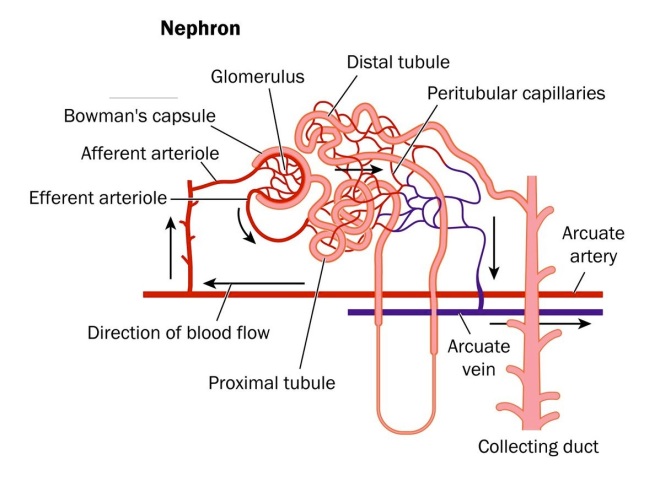
The tagging in this experiment is complicated, but quite technically brilliant. The kidney is composed of myriads of tiny functional units called nephrons. Each nephron is fed by a tiny knot of blood vessels called a glomerulus.  The structure of a nephron is shown below.  

Nephron-image

The blood supply to the kidney comes from branches off the descending aorta knows as renal arteries.  After entering the kidneys, the renal arteries branch multiple times until they become tiny vessels that feed into each nephron known as afferent arterioles.  The afferent arterioles forms a dense network of knot-like vessels that form the glomerulus and the portion of the nephron that interacts with the glomerulus is known as the Bowman’s capsule..  The blood vessels of the glomerulus are very special because they are exceptionally porous.  However, the Bowman’s capsule has a series of cells with foot-like extensions that coat the glomerulus called “podocytes.”  An especially beautiful picture of podocytes wrapped around a glomerular vessel is shown below.

normal-kidney-podocyte

The podocytes cover the pores of the glomerulus and only allows water and things dissolved in water through the pores.  Proteins do not make it through – they are too heavily charged.  Cells also do not make it through – they are too big.  But water, sodium ions, potassium ions, hydrogen ions, some drugs, metabolites, waste products, and things like that all make it through the podocyte-guarded pores.  For this reason, if you have excessive protein or some blood cells in your urine, it is usually an indication that something is wrong.  

Now, rest of the tubing attached to the nephron serve to reabsorb all the things you do not want to get rid of and not absorb all the things you do want to get rid of.  The amount of water you eliminate depends on your degree of hydration and is controlled by a hormone called antidiuretic hormone, which is release by the posterior lobe of your pituitary gland when you are dehydrated.  In the presence of ADH, the posterior tubing reabsorbs more water, and in lower concentrations of this hormone, it reabsorbs far less.  

Now that we know something about the kidney, here’s how Humphreys and others genetically marked the kidneys of their mice.  The sodium-dependent inorganic phosphate transporter (SLC34a1) is only expressed in mature proximal tubule cells.  Tetsuro Kusaba, the first author on this paper, and his colleagues inserted a CreERT2 cassette into this gene.  If you are lost at this point all you need to remember is this: the CreERT2 cassette is inserted into a gene that is ONLY expressed in specific kidney cells.  The Cre gene encodes a recombinase that clips out specific bits of DNA from a chromosome.  Kusaba and others crossed these engineered mice with another strain of mice that had the gene for a bright red dye inserted into another gene, but this dye could not be expressed because another piece of DNA was in the way.  When these hybrid mice were fed a drug called tamoxifen, it activated the expression of the Cre protein, but only in the proximal tubule cells of the kidney and this Cre protein clipped out the piece of DNA that was preventing the red dye gene from being expressed.  Therefore, these mice had a particular part of their nephrons, the proximal tubules glowing bright red.  This is a stroke of shear genius and it genetically marks these cells specifically and strongly.  

Next, Kusaba and colleagues used unilateral ischemia reperfusion injury (IRI) to damage the kidneys.  In IRI, the blood supply is stopped to one kidney but not the other for a short period of time (26 minutes).  This causes cell death and kidney damage.  The other kidney is not damaged and serves as a control for the experiment.  

Examination of the damaged kidneys showed that  red-glowing cells were found in areas other than the proximal tubules.  The only way these cells could have ended up in these places was if the differentiated cells divided and helped repair the damaged parts of the nephrons.  

Other research groups have seen similar results, but interpreted them as evidence of stem cell populations in the kidney.  However, Humphreys groups discovered something even more fascinating.  These “stem cell-markers” in the kidney are actually markers of kidney damage and regeneration and all cells in the kidney express them.  In Humphreys words, “What was really interesting is when we looked at the appearance and expression patterns of these differentiated cells, we found that they expressed the exact same ‘stem cell markers’ that these other groups claimed to find in their stem cell populations.  And so, if a differentiated cell is able to express a ‘stem cell marker’ after injury, then what our work shows is that that’s an injury marker – is doesn’t define a stem cell.”  

Indeed, several genes that have been taken to be signs of a kidney stem cell population (CD133, CD24, vimentin, and KIM-1) were expressed in red-glowing cells.  A stem cell population should not be fully differentiated and therefore, should not be able to express the red dye.  However, red-glowing cells clearly expressed these found genes after injury.  This rather definitely shows that it is the fully differentiated cells that are doing the regeneration in the kidney and not a resident stem cell population.  This does not prove that there is no resident stem cell population in the kidney, but only that the lion’s share of the regeneration is done by differentiated cells, and that under these conditions, no stem cell population was detected.  

This new interpretation of kidney repair suggests that cells can reprogram themselves in a way that resembles the way mature cells are chemically manipulated to revert to an induced pluripotent state.  

See Tetsuro Kusaba, Matthew Lalli, Rafael Kramann, Akio Kobayashi, and Benjamin D. Humphreys. Differentiated kidney epithelial cells repair injured proximal tubule. PNAS (October 14, 2013); doi:10.1073/pnas.1310653110.  

Scientists Generate “Mini-kidney” Structures from Human Stem Cells


Kidney Disease represents a major and unsolved health issue worldwide. Once damaged by disease, kidneys rarely recover their original level of function, and this highlights the urgent need for better knowledge of kidney development and physiology.

Now, a team of researchers led by scientists at the Salk Institute for Biological Studies has developed a novel platform to study kidney diseases. This new platform should open new avenues for the future application of regenerative medical strategies to restore kidney function.

For the first time, the Salk researchers have generated three-dimensional kidney structures from human stem cells. These findings were reported November 17, 2013 in Nature Cell Biology, and they suggest new ways to study the development and diseases of the kidneys and to discover and test new drugs that target human kidney cells.

Scientists had created precursors of kidney cells using stem cells as recently as this past summer, but the Salk team was the first to coax human stem cells into forming three-dimensional cellular structures similar to those found in our kidneys.

“Attempts to differentiate human stem cells into renal cells have had limited success,” says senior study author Juan Carlos Izpisua Belmonte, a professor in Salk’s Gene Expression Laboratory and holder of the Roger Guillemin Chair. “We have developed a simple and efficient method that allows for the differentiation of human stem cells into well-organized 3D structures of the ureteric bud (UB), which later develops into the collecting duct system.”

The Salk findings demonstrate for the first time that pluripotent stem cells capable of differentiating into the many cells and tissue types that make up the body can be induced to differentiate into those cells found in the ureteric bud, which is an early developmental structure of the kidneys. Furthermore, these same cells can differentiate further into three-dimensional structures in organ cultures. Ureteric bud cells form the early stages of the human urinary and reproductive organs during development and later develop into a conduit for urine drainage from the kidneys. Izpisua Belmonte’s research group accomplished this with both human embryonic stem cells and induced pluripotent stem cells (iPSCs), human cells from the skin that have been reprogrammed into their pluripotent state.

Kidney development

After generating iPSCs that demonstrated pluripotent properties and were able to differentiate into mesoderm, the embryonic germ cell layer from which the kidneys develop, the Salk Institute team used growth factors known to be essential during the natural development of our kidneys to culture both iPSCs and embryonic stem cells.  The combination of signals from these growth factors, molecules that guide the differentiation of stem cells into specific tissues, committed the cells to become progenitors that exhibit clear characteristics of renal cells in only four days.

The researchers then guided these cells to further differentiate into organ structures similar to those found in the ureteric bud by culturing them with kidney cells from mice. This demonstrated that the mouse cells were able to provide the appropriate developmental cues to allow human stem cells to form three-dimensional structures of the kidney.

Izpisua Belmonte’s team also tested their protocol on iPSCs from a patient clinically diagnosed with polycystic kidney disease (PKD), a genetic disorder characterized by multiple, fluid-filled cysts that can lead to decreased kidney function and kidney failure. They found that their methodology could produce kidney structures from patient-derived iPSCs.

Polycystic kidneys
Polycystic kidneys

Because of the many clinical manifestations of the disease, neither gene- nor antibody-based therapies are realistic approaches for treating PKD. The Salk team’s technique might help circumvent this obstacle and provide a reliable platform for pharmaceutical companies and other investigators studying drug-based therapeutics for PKD and other kidney diseases.

“Our differentiation strategies represent the cornerstone of disease modeling and drug discovery studies,” says lead study author Ignacio Sancho-Martinez, a research associate in Izpisua Belmonte’s laboratory. “Our observations will help guide future studies on the precise cellular implications that PKD might play in the context of kidney development.”

A New Way to Treat Kidney Disease and Heart Failure


St. Michael’s Hospital in Toronto, Ontario is the site of new research that uses bone marrow stem cells to treat chronic kidney disease and heart failure in rats.

Darren Yuen and Richard Gilbert of St. Michael’s Hospital were the first to show in 2010 that enriched stem cells improved heart and kidney functions in rats afflicted with both diseases. Their work generated concerns about the side effects of returning such stem cells to the body.

Since 2010, Yuen and Gilbert have found that enriched bone marrow stem cells secrete stromal cell–derived factor-1α (SDF-1α), a chemokine that is made by ischemic tissue but is rapidly degraded by dipeptidyl peptidase-4 (DPP-4), in culture dishes.  Injection of SDF-1α into rats has many of the same positive effects as when the stem cells themselves are injected into rats.  Even more remarkably, if a drug that inhibits the enzyme DPP-4 is given (sitagliptin) produced many improvements as well.

“We’ve shown that we can use these ‘hormones’ to replicate the beneficial effects of the stem cells in treating animals with chronic kidney disease and heart failure,” said Yuen, who practices as a nephrologist. “In our view, this is a significant advance for stem cell therapies because it gets around having to inject stem cells.”

Yuen said that they do not yet know what kind of hormone the cells are secreting, but identifying the hormone would be the first step toward the goal of developing a synthetic drug.

Chronic kidney disease (CKD) is much more prevalent than was once believed, with recent estimates suggesting that up to five percent of the Canadian population may be affected with this condition.

The number of people with CKD and end-stage renal failure is expected to rise as the population ages and more people develop Type 2 diabetes. People with kidney disease often develop heart disease, and many of them die from heart failure rather than kidney failure.

Regenerating Injured Kidneys with Exosomes from Human Umbilical Cord Mesenchymal Stem Cells


Zhou Y, Xu H, Xu W, Wang B, Wu H, Tao Y, Zhang B, Wang M, Mao F, Yan Y, Gao S, Gu H, Zhu W, Qian H: Exosomes released by human umbilical cord mesenchymal stem cells protect against cisplatin-induced renal oxidative stress and apoptosis in vivo and in vitro. Stem Cell Res Ther 2013, 4:34.

Ying Zhou and colleagues from Jiangsi University have provided helpful insights into how adult stem cell populations – in particular, mesenchymal stem cells (MSCs) isolated from human umbilical cord (hucMSCs) – are able to regulate tissue repair and regeneration. Adult stem cells, including MSCs from different sources, confer regenerative effects in animal models of disease and tissue injury. Many of these cells are also in phase I and II trials for limb ischemia, congestive heart failure, and acute myocardial infarction (Syed BA, Evans JB. Nat Rev Drug Discov 2013, 12:185–186).

Despite the documented healing capabilities of MSCs, in many cases, even though the implanted stem cells produce genuine, reproducible therapeutic effects, the presence of the transplanted stem cells in the regenerating tissue is not observed. These observations suggest that the predominant therapeutic effect of stem cells is conferred through the release of therapeutic factors. In fact, conditioned media from adult stem cell populations are able to improve ischemic damage to kidney and heart, which confirms the presence of factors released by stem cells in mediating tissue regeneration after injury (van Koppen A, et al., PLoS One 2012, 7:e38746; Timmers L, et al., Stem Cell Res 2007, 1:129–137). Additionally, the secretion of factors such as interleukin-10 (IL-10), indoleamine 2,3-dioxygenase (IDO), interleukin-1 receptor antagonist (IL-1Ra), transforming growth factor-beta 1 (TGF-β1), prostaglandin E2 (PGE2), and tumor necrosis factor-alpha-stimulated gene/protein 6 (TSG-6) has been implicated in conferring the anti-inflammatory effects of stem cells (Pittenger M: Cell Stem Cell 2009, 5:8–10). These observations cohere with the positive clinical effects of MSCs in treating Crohn’s disease and graft-versus-host disease (Caplan AI, Correa D. Cell Stem Cell 2011, 9:11–15).

Another stem cell population called muscle-derived stem/progenitor cells, which are related to MSCs, can also extend the life span of mice that have the equivalent of an aging disease called progeria. These muscle-derived stem/progenitor cells work through a paracrine mechanism (i.e. the release of locally acting substances from cells; see Lavasani M, et al., Nat Commun 2012, 3:608). However, it is unclear what factors released by functional stem cells are important for facilitating tissue regeneration after injury, disease, or aging and the precise mechanism through which these factors exert their effects. Recently, several groups have demonstrated the potent therapeutic activity of small vesicles called exosomes that are released by stem cells (Gatti S, et al., Nephrol Dial Transplant 2011, 26:1474–1483; Bruno S, et al., PLoS One 2012, 7:e33115; Lai RC, et al., Regen Med 2013, 8:197–209; Lee C, et al., Circulation 2012, 126:2601–2611; Li T, et al., Stem Cells Dev 2013, 22:845–854). Exosomes are a type of membrane vesicle with a diameter of 30 to 100 nm released by most cell types, including stem cells. They are formed by the inverse budding of the multivesicular bodies and are released from cells upon fusion of multivesicular bodies with the cell membrane (Stoorvogel W, et al., Traffic 2002, 3:321–330).

Exosomes are distinct from larger vesicles, termed ectosomes, which are released by shedding from the cell membrane. The protein content of exosomes depends on the cells that release them, but they tend to be enriched in certain molecules, including adhesion molecules, membrane trafficking molecules, cytoskeleton molecules, heat-shock proteins, cytoplasmic enzymes, and signal transduction proteins. Importantly, exosomes also contain functional mRNA and microRNA molecules. The role of exosomes in vivo is hypothesized to be for cell-to-cell communication, transferring proteins and RNAs between cells both locally and at a distance.

To examine the regenerative effects of MSCs derived from human umbilical cord, Zhou and colleagues used a rat model of acute kidney toxicity induced by treatment with the anti-cancer drug cisplatin. After treatment with cisplatin, rats show increases in blood urea nitrogen and creatinine levels (a sign of kidney dysfunction) and increases in apoptosis, necrosis, and oxidative stress in the kidney. If exosomes purified from hucMSCs, termed hucMSC-ex are injected underneath the renal capsule into the kidney, these indices of acute kidney injury decrease. In cell culture, huc-MSC-exs promote proliferation of rat renal tubular epithelial cells in culture. These results suggest that hucMSC-exs can reduce oxidative stress and programmed cell death, and promote proliferation. What is not clear is how these exosomes pull this off. Zhou and colleagues provide evidence that hucMSC-ex can reduce levels of the pro-death protein Bax and increase the pro-survival Bcl-2 protein levels in the kidney to increase cell survival and stimulate Erk1/2 to increase cell proliferation.

Another research group has reported roles for miRNAs and antioxidant proteins contained in stem cell-derived exosomes for repair of damaged renal and cardiac tissue (Cantaluppi V, et al., Kidney Int 2012, 82:412–427). In addition, MSC exosome-mediated delivery of glycolytic enzymes (the pathway that degrades sugar) to complement the ATP deficit in ischemic tissues was recently reported to play an important role in repairing the ischemic heart (Lai RC, et al., Stem Cell Res 2010, 4:214–222). Clearly, stem cell exosomes contain many factors, including proteins and microRNAs that can contribute to improving the pathology of damaged tissues.

The significance of the results of Zhou and colleagues and others is that stem cells may not need to be used clinically to treat diseased or injured tissue directly. Instead, exosomes released from the stem cells, which can be rapidly isolated by centrifugation, could be administered easily without the safety concerns of aberrant stem cell differentiation, transformation, or recognition by the immune system. Also, given that human umbilical cord exosomes are therapeutic in a rat model of acute kidney injury, it is likely that stem cell exosomes from a donor (allogeneic exosomes) would be effective in clinical studies without side effects.

These are fabulously interesting results, but Zhou and colleagues have also succeeded in raising several important questions. For example: What are the key pathways targeted by stem cell exosomes to regenerate injured renal and cardiac tissue? Are other tissues as susceptible to the therapeutic effects of stem cell exosomes? Do all stem cells release similar therapeutic vesicles, or do certain stem cells release vesicles targeting only specific tissue and regulate tissue-specific pathways? How can the therapeutic activity of stem cell exosomes be increased? What is the best source of therapeutic stem cell exosomes?

Despite these important remaining questions, the demonstration that hucMSCderived exosomes block oxidative stress, prevent cell death, and increase cell proliferation in the kidney makes stem cell-derived exosomes an attractive therapeutic alternative to stem cell transplantation.

See Dorronsoro and Robbins: Regenerating the injured kidney with human umbilical cord mesenchymal stem cell-derived exosomes. Stem Cell Research & Therapy 2013 4:39.

Radio Interview About my New Book


I was interviewed by the campus radio station (89.3 The Message) about my recently published book, The Stem Cell Epistles,

Stem Cell Epistles

It has been archived here. Enjoy.