BMP-2 Treatment Limits Infarct Size in After a Heart Attack in Mice


Bone Morphogen Protein 2 (BMP2) is a powerful signaling molecule that is made during development, healing, and other significant physiological events. During the development of the heart, BMP2 modulates the activation of cardiac genes. In culture, BMP2 can protect heart muscle cells from dying during serum starvation. Can BMP2 affect hearts that have just experienced a heart attack?

Scientists from the laboratories of Karl Werdan and Thomas Braun at the Max Planck Institute or Heart and Lung Research in Bad Nauheim, Germany have addressed this question in a publication in the journal Shock.

In this paper, Henning Ebelt and his colleagues Gave intravenous BMP2 to mice after a heart attack. CD-1 mice were subjected to LAD-ligation to induce a heart attack (LAD stands for left anterior descending coronary artery, which is tied shut to deprive the heart muscle of oxygen). 1 hour after the heart attack, mice were given 80 microgram / gram of body weight of intravenous recombinant BMP2. The hearts of some animals were removed 5-7 days after the heart attack, but others were examined 21 days after the heart attack to determine the physiological performance of the hearts. Control animals were given intravenous phosphate buffered saline.

Coronary arteries

The extirpated hearts were analyzed for cell death, and the size of their heart scars. Also, protein expression analyses showed the different proteins expressed in the heart muscle cells as a result of BMP2 treatment. Also, the effects of BMP2 on cultured heart muscle cells was ascertained.

The results showed that BMP2 could protect cultured heart muscle cells from dying in culture if they when they were exposed to hydrogen peroxide. Hydrogen peroxide mimics stressful conditions and under normal circumstances, cultured heart muscle cells pack up and die in the presence of hydrogen peroxide (200 micromolar for those who are interested). However, if cultured with 80 ng / mL BMP2, the survival of cultured heart muscle cells greatly increased.

When it came to the hearts of mice that were administered iv BMP2, the BMP2-administered mice survived better and had a smaller infarct size (almost 50% of the heart in the controls and less than 40% in the BMP2-administered hearts). When the degree of cell death was measured in the mouse hearts, those hearts from mice that were administered BMP2 showed less cell death (as determined by the TUNEL assay). BMP2 also increased the beat frequency and contractile performance of isolated heart muscle cells.

FInally, the physiological parameters of the BMP2-treated animals were slightly better than in the control animals. The improvements were consistent, but not overwhelming.

Interestingly, when the proteins made by the hearts of BMP2- and PBS-administered animals were analyzed, there were some definite surprises. BMP2 normally signals to cells by binding a two-part receptor that sticks phosphates on itself, and in doing so, recruits “SMAD” proteins to it that end up getting attached to them. The SMAD proteins with phosphates on them stick together and go to the nucleus where they activate gene expression.

BMP signaling

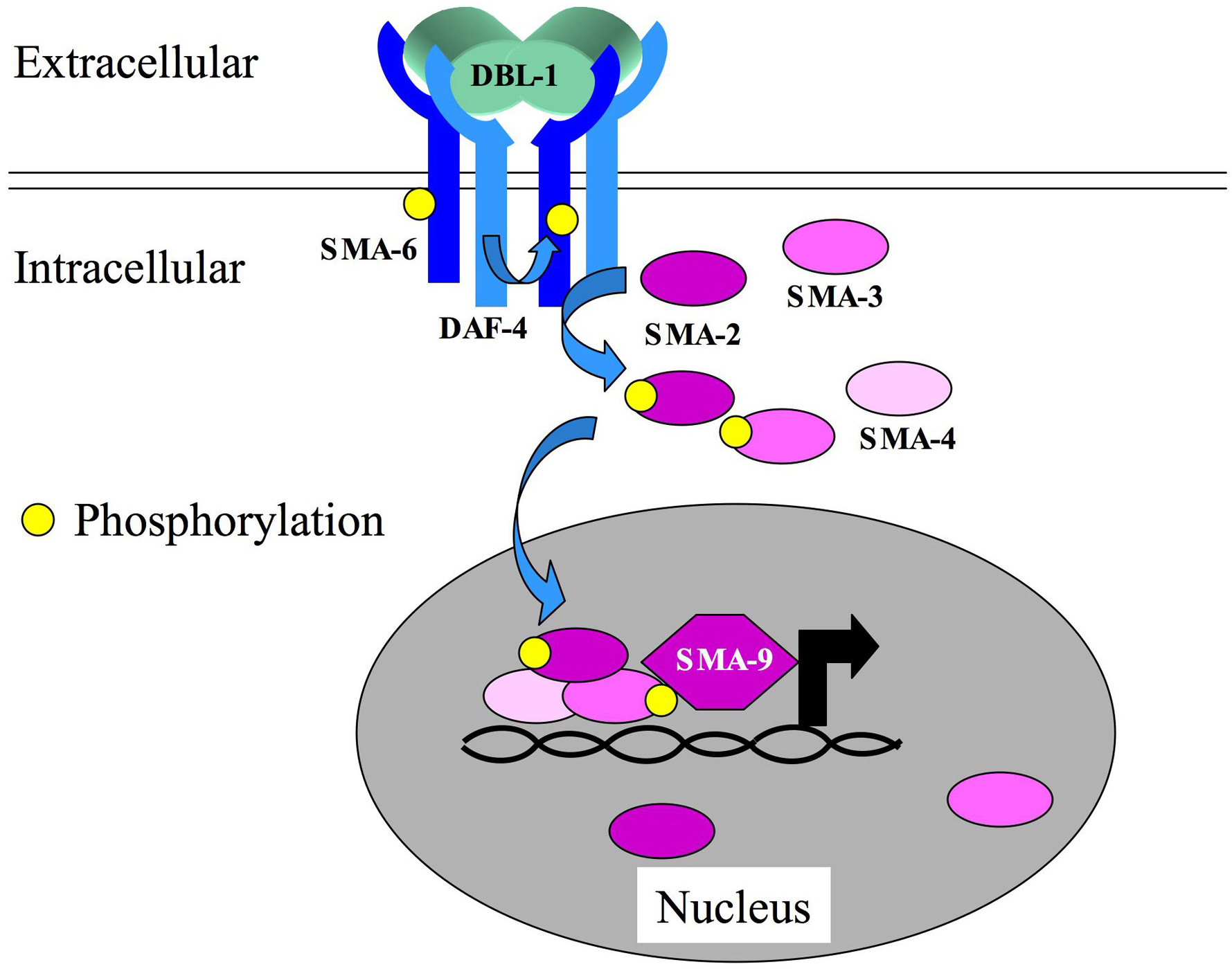
However, the heart muscle cells of the BMP2-administered mice did not contain heavily phosphorylated SMAD2, even though they did show phosphorylated SMAD1, 5, & 8.  I realize that this may sound like Greek to you, but it means this:  Different members of the BMP superfamily signal to cells by utilizing different combinations of phosphorylated SMADs.  The related signaling molecule, TGF-beta (transforming growth factor-beta), increases scar formation in the heart after a heart attack.  TGF-beta signals through SMAD2.  BMP2 does not signal through SMAD2, and therefore, elicits a distinct biological response than TGF-beta.

These results show that BMP2 administration after a heart attack decreases cell death and decreases the size of the heart scar.  There might be a clinical use for BMP2 administration after a heart attack.

See Henning Ebelt, et al., Shock 2013 Apr;39(4):353-60.

Major Clinical Trial Finds Bone Marrow Stem Cell Treatments Provide No Benefits After a Heart Attack


A large and very well designed and carefully controlled clinical trial known as TIME has failed to demonstrate any benefit for infusions of bone marrow stem cells into the heart 3-7 days after a heart attack.  This study comes on the heals of a similar clinical study known as LateTIME, which stands for Late Timing In Myocardial infarction Evaluation, and tested the effects of bone marrow stem cells infusions into the heart of heat attack patients 2-3 weeks after a heart attack.

LateTIME enrolled 87 heart attack patients, and harvested their bone marrow stem cells.  The stem cells were delivered into the hearts through the coronary arteries, but some received a placebo.  All patients had their ejection fractions measured, their heart wall motions in the damaged areas of the heart and outside the damaged areas and the size of their infarcts.  There were no significant changes in any of these characteristics after six months. Because another large clinical study known as the REPAIR-AMI study showed significant differences between heart attack patients that had received the placebo and those that had received bone marrow stem cells 3-7 days after a heart attack, this research group, known as the Cardiovascular Cell Therapy Research Network (CCTRN), sponsored by the National Institutes of Health, decided the test their bone marrow infusions at this same time frame.

TIME was similar in design to LateTIME.  This study enrolled 120 patients that had suffered a heart attack and all patients received either an infusion of 150 million bone marrow stem cells or a placebo within 12 hours of bone marrow aspiration and cell processing either 3 days after the heart attack to 7 days.  The researchers examined the changes in ejection fraction, movement of the heart wall, and the number of major adverse cardiovascular events plus the changes in the infarct size.

The results were resoundingly negative.  At 6 months after stem cell infusion, there was no significant increase in ejection fractions versus the placebo and no significant treatment effect on the function of the left ventricle in either the infarct or the border zones.  These findings were the same for those patients that received bone marrow stem cell infusions 3 days after their heart attack or 7 days after their heart attacks.  Fortunately, the incidence of major adverse events were rare among all treatment groups.

Despite the negative results for these clinical trials, there are a few silver linings.  First of all, the highly controlled nature of this trial sets a standard for all clinical trials to come.  A constant number of stem cells were delivered in every patient, and because the stem cells were delivered soon after they were harvested, there were no potential issues about bone marrow storage.

Jay Traverse, the lead author of this study, made this point about this trial:  “With this baseline now set, we can start to adjust some of the components of the protocol to grow and administer stem cell [sic] to find cases where the procedure may improve function.  For example, this therapy may work better in different population groups, or we might need to use new cell types or new methods of delivery.”

When one examines the data for this study, it is clear that some patients definitely improved dramatically, whereas others did not.  Below is a figure from the Traverse et al paper that shows individual patient’s heart function data 6 months after the stem cell infusions.

BMC indicates bone marrow mononuclear cell; MI, myocardial infarction.

From examining these data even cursorily, it is clear that some patients improved dramatically while others tanked.  Traverse is convinced that bone marrow stem cell infusions help some people, but not others (just like any other treatment).  He is convinced that by mining these data, he can begin to understand who these patients are who are helped by bone marrow stem cell transplants and who are not.  Also, the stem cells of these patients have been stored.  Hopefully, further work with them will help Traverse and his colleagues clarify what, if anything, about the bone marrow of these patients makes them more likely to help their patients and so on.

There are some possible explanations for these negative results.  Whereas the positive REPAIR-AMI used the rather labor-intensive Ficoll gradient protocols for isolating mononculear cells from bone marrow aspirates, the TIME trials used and automated system for collecting the bone marrow mononuclear cells.  Cells isolated by the automated system have neither been tested in an animal model of heart attacks, nor established as efficacious in a human study of heart disease.  Therefore, it is possible that the bone marrow used in this study was largely dead.  Secondly, the cell products were kept in a solution that had a heparin concentration that is known to inhibit the migratory properties of mononuclear cells (See Seeger et al., Circ Res 2012 111(7): 1385-94).  Therefore, there is a possibility that the bone marrow used in this study was no good.  Until the bone marrow stem cells collected by this method are confirmed to be efficacious, judgment must be suspended.